Research Article
Austin J Clin Ophthalmol. 2018; 5(1): 1085.
The Ocular Pathology of the Child: About 751 Cases in Teaching University Hospital of Brazzaville, Congo
Makita C*, Nganga Ngabou GCF, Koulimaya RC and Diatewa B
Department of Ophthalmology, Teaching University Hospital of Brazzaville, Congo
*Corresponding author: Chantal Makita, Department of Ophthalmology, Teaching University Hospital of Brazzaville, BP: 14626, Brazzaville-Congo, Congo
Received: January 25, 2018; Accepted: February 22, 2018; Published: March 01, 2018
Abstract
Background: The objective of this study is to determine the profile of ocular pathologies observed in children under 15 years old.
Methods: This retrospective study included4307children aged from 0 to 15 years old, consulted in Ophthalmology Department of Teaching University Hospital of Brazzaville (Congo) from January 2014 to December 2016.
Results: During this period, 751 children had unilateral or bilateral ocular involvement. They were 409 boys (54.5%) and 342 girls (45.5%) for a ratio of 1.19. The middle age was about 8.2±4.6 years (range: 6 days-15 years). The age group from 6 to 15 years was the most representative with a total of 591 children (78.7%) and 160 from 0 to 5 years (21.3%). The clinical symptoms involving a consultation were dominated by the decline in visual acuity (34.8%), followed by itching (20.9%), pain (10.9%), and redness (9.3%). The main pathologies were represented by ametropia (38.2%) among which astigmatism accounted for a prevalence of 29.9% of all types, followed by conjunctival pathology represented largely by 27.9% of tropical endemic limbo-conjunctivitis (LCET) and 14.4% of infectious conjunctivitis, orbitopalpebral diseases (7.4%) and corneal affections (2.8%). Binocular blindness was reaching 1.2% of children (n = 9), low vision accounted for 0.8% (n = 6).
Keywords: Child; Ametropia; Tropical Endemic Limbo-Conjunctivitis; Blindness
Background
The ocular pathology of the child occupies an important place in our daily activity, considering the considerable number of the children consultants and the handicap that it can involve on a visual system still immature. Some African studies report that the pathologies of the child are much diversified [1-3]. Vices of refraction are the most frequent, followed by conjunctival affections represented by endemic conjunctival limbo of the tropics, traumatisms and cataracts are classified as causes of childhood blindness [4-6]. Many of them are preventable and can be prevented and treated [7]. With the majority of children in school, neglected eye conditions can be the cause of poor school performance compromising their future. The World Health Organization estimates that the prevalence of childhood blindness is 0.3 to 1000 [8]. It is for this purpose that we undertook this work to determine the profile of ocular pathologies observed in children aged less than 15 years.
Methodology
Data sources
This descriptive and analytical study is conducted in Ophthalmology Department of Teaching University Hospital of Brazzaville in Congo over a period of 3 years, from January 2014 to December 2016. During this period, 4307 patient files were collected externally by three physicians. The inclusion criteria included the questioning which allowed to specify, age, sex, symptoms, antecedents, and ophthalmological examination made of an inspection, measurement of visual acuity by far when this was possible from anterior segment biomicroscopy, air tonometer tone, automatic cycloplegia refractometry and indirect ophthalmoscopy fundus with Volk 90 glass. A standard radiograph, an ultrasound, and a scanner were realized if possible. The exclusion criteria were incomplete files. A total of 751 children was selected, aged from 0 to 15 years, hospital frequency equal to 17.43.
Study variables
Low vision is defined by the WHO as inferior visual acuity. At 6/18 but greater than or equal to 3/60 in the best eye with the best correction. Binocular blindness has been defined as any child with visual acuity less than 3/60 of the best eye with correction [8].
Methods of data analysis
The analysis of the data was carried out with the software Epi info, version 2012. The p-values were considered as significant when the threshold of significance was less than or equal to 0.05.
Analysis and Results
During this period, 751 children aged 0-15 years presented ocular pathology. There were 409 boys (54.5%) and 342 girls (45.5%), sexratio equal to 1.19. The mean age was 8.±4.6 years (range: 6 days-15 years). The age group from 6 to 15 years was the most representative; the children aged 0 to 5 years were most found: 591 children (78.7%) and 160 children (21.3%) (Table 1).
    Tranches d’âge
      (ans)
    Féminin
    Masculin
    Total


    N
    (%)
    N
    (%)
    N
    (%)


    0- 5
    53
    (7,1)
    107
    (14,2)
    160
    (21,3)


    6- 10
    122
    (16,2)
    172
    (22,9)
    294
    (39,1)


    11-15
    167
    (22,2)
    130
    (17,4)
    297
    (39,6)


    Total
    342
    (45,5)
    409
    (54,5)
    751
    (100)
Table 1: Distribution by sex and age group.
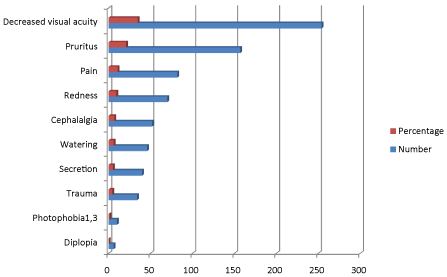
The clinical symptoms motivating the consultation were dominated by decreased visual acuity (34.8%), followed by pruritus (20.9%), pain (10.9%) and redness (9.3%) (Figure 1). It should be noted that a child could have multiple complaints and eye damage was uni or bilateral.

Figure 1: Reasons for consultation.
The main pathologies are shown in Table 2. Ametropia was present in 287 children (38.2%), including 162 boys (21.6%) and 125 girls (16.6%) (p<0.05). Astigmatism was found in 225 (29.9%) cases of all types, followed by myopia and hyperopia 42 cases (5.6%) and 20 cases (2.7%) respectively.
    Anatomical structures
    diseases
    Numbers
    Percentage


    Attachments
    LCET
      Conjunctivitis
      chalazion
      blepharitis
      Traumatic edema palpebral
      Orbital cellulitis
      Subconjunctival hemorrhage
      Palphaloplasty
      Bullous drug
      Pelvic foreign body
    210
      108
      16
      14
      8
      8
      5
      4
      4
      2
    27.9
      14.4
      2.1
      1.8
      1
      1
      0.6
      0.5
      0.5
      0.2


    Previous segment
    Corneal wound
      Cataract
      Corneal opacity
      keratitis
      uveitis
      Cornealdystrophy
      hyphema
    8
      8
      6
      5
      4
      2
      2
    1
      1
      0.8
      0.6
      0.5
      0.2
      0.2


    Posterior segment
    Tumor
      Glaucoma
      Opticneuropathy
    10
      7
      2
    1.3
      0.9
      0.2


    Other
    ametropia
      Strabismus
      Albinism
      Cortical blindness
      microphthalmos
      ICHT
      Normal ophthalmicexamination
    287
      14
      3
      2
      2
      1
      9
    38.2
      1.8
      0.4
      0.2
      0.2
      0.1
      1.1


    TOTAL

    751
    100
Table 2: Distribution by sex and age group.
Orbitopalpebral disorders were found in 56 cases (7.4%).It was about chalazion 16 cases (2.1%), blepharitis 12 cases (1.8%), traumatic edema palpebral 8 cases (1%), orbital cellulitis 8 cases (1%), 4 palpebral wounds (0.5%), 4 cases of bullous toxin (0.5%) and 2 cases of palpebral foreign bodies (0.2%).
The conjunctival pathology was composed of the LCET found in 210 cases (27.9%): 114 boys (15.1%) and 66 girls (8.8%) (p<0.05); the age range of 6-10 years with 112 cases (14.9%) was most frequent. Infectious conjunctivitis was found in 108 cases (14.4%) dominated by acute bacterial conjunctivitis 70 cases (9.3%), subconjunctival hemorrhage in 5 cases (0.6%).
We found 21 cases of corneal disorders (2.8%) made of keratitis in 5 cases (0.6%), corneal opacities 6 cases (0.8%), corneal wounds 8 cases (1%) and corneal dystrophy 2 cases (0.2%).
Among crystalline and uveal pathologies, we found: 6 cases of traumatic cataracts and 2 cases of bilateral congenital cataracts (1%); four cases of uveitis including 2 cases of indeterminate uveitis (0.2%), one case of traumatic uveitis and one case of uveitis in juvenile arthritis (0.1%).
Glaucoma was present in 7 cases (09%), including 5 juvenile cases and 2 congenital cases.
Retinoblastoma was found in 8 cases (1%), optic neuropathies 4 cases 0.5%, strabismus 14 cases 1.8% dominated by bilateral convergents (1.3%), microphthalmia found in embryofoetopathies 4 cases (0.5%), oculocutaneous albinism 3 cases (0.4%), cortical blindness 1 case (0.1%) as well as intracranial hypertension 1case (0.1%).
The frequency of binocular blindness was 9 cases (1.2%) and low vision 6 cases (0.8%). The causes of binocular blindness were bilateral congenital corneal dystrophy (2 cases); optic neuropathy (2 cases) retinoblastoma in its bilateral form (2 cases); bullous toxidermia (2 cases); cortical blindness after meningitis (1 case); while the low vision was due to bilateral congenital cataract (2 cases), embryofoetopathy (2 cases) and oculocutaneous albinism (2 cases).
Discussion
Ophthalmological conditions occurring in children occupy a significant place in daily practice. They interest all the tunics of the eye. Indeed, ametropia is the most common pathologies reported by most authors [1-3]. For some, these refractive errors constitute the first reason for ophthalmological consultation in schoolchildren [3,9]. The hospital frequency of ametropia of 38.2% observed in our environment is comparable to that found by Omgbwa in Cameroon (43.1%) [1]. the prevalence of refractive errors varies among studies. It is high for Ayed et al [2] in Tunisia and for Maul et al [10] in Chile respectively estimated at 57.2%, 56.3%. On the other hand, it is low, of 8.1% reported by Nepal et al [11] in Nepal and 10.6% by Sounouvou [12] in Benin. This disparity in frequency could be explained by ethnic and genetic factors rather than by environmental factors [2]. We did not note any predominance of sex as well as in Cameroon and Tunisia [1,2]. Astigmatism is the most common ametropia (29.9%), followed by myopia (5.6%) and hyperopia (2.7%). These results are comparable to those in Benin [12] where astigmatism dominates with a frequency of (91.9%) followed by myopia (4.5%) and hyperopia (3.6%).
LCET is the second pathology encountered. Its frequency is 27.9%, similar to that obtained in a previous study where it was estimated at 30.3% [13], comparable to that reported by KOKI et al. (31.5%) [14] in Cameroon and Chenge et al in Democratic Republic of Congo (32.9%) [15] for the same age group. This relative frequency is variously appreciated by some authors such as Dialloet al (80 to 90%) [16], Ayenaet al (12.2%) [17]. Several authors testify that the LCET is a disease of young children [1,13-17] and poses a real challenge for ophthalmologists in sub-Saharan regions because of relapses and sequelae.
Infectious conjunctivitis is present in 108 cases (14.4%), dominated by acute bacterial conjunctivitis with 70 cases (9.3%), those due to chlamydia trachomatis (n=8) and gonococcus (n=2).
Orbitopalpebral pathology included chalazion in the 6 to 15 age group, followed by eyelid trauma, blepharitis, and cellulitis. Omgbwa et al in Cameroon [1] reported a prevalence of 2.8% for chalazion and 1.9% for cellulite.
Corneal pathology was dominated by corneal wounds (1.3%). Mayouegoet al noted that the cornea was the predominant seat of traumatic lesions [18]. The opacities and corneal dystrophies in our series were due to measles lesions.
Cataract is an important cause of blindness and visual impairment [6,19]. She was more traumatic than congenital; even operated certain authors claim to be confronted with problems of amblyopia [20,21].
Uveitis was rare in our study (0.5%); Omgbwa et al in Cameroon found a frequency of 2.8% [1]. They testify that these with severe prognosis were related to post-traumatic endophthalmitis hence the need for early education of children on ocular trauma and promote their rapid and adequate management to reduce the sequelae.
Glaucomatous pathology was observed in 0.9% of cases with a predominance of juvenile glaucoma in 0.7%. BellaHiag in Cameroon in a larger series notes a frequency of 5.8% [22].
The strabismus (1.8%) in our series is also observed in 1.22% by EbanaMvogo [23]. Retinoblastoma is common as reported in a previous study in children aged 0 to 14 years with a frequency of 25.3% [24].
The main causes of binocular blindness were optic neuropathy, bilateral and congenital corneal dystrophy, retinoblastoma in its bilateral form, bullous toxidermia, and cortical blindness after meningitis. We found the same causes of binocular blindness as Omgbwa in Cameroon [1] apart from retinoblastoma. In some countries, such as Mongolia [25] and Malaysia [26], lens pathology remains the leading cause of blindness, with 35.6% and 23%, respectively. As for low vision, bilateral congenital cataract and albinism are the main causes found in our study as in that of Omgbwa in Cameroon [1].
Conclusion
The pathology of the child is very diverse in our environment. It is dominated by refractive errors, conjunctivitis with LCET as the lead. Traumatic pathology is not uncommon. The high prevalence of ametropia and LCET show the need for adequate management to improve the ocular health of the child.
References
- OmgbwaEballé A, Assupta Bella L, Owona D, Mbomé S, EbanaMvogo. La pathologie de l’enfant de 6 à 15 ans: étude hospitalière de Yaoundé. Cahiers Santé. 2009; 19: 61-66.
- Ayed T, Sokkah M, Charfi O, El matri Epidémiologie des erreurs réfractives chez des enfants scolarisés socio-économiquement défavorisés en Tunisie. J Fr Ophtalmol. 2002; 25: 712-717.
- OdoulamiYehouessi L, Tchiabi S, et al. La réfraction de l’enfant scolarisé au CNHU de Cotonou. Mali Méd. 2005; 2: 24-27.
- AdamaMensan, Fany A, Christiane Adjorlolo C, Toure ML, KassieuGbe M, Milhuedo KA, et al. Epidémiologie des traumatismes oculaires de l’enfant à Abidjan. Cahiers d’Etude de la Recherche Francophone. 2004; 14: 239-243.
- Ahnoux-Zabsonre AA, Keita C, Safede K. Traumatismes oculaires graves de l’enfant au Centre Hospitalier Universitaire de Cocody d’Abidjan en 1994. J Fr Ophtalmol. 1997; 11: 489-495.
- ParishitGogate, Clare Gilbert. La cécité infantile: panorama mondiale. Revue de Santé Oulaire Communautaire. 2008; 5: 37-39.
- Rahi JS, Gilbert CE, Foster A, Minassian D. Measuring the burden of childhood blindness. Br J Ohthalmol. 1999; 83: 387-388.
- World Health Organisation. Préventing blindness in children. Report of WHO/APB scientific meeting Hyderabad, India 13-17 April 1999 Geneva Switzerland. World Health Organisation; 2000.
- JeddiBlouza A, Lukil I, Mhenni A, Khayati L, Mallouche N, Zouari B. Prise en charge de l’hypermétropie de l’enfant. J FrOphtalmol. 2007; 30: 255-259.
- Maul E, Barrosso ST, Munoz SR, Sperduto RD, Ellwein LB. Refractive error study in children: results from La Florida, Chile. Am J Ophtalmol. 2000; 129: 445-454.
- Népal BP, Koirala S, Adhikary S, Sharma AK. Ocular morbidy in school childreen :results from Méchi Zone Népal. Am JOphtalmol. 2000; 129: 436-444.
- Sounouvou I, Tchiabi S, Doutetien C, Sonon F, Yehouessi L, Bassabi SK. Amétropies en milieu scolaire primaire à Cotonou (Bénin). J Fr Ophtalmol. 2008; 31: 771-775.
- Makita C, NgangaNgabou C, Diatewa B. La limbo-conjonctivite endémique des tropiques à propos de 558 cas. MédAfr Noire. 2015; 62: 609-613.
- Koki G, OmgbwaEballe A, Epée E, Njuenwet Njapdunke SB, SouleymanouWadjiri Y, Bella Asumpta L, et al. La limbo-conjonctivite endémique des tropiques au nord du Cameroun. J Fr Ophtalmol. 2011; 34: 113-117.
- Chenge B, Makumyuamviri AM, Kaimbo Wa Kaimbo D. La limbo-conjonctivite endémiques des tropiques à Lumbubashi, République Démocratique du Congo. Bull Soc Belge Ophtalmol. 2003; 290: 9-16.
- Diallo JS. La limbo-conjonctivite endémiques des tropiques. Rev IntTrachPathoOcul Trop Subtrop.1976; 3-4: 71-80.
- Ayena KD, Banla M, Agbo ADR, Ngeni OB, Balo K. Aspects épidémiologiques et cliniques de la limbo-conjonctivite endémique des tropiques en milieu rural au Togo. MédAfr Noire. 2008; 55: 319-324.
- MayouegoKouam J, Epée E, Azaria S, Enyama D, OmgbwaEballe A, EbanaMvogo C, et al. Aspects épidémiologiques cliniques et thérapeutiques des traumatismes oculaires de l’enfant dans un service d’urgences ophtalmologiques en Île-de-France. J FrOphtalmol. 2015; 38: 743-751.
- Robaei D, Ojami E, Kifley A, Huynh S, Mitcheli P. Visual acuity and the causes of visual loss in a population- based sample of 6 year-old Australian children. Ophtalmology. 2005; 112: 1275-1282.
- Deby F, Kodjkian L, Roche O, Donate D, Kouassi N, BurillanC,Denis P. Traumatismes oculaires perforants de l’enfant : étude rétrospective de 57cas. J FrOphtalmol. 2006; 29: 20-23.
- Thompson CG, Kumar N, Bilson FA, Martin F. The aetiology of perforating ocular injuries in children’s. Br Ophtalmol. 2002; 86: 920-922.
- Bella Hiag AL, EbanaMvogo C, Ngosso A, Ellong A. Etude de la pression intraoculaire dans une population des jeunes camerounais. J Fr Ophtalmol. 1996; 10: 585-590.
- EbanaMvogo C, Bella Hiag AL, Epesse M. Le strabisme au Cameroun. J Fr Ophtalmol. 1996; 19: 705-709.
- NsondéMalanda J, MakitaBagamboula C, Bambara TA, Makosso E, GombéMbalawa C. Le traitement du rétinoblastome au CHU de Brazzaville. Carcinologie Clinique en Afrique. 2011; 10: 34-39.
- Bulgan T, Gilbert CE. Prévalence and cause of severe visual impairment and blindness in children in mongolia. OphtalmicEpidémiolol. 2002; 9: 271-281.
- Meddy SC, Tan BC. Causes of childhood blindness in Malaysia: result from a national study of blind school students. IntOphthalmol. 2002; 24: 53-59.