
Research Article
Austin J Clin Ophthalmol. 2018; 5(2): 1092.
Age and Gender Related Changes in Intraocular Pressure among Patients Attending a Peripheral Eye Clinic in Port Harcourt, Nigeria
Ejimadu CS¹, Chinawa NE² and Fiebai B¹*
¹University of Port Harcourt Teaching Hospital, Nigeria
²Mercy Eye Hospital, Abak, Nigeria
*Corresponding author: Fiebai B, Department of Ophthalmology, University of Port Harcourt Teaching Hospital, Port Harcourt, Rivers State, Nigeria
Received: March 20, 2018; Accepted: April 17, 2018; Published: April 24, 2018
Abstract
Purpose: To determine the age and gender related changes in intraocular pressure among patients attending a private eye clinic in Port Harcourt Nigeria.
Methods: This was a cross sectional study done in a private hospital in Port Harcourt. The first one hundred patients attending the hospital in 2013 and met the inclusion criteria were recruited for the study. Those with corneal diseases/ lesions were excluded. Age and sex were obtained from the patient’s history while intraocular pressure (IOP) was measured using Perkins applanation tonometer (MK2 Model). Intraocular measurements were taken between 9am and 12pm.
Results: There were 100 subjects in this study comprising 59 males and 41 females (M: F=3:2). The mean age was 45.83±20.43years. The mean IOP was 16.18±6.13 for right eye and 16.82±8.22 mmHg for left eye.
IOP was higher in right and left male eyes compared to females but these were not statistically significant (p-value = 0.133 for right eye and; p-value = 0.267 for left eye). There was a weak positive correlation between right eye IOP and age (r=0142; p-value=0.174) and left eye IOP and age (r=0.202; p-value=0.0505). There was fluctuation of IOP in different age groups.
Conclusions: There was no statistical significance difference in IOP in both genders. There is a weak positive correlation between IOP and age.
Keywords: Age; Gender; Glaucoma; Intra Ocular Pressure
Abbreviations
ANOVA: Analysis of Variance; IOP: Intraocular pressure; S.D: Standard deviation
Introduction
Intraocular pressure (IOP) is the fluid pressure inside the eye. Elevated intraocular pressure is a major risk factor for the development of primary open-angle glaucoma [1], and even in normal tension glaucoma the reduction of IOP may slow the progression of visual field loss [2]. It is the only proven treatable risk factor. People with a high IOP with no proof of having primary open-angle glaucoma are considered at risk of developing optic nerve damage, even if they do not suffer from any ocular disease [3].
Numerous factors have been known to influence IOP. These include age and sex. IOP distribution and associated ocular features and its correlation with age are of clinical interest. The relationship between IOP and age varies in different ethnicities. Studies conducted in Western countries [4,5] Iran [6], and Barbados [7] show a positive correlation between IOP and age. On the contrary, most of the East Asia studies reported a negative correlation between IOP and increasing age [8,9]. Other studies show no association [10,11].
Sex-related differences in the distribution of IOP and its changes with age have also been inconsistent across studies. In a study [12] by Mohammed J et al there was a highly significant difference between the mean IOP in males (15.2mmHg) and that in females (16.5mmHg) with the SD of ± 2.43 and ± 3.28 respectively. There was no significant difference in age between the male and female group in this study. Similar association was found in the Barbados Eye study [13], the Rotterdam study [14], the Los Angeles Latino Eye Study [15], and the Beaver Dam Eye Study [16], where men had lower IOP. On the contrary higher IOP was reported for men in the Egna-Neumarkt [17] and the Gutenberg Health [18] studies while the Framingham Eye study [19] and the Health and Nutrition Examination Survey [20] reported no association between sex and IOP.
Methodology
This was a cross sectional study done in a private hospital in Port Harcourt. The first one hundred patients attending the hospital in 2013 and met the inclusion criteria were recruited for the study. Those with corneal disorders were excluded. Informed consent was obtained and the whole exercise followed the tenet of declaration of Helsinki. Age and sex were obtained from the patient’s history while intraocular pressure was measured using Perkins applanation tonometer (MK2 Model). Three readings were taken between 9am and 12pm and the patient’s average value calculated. Patients were in sitting position and had their eye anaesthetized with topical anesthetic agent (1% tetracaine) and then 2% fluorescein dye was instilled before taking the pressures. Intraocular measurements were taken between 9am and 12pm.
Sample size calculation
The minimum sample size was calculated using the formula for quantitative variables. Based on the 95% significance level, an IOP standard deviation of 2.69 from a similar Nigerian study and a precision of 0.55, a minimum sample size of 91 was obtained. However, the study comprised of 100 respondents and a total of 200 eyes were examined.
Statistical analysis
The Statistical Package of Social Sciences (SPSS) version 20 was used for analysis. Descriptive statistics involved means and standard deviation for numerical variables while frequencies and proportions were used for categorical variables. A paired t test was used to compare the differences in mean IOP across left and right eyes. Independent t test was used to compare the differences in mean IOP across two independent categories e.g. gender while Analysis of Variance (ANOVA)/F-test was used for the comparisons across more than two independent categories. Pearson’s correlation coefficient and simple linear regression analysis was used in examining relationship between age and IOP. Statistical significance was set at 0.05.
![]()
    Variables (N=100)
    Frequency
    Percentage


    Age category


    <20 years
    12
    12.0


    20 – 29 years
    6
    6.0


    30 – 39 years
    18
    18.0


    40 – 49 years
    22
    22.0


    50 – 59 years
    15
    15.0


    =60 years
    27
    27.0


    Mean age ± S.D = 45.83±20.43years
      Range = 8 – 100 years


    Sex


    Male
    59
    59.0


    Female
    41
    41.0


    S.D: Standard deviation.
Table 1: Age and sex distribution of respondents of the study.
![]()
    VA (N=100)
    n
    %


    Right eye


    Normal (better than 6/18)
    39
    39.0


    Mild (6/18)
    16
    16.0


    Moderate (6/24, 6/36, 6/60)
    32
    32.0


    Severe (3/60)
    1
    1.0


    Blindness (worse than 3/60)
    12
    12.0


    Left eye


    Normal (better than 6/18)
    36
    36.0


    Mild (6/18)
    22
    22.0


    Moderate (6/24, 6/36, 6/60)
    31
    31.0


    Severe (3/60)
    0
    0.0


    Blindness (worse than 3/60)
    11
    11.0
Table 2: Presentation of right and left eye VA of respondents.
![]()
    Right Eye
    Left Eye


    Mean ± S.D
    Mean ± S.D
    t*
    P-value


    Intra Ocular Pressure (mmHg)
    16.18±6.13
    16.82±8.22
    0.768
    0.444
Table 3: Mean IOP in right and left eye of respondents.
Result
There were 100 subjects in this study comprising 59 males and 41 females (M:F=3:2). The mean age was 45.83±20.43 years. The mean IOP was 16.18±6.13 for right eye and 16.82±8.22 mmHg for left eye.
IOP was higher in right and left male eyes compared to females but these were not statistically significant (p-value = 0.133 for right eye and; p-value = 0.267 for left eye). There was a weak positive correlation between right eye IOP and age (r=0142; p-value=0.174) and left eye IOP and age (r=0.202; p-value=0.0505). There was fluctuation of IOP in different age groups.
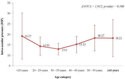
Figure 1: Intra ocular pressure of Right eye across age categories.
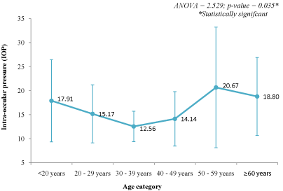
Figure 2: Intraocular pressure in the Left eye across age categories.
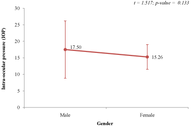
Figure 3: Intraocular pressure in the Right eye of male and females.
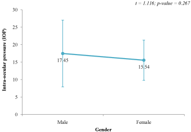
Figure 4: Intraocular pressure in the left eye of males and females.
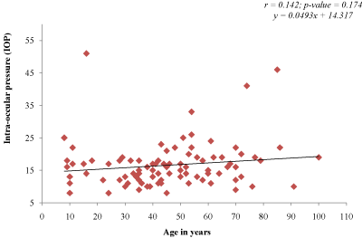
Figure 5: Correlation between IOP and age in the Right eye.
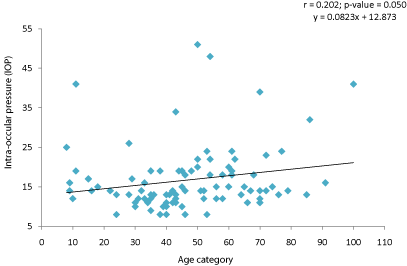
Figure 6: Correlation between IOP and age in the Left eye.
Discussion
Glaucoma is the commonest cause of irreversible blindness globally and in Nigeria. Several risk factors have been attributed to Glaucoma but of all these, the only modifiable risk factor is intra ocular pressure. It becomes necessary to study age and gender factors as they relate to intraocular pressure in our environment.
The mean IOP of 16.18±6.13 in males and 16.82±8.22 was within the normal range for the general population. However, it was observed that 12% of the right eyes and 11% of the left eyes were already blind at presentation. This depicts the poor health seeking behavior in our environment such that patients present late for medical attention. There was no statistical significant difference in the number of male and female patients. This could be due to increased awareness and education bearing in mind that this hospital was in the city thus the effect of cultural barrier and illiteracy were minimal.
Several studies have shown conflicting results in the association of age and intraocular pressure. While some found a positive association [4-7]. Others found a negative association [8,9] while some found no association [10,11]. From our study, there was a weak positive correlation between age and intraocular pressure. However, there was a statically significant difference across different age groups with a decline from ages <20-39, an increase from 40-59 and a slight decline beyond 60 years. Aging is associated both with reduced production of aqueous humor [21], which leads to a reduction of IOP, and with structural changes in the trabecular meshwork, which increase the resistance to aqueous humor outflow, increasing IOP [22]. The net change in IOP may be determined by the balance between these processes, which may differ in different ethnicities and across age groups.
There was an increase in IOP in males than females though this increase is not statistically significant. Several studies have shown conflicting results; while some showed higher IOP in males [17,18] others showed higher values in females [12-16] and some showed no association [19-20]. It has been hypothesized that the higher IOP in men could be due to a higher prevalence of cardiovascular risk factors in men [15,18]. Hormonal differences and the effect of menopause may also explain some gender differences in IOP [23]. Estrogen may affect the inflow of aqueous humor, the ciliary body, and the trabecular meshwork [24]. An Indian study showed that the IOP in postmenopausal women was higher compared with premenopausal women and attributed this difference to the higher levels of testosterone and the decrease in estrogen and progesterone levels with the onset of menopause [25].
Conclusion
There was no statistical significance difference in IOP in both genders. There is a weak positive correlation between IOP and age.
References
- Leske MC. The epidemiology of open-angle glaucoma: a review. Am J Epidemiol. 1983; 118: 166–191.
- Bonomi L, Marchini G, Marraffa M. Prevalence of glaucoma and intraocular pressure distribution in a defined population. The Egna-Neumarkt Study. Ophthalmology. 1998; 105: 209–215.
- Sanaa AY, Elham RA. Age, gender and refractive error association with intraocular pressure in healthy Saudi participants: A cross-sectional study. Saudi J Ophthalmol. 2016; 30: 44–48.
- Al-Baghli NA, Al-Ghamdi AJ, Al-Turki KA, El-Zubaier AG. Hypertension in the eastern province of Saudi Arabia: results of a screening campaign. J Fam Community Med. 2008; 15: 95–101.
- Klein BEK, Klein R, Linton KLP. Intraocular pressure in an American community: the Beaver Dam eye study. Invest Ophthalmol Vis Sci. 1992; 33: 2224–2228.
- Hashemi H, Kashi AH, Fotouhi A, Mohammad K. Distribution of intraocular pressure in healthy Iranian individuals: the Tehran eye study. Br J Ophthalmol. 2005; 89: 652–657.
- Wu S, Leske MC. Associations with intraocular pressure in the Barbados eye study. Arch Ophthalmol. 1997; 115: 1572–1576.
- Shiose Y, Kawase Y. A new approach to stratified normal intraocular pressure in a general population. Am J Ophthalmol. 1986; 101: 714–721.
- Nomura H, Shimokata H, Ando F, Miyake Y, Kuzuya F. Age-related changes in intraocular pressure in a large Japanese population: a cross-sectional and longitudinal study. Ophthalmology. 1999; 106: 2016–2022.
- Rochtchina E, Mitchell P, Wang JJ. Relationship between age and intraocular pressure: the Blue Mountains Eye Study. Clin Experiment Ophthalmol. 2002; 30: 173–175.
- Weih LM, Mukesh BN, McCarty CA, Taylor HR. Association of demographic, familial, medical, and ocular factors with intraocular pressure. Arch Ophthalmol. 2001; 119: 875–880.
- Jeelani M, Taklikar RH, Taklikar A, Itagi V, Bennal AS. Variation of intraocular pressure with age and gender. Natl J Physiol Pharm Pharmacol. 2014; 4: 57-60.
- Leske MC, Connell AM, Wu SY, Hyman L, Schachat AP. Distribution of intraocular pressure. The Barbados Eye Study. Arch Ophthalmol . 1997; 115: 1051–1057.
- Dielemans I, Vingerling JR, Algra D, HofmanA, Grobbee DE, de Jong PT. Primary open-angle glaucoma, intraocular pressure, and systemic blood pressure in the general elderly population. The Rotterdam Study. Ophthalmology. 1995; 102: 54–60.
- Memarzadeh F, Ying-Lai M, Chung J, Azen SP, Varma R. Blood pressure, perfusion pressure, and open-angle glaucoma: the Los Angeles Latino Eye Study. Invest Ophthalmol Vis Sci. 2010; 51: 2872–2877.
- Klein BE, Klein R, Linton KL. Intraocular pressure in an American community. The Beaver Dam Eye Study. Invest Ophthalmol Vis Sci. 1992; 33: 2224–2228.
- Bonomi L, Marchini G, Marraffa M. Prevalence of glaucoma and intraocular pressure distribution in a defined population. The Egna-Neumarkt Study. Ophthalmology. 1998; 105: 209–215.
- Hoehn R, Mirshahi A, Hoffmann EM. Distribution of intraocular pressure and its association with ocular features and cardiovascular risk factors: The Gutenberg Health Study. Ophthalmology. 2013; 120: 961–968.
- Kahn HA, Leibowitz HM, Ganley JP. The Framingham Eye Study. I. Outline and major prevalence findings. Am J Epidemiol. 1977; 106: 17–32.
- Hiller R, Sperduto RD, Krueger DE. Race, iris pigmentation, and intraocular pressure. Am J Epidemiol. 1982; 115: 674–683.
- Brubaker RF, Nagataki S, Townsend DJ, Burns RR, Higgins RG, Wentworth W. The effect of age on aqueous humor formation in man. Ophthalmology. 1981; 88: 283–288.
- Miyazaki M, Segawa K, Urakawa Y. Age-related changes in the trabecular meshwork of the normal human eye. Jpn J Ophthalmol. 1987; 31: 558–569.
- Qureshi IA. Intraocular pressure: a comparative analysis in two sexes. Clin Physiol. 1997; 17: 247–255.
- Gupta PD, Johar K, Sr Nagpal K, Vasavada AR. Sex hormone receptors in the human eye. Surv Ophthalmol. 2005; 50: 274–284.
- Panchami, Pai SR, Shenoy JP, Shivakumar J, Kole SB. Postmenopausal intraocular pressure changes in South Indian females. J Clin Diagn Res. 2013; 7: 1322–1324.