
Research Article
Austin J Clin Ophthalmol. 2019; 6(2): 1105.
Comparative and Prospective Study of Kerato-Allergic Conjunctivitis Associated with Keratoconus
Bencharki Y*, Bouzoubaa T, Tamym B, Salek H, Baddou T and Berraho A
Service d’Ophtalmologie B, Hôpital Des Spécialités de Rabat, Morocco
*Corresponding author: Bencharki Y, Service d’Ophtalmologie B, Hôpital Des Spécialités de Rabat, Morocco
Received: October 02, 2019; Accepted: November 01, 2019; Published: November 08, 2019
Abstract
Introduction: Chronic ocular friction during allergic keratoconjunctivitis in particular vernals could be one of the most important environmental factors that lead to development of keratoconus on genetically predisposed sites, severity and progression of keratoconus has been reported in these patients as well as earlier surgery.
The main goal of our work is to conduct a comparative and prospective study between allergic keratoconjunctivitis associated with keratoconus on a series of 60 patients followed at the Ophthalmology B department of the Rabat Specialty Hospital.
Materials and Methods: Our comparative and prospective study focused on 120 eyes of 60 patients with keratoconus associated with allergic peranual conjunctivitis or a vernal keratoconjunctivitis versus a control group presenting a keratoconus without ocular allergy, consultant of their own for a decrease in visual acuity or symptoms related to their allergic conjunctivitis, keratoconus have been confirmed by the realization of a corneal topography and followed in the Ophthalmology B department at Rabat Speciality Hospital over a period of 24 months ranging from September 2016 to August 2018.
Results: Our study has highlighted the role of allergic keratoconjunctivitis on the progression and severity of Keratoconus in particular during KCV with more advanced and severe Keratoconus. Indeed, in our study, in patients with vernal keratoconjunctivitis we find a predominance of the male sex in younger subjects with a mean age of our patients with this association of 12 years with respectively 07 and 05 years younger than those who had an allergy-free Keratoconus and those with the combination perenual allergic conjunctivitis and keratoconus. KCVs also had more severe keratoconus, depending on the median of spherical equivalent which was significantly higher in group B (Vernal Keratoconjunctivitis and Keratoconus) than the other 02 groups, Moreover there was no significant difference in the value of the cylinder according to the groups, the better corrected visual acuity was significantly lower, corneal thickness thinner central and higher IOP frequency in group B compared to group A and C. The values of keratometry were significantly higher in the group B compared to groups A and C with a proportion of 62.5% of stage Keratoconus 4 according to Amsler Krumeich’s classification for group B, compared to the group A and C with respectively 19.3% and 18.2% of stage 4.
Conclusion: The association between different forms of ocular allergy and the Keratoconus have been frequently found in recent years by many authors. In our study, grade 4 keratoconus was found significantly more important in the KCV than in the other 02 groups.
Our study has highlighted the role of allergic conjunctivitis Peranuelle and Vernal Keratoconjunctivitis on the progression and severity of Keratoconus especially during the KCV with Keratoconus more evolved and more severe than this with regard to the visual acuity of the patients, their keratometric values, the weak central corneal thickness that sometimes prevents the achievement of physical therapy such as cross-linking and finally high IOP values mostly secondary to uncontrolled and abusive use of local corticosteroid therapy in these patients. It is crucial to explain to patients with a risk the importance of no longer rubbing their eyes. This simple prescription could have the potential to decrease the incidence of this pathology and slow down its progression alone or in combination with other therapies that have shown their effectiveness in recent years as the reticulation of corneal collagen.
Keywords: Keratoconus; Vernal Conjunctivitis; Cornea; Allergy
Introduction
Allergic conjunctivitis includes a wide variety of symptoms, ranging from simple itching to more severe eye pruritus, particularly during Vernal Keratoconjunctivitis (VKC). Keratoconus on the other hand is an evolutionary bilateral disorder, non-inflammatory, associated with thinning and corneal protrusion inducing myopia and irregular astigmatism especially during puberty.
The pathogenesis commonly proposed includes the release of inflammatory mediators due to ocular friction induced by pruritus that can alter corneal collagen and lead to corneal ectasia. Tissue damage could be due to chronic epithelial trauma resulting from prolonged and slow release of Metalloproteinases (MMP) [1,2]. His association with the different types of allergic conjunctivitis especially vernal keratoconjunctivitis is common. The incidence of Keratoconus is estimated to be between 50 and 230 per 100 000 population and its prevalence at 54.5 per 100,000 (about 1/2000) [3] in Caucasian populations these figures are probably underestimated because of the subclinical forms.
Chronic ocular friction could be an important environmental factor that leads to the development of keratoconus on genetically predisposed sites. Some forms of ocular allergies, especially vernal keratoconjunctivitis, are difficult to manage, and needs a prolonged use of local corticosteroid therapy, with all the associated iatrogenic complications, the use of Ciclosporine remains limited in our context because of its lower availability and its high cost. [4] In atopic patients, severity and faster progression of keratoconus have been reported as well as earlier use of surgery.
The main goal of our work is to conduct a comparative and prospective study between allergic kerato-conjunctivitis associated with keratoconus on a series of 60 patients.
Material and Methods
Our comparative and prospective study focused on 120 eyes of 60 patients with keratoconus associated with allergic peranual conjunctivitis (group A) or vernal keratoconjunctivitis (group B) versus a control group (group C) with keratoconus without ocular allergy, the follow up was performed in our department over a period of 24 months ranging from September 2016 to August 2018.
The diagnosis of VKC was confirmed by the presence of severe pruritus resistant to medical treatment, the presence of giant papillae at the eversion of the upper eyelids, the conjunctival hyperemia and significant and permanent photophobia, in some cases the presence of Trantas nodules on limb or limbic infiltrates. VKCs have been classified according to Cameron’s classification in limbic, palpebral or mixed form. The diagnosis of Keratoconus was made by biomicroscopic examination in search of Munson’s sign revealing a corneal protrusion, Vogt’s streaks, Fleischer or corneal opacities when these signs were obvious or by the realization of a PENTACAM corneal topography with measurement of central corneal thickness highlighting corneal thinning with high Keratometry values (›47 D) as well as on maps of especially posterior elevation in favor of the diagnosis of Keratoconus. We found the best corrected visual acuity comparatively in the 03 groups and converted to a logarithm (log Mar) and measuring IOP as a function of the central corneal thickness according to the IOP Converter application or by using the formula: Corrected PIO: Measured IOP - (CCT-545)/50 SPSS statistical software software version 11.5 was used.
We studied the following parameters: age, sex, associated factors, existence or family history, the uni or bilateral character of the infringement, the motive for consultation, the clinical examination, in particular the best corrected visual acuity, the eversion of eyelids looking for moderate or giant tarsal papillae, the detailed examination of the of the remainder of the anterior and systematic segment of the posterior pole, the characteristics keratoconus clinics, pachymetriccorrected eye tone measurement CCT (corneal central thickness), values of keratometry complementary examinations particularly Corneal Topography (PENTACAM), medical treatment, optics, physical therapy (Cross - Linking), surgical procedures, and short and long term follow-up (Figure 1 and 2).
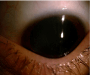
Figure 1: Patient of our series presenting a sign of Munsen.
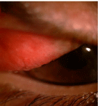
    Figure 2: Vernal keratoconjunctivitis associated with a Keratoconus Stage 4
with giant papillae at the eversion of the upper eyelid.
The exclusion factors were: patients with a history of trauma, patients with associated retinal pathologies, non-exploitable files and patients lost to follow-up.
Results
Median age of presentation significantly younger in group B (Keratoconjunctivitis associated with a Keratoconus) compared to other groups) ‹0.001). Significant age difference also between the group A (Keratoconus alone without associated allergy) and C (Perennial allergic conjunctivitis associated with a Keratoconus). On the other hand there was a slight male predominance of 56%. The sex ratio was 0.78. The majority of our patients were of low socioeconomic status (88%), with only a minority of the average socio-economic level (12%). 38% of our patients were children of school age, 36% of high school students and 26% of students. The decline in visual acuity was the cause of school failure.
regarding eye antecedents, Allergic conjunctivitis was present in 40 patients ( 66.6% of cases).In the context of allergic conjunctivitis, 02 patients developed cortisone glaucoma (3.3% of cases) by selfmedication with corticosteroids and corticosteroid cataract was present in 01 patients (1.6%), 02 patients were already grafted in one eye (3.3%). Therefore an associated general pathology was present in 31% of patients as general allergy present in 66.6% of patients: Rhinitis in 80% cases, 18% of asthma and 02% of association urticaria and eczema. Diabetes was found in 1.6% of cases. Moreover, the analysis found 16.6% of known familial cases (10 patients), particularly in the siblings.
Keratoconus was bilateral in 47 patients or 78.3% and 13 patients had unilateral infringement of 21.6%. At the time of the first consultation, 12.5% of the eyes had lower visual acuity equal to 1/10, 22 patients had no correction, 36 patients were wearing glasses and 2 patients were already wearing rigid contact lenses. Regarding refraction, myopic astigmatism was found in 75% of eyes, its value was -3.17 diopters.
For the spherical equivalent, it was significantly higher in group B (Vernal Keratoconjunctivitis and Keratoconus) than the other 02 groups (= 0.003). In addition, there was no significant difference in the value of the cylinder according to the groups and the median of the best visual acuity was significantly lower (= 0.013) in group B. The values of keratometry were significantly lower (‹0.001) in groups A and C with respect to group B.
At the examination of eyelids and conjunctiva 66.6% of patients had tarsal papillae on eyelid eversion, 33.3%of our patients had a vernal keratoconjunctivitis with taste buds 2% had vernal plaques. At the corneal level, the following abnormalities were noted: corneal hydrops in 04 eyes (6.6%), Fleischer ring in one patient (0.8%), Vogt streaks in 03 eyes (2.5%) and subepithelial opacities in 06 eyes (5%), and also Nodules of Trantas in limb in 04 eyes (20%).
The mean ocular tone of our patients was 14mmHg, ranging from 10 to 24. There was no significant difference in IOP between the groups (= 0.44), however, 04 patients (6.6%) had corrected IOP greater than 21mmHg in group B.
Moreover there was no significant difference in the value of the cylinder according to groups, the best corrected visual acuity was significantly lower, thinner central corneal thickness and a frequency of IOP higher in group B compared to group A and C. keratometry values were significantly higher in the group B with a proportion of 62.5% of stage 4 keratoconus according to the Amsler Krumeich classification Against 19.3% and 18.4% for the group A and C respectively.
Discussion
The association between different forms of ocular alergy and keratoconus has been frequently found in recent years. The study conducted by The Dundee University scottish keratoconus [6,7] which is a prospective study of 200 cases of patients suffering from keratoconus has found pathologies related to atopy including asthma in 23% of cases. eczema in 14% of cases and high fever in 30% of cases. An Indian study by Agrawal et al [8], evaluated 274 patients with keratoconus and revealed a high prevalence of allergy in these patients.
In our study grade 4 keratoconus was found more significantly in the KCVs than in the other 2 groups. All of these elements suggest that these patients should be considered as a group to share for better support. In fact, keratoconus is not a dystrophy, but an affection of mechanical origin. However, our study has certain limits, including eye rubs in children who cannot be seen by their parents; the difficulty of tracking these patients and the dangers of self-medication and the misuse of corticosteroids especially in our context.
Conclusion
Allergic keratoconjunctivitis has a role in progression and severity of keratoconus. It is crucial to explain to patients with the disease or at risk to avoid rubbing their eyes. This simple prescription could reduce the incidence of the pathology and slow down it progression.
References
- Hogan MJ, Alvarado JA, Weddel JE. The Cornea, in Histology of the human eye: an Atlas and Textbook. W.B. Saunders: Philadelphia. 1971; 55-111.
- Renard G, Lemasson C, Sarraux H. Anatomie de l’oeil et de ses annexes. Paris: édition Masson et Cie. 2001.
- Amsler M. Kératocône classique et kératocône fruste: arguments unitaires. Ophthalmologica. 1946; 111: 96.
- Harrison RJ, Klouda PT, Easty DL, Manku M, Charles J, Stewart CM. Association between keratoconus and atopy. Br JOphthalmol. 1989; 73: 816- 822.
- Riga D. L’épithélium cornéen. Masson. 1993: 8.
- Weed KH, MacEwen CJ, Giles T, Low J, McGhee CN. The Dundee University Scottish Keratoconus study: demographics, corneal signs, associated diseases, and eye rubbing.Eye (Lond). 2008; 22: 534-541.
- Weed KH, Macewen CJ, McGhee CN. The Dundee University Scottish Keratoconus Study II: a prospective study of optical and surgical correction. Ophthalmic Physiol Opt. 2007; 27: 561-567.
- Agrawal VB. Characteristics of keratoconus patients at a tertiary eye center in India. J Ophthalmic Vis Res. 2011; 6: 87-91.