
Clinical Image
Austin J Clin Ophthalmol. 2020; 7(1): 1107.
Angiographic Aspect of Complicated Angioid Streaks Associated with Diabetic Retinopathy
Karmoun S*, Aboulanouar A, Hajji Z, Tamym B and Berraho A
Service d’ophtalmologie B, Hôpital des spécialités de Rabat, Morocco
*Corresponding author: Karmoun Souhaila, Service d’ophtalmologie B, Hôpital des spécialités de Rabat, Morocco
Received: April 16, 2020; Accepted: June 23, 2020; Published: June 30, 2020
Clinical Image
We report the case of angioid streaks complicated by choroidal neovascularization associated with diabetic retinopathy, in 53-yearold patient, recently discovered diabetes, seen in an ophthalmological consultation as part of his diabetes.
The ophthalmological examination found: visual acuity : 7/10 P4 in the right eye and 2/10 P10 in the left eye. The examination of the fundus finds in both of eyes the appearance of streaks in a spoke from the papilla, complicated in the left eye with a macular lesion probably dystrophic, we also note presence of a moderate non-proliferative diabetic retinopathy in the right eye and minimal in the left eye.
In fluorescein angiography, an inhomogeneous hyperfluorescence was objectified from the early times on two eyes concerning the posterior pole persistent in time (Figure 1 and 2), with on the left eye hyperfluorescence taking all the persistent macular area in late times : This is a juxta-foveolar choroidal neovacularization confirmed with optical coherence tomography (Figure 3 and 4).
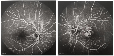
Figure 1 and 2: Inhomogeneous hyperfluorescence from the early times in both of eyes in posterior pole persistent in time.
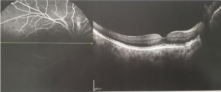
    Figure 3: Image of the right eye showing the rupture of Bruch’s membrane
with rupture of the pigmented epithelium opposite.
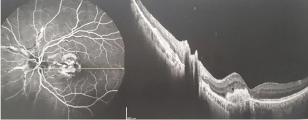
    Figure 4: Hyperfluorescence taking up all the persistent macular area at late
times complicated by juxta-foveolar choroidal neovacularization confirmed by
optical coherence tomography.
We also note the presence of microaneurysms in the 4 quadrants in the right eye and interesting 2 quadrants in the left one of which the drop in visual acuity is due to the macular lesion.
Angioid streaks can cause subretinal hemorrhage due to choroidal fibrovascular growth. These hemorrhages can disappear spontaneously or lead to choroidal neovascularization. The association of angioid streaks and diabetes is described in the literature. The patient finds himself threatened with retinal and choroidal neovascularization, hence the interest in close clinical and angiographic monitoring for early diagnosis and adequate management.