
Case Report
Austin J Clin Ophthalmol. 2020; 7(1): 1108.
High Myopia Associated with Advanced Glaucoma due to Microspherophakia: A Case Report
Karmoun S*, Aboulanouar A, Hajji Z, Elmarzouqi B, Elhassan A and Berraho A
Service d’ophtalmologie B, Hôpital des spécialités de Rabat, Morocco
*Corresponding author: Karmoun Souhaila, Service d’ophtalmologie B, Hôpital des spécialités de Rabat, Morocco
Received: April 17, 2020; Accepted: June 23, 2020; Published: June 30, 2020
Abstract
We report a case of a 24-year-old girl presenting to our hospital with blurred vision in both eyes. Ophthalmic examination revealed high myopia and angleclosure glaucoma related to pupillary block caused by small, spherical lenses. Treatment approaches to glaucoma in patients with microspherophakia are discussed in this case report.
Keywords: Microspherophakia; Glaucoma; High myopia
Introduction
Microspherophakia is a relatively rare condition in which the lens takes a spherical shape with an increased antero-posterior diameter and a reduction in the equatorial diameter [1].
The pathogenesis of this condition is thought to be linked to defective development of the lens zonules [2]. The spherical lens can then lead to pupillary blockage and secondary angle-closure glaucoma.
Lens myopia and lens dislocation are other common signs of microspherophakia [3].
The treatment of these patients is difficult and there is no consensus on the therapeutic approach, especially in patients with secondary angle-closure glaucoma.
We report a case of microspherophakia, presenting high myopia with bilateral secondary glaucoma.
Case Presentation
24-year-old young woman, chef as profession, presented to our hospital for the management of glaucoma refractory to medical treatment. The patient has been wearing optical correction glasses since childhood and receiving anti-glaucomatous treatment and still has a progressive decrease in visual acuity (Table 1) (Figure 1-4).
    Ophthalmic    examination
    Right eye
    Left eye


    Visual    acuity
    2/10    (-11 ; (- 1,50 à 153°))
    1/10 (-13    (-1,50 à 59°))


    Ocular    pressure
    26mmhg with oral Acetazolamide    and triple therapy (Dorzolamide 20mg +  Brimonidine 2mg + Timolol 5mg )
    28mmhg with oral Acetazolamide    and triple therapy (Dorzolamide 20mg +     Brimonidine 2mg + Timolol 5mg)


    Cornea
    Transparent,    pachymetry at 555μm
    Transparent,    pachymetry at 550


    Anterior    chamber
    Reduced depth, presence of a    remnant of epi-pupillary membrane above
    Reduced depth, presence of a    remnant of epi-pupillary membrane above


    Iris
    Posterior synechiae at 5    o'clock
    Posterior Synechiae between 6    and 7 o’clock with poor dilation


    After    dilation


    Lens
    Clear, domed in anterior    chamber with visualization of the zonules. Reduction of vertical diameter and the sphericity of the    lens (Figure 1)
    Clear, domed in anterior    chamber with visualization of the zonules. Reduction of the vertical diameter    and the sphericity of the lens (Figure 2)


    Vitreous
    Fibrillar
    Fibrillar


    Fundus    examination
    Papillary excavation at 8/10    with blurred contour, Macula clear, no sign of myopic choroidosis (fig 3)
    Subtotal papillary excavation    with blurred contour, Macula clear , no sign of myopic choroidosis (fig4)


    Gonioscopy
    360 °    closed angle, no goniosynechia
    360 °    closed angle, no goniosynechia


    UBM
    Anterior chamber depth at    1.17mm; irido-corneal angle closed on the 4 quadrants; lens arrow at 2250    microns, no iris plateau configuration
    Anterior chamber depth at    1,39mm ; irido-corneal angle closed on the 4 quadrants ; lens arrow    at 1930 microns, no iris plateau configuration
Table 1:
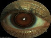
Figure 1: Photo of the right eye.
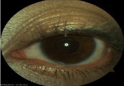
Figure 2: Photo of the left eye.
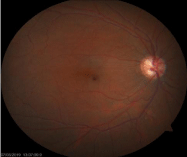
Figure 3: Papillar excavation 8/10 with blurred edges, Macula clear.
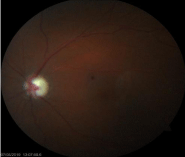
Figure 4: Papillar excavation 9/10 with blurred edges, Macula clear.
The patient benefited from phakophagia with irrigation and aspiration in the right eye and an implantation by collapsible intraocular lens (IOL) of power 26D introduced into the capsular bag followed by phacoemulsification of the left eye with implantation of collapsible IOL of 28, 5D (Figure 5-7). Both of interventions were done under general anesthesia spaced 2 months.

Figure 5-7: Operating times in left eye showing the aspiration of the lens nucleus, and the placement of foldable IOL in the lens bag.
The postoperative evolution was marked by a reduction in ocular pressure to 12mmhg in right eye and 14mmhg in the left without any treatment.
Visual acuity at 3 months post-surgery was 4/10 (-2.5 at 160°) P4 in OD and 8/10 (-1.50 (-0.50 at 95°)) P2 with an addition of +2.50.
On clinical examination, a secondary cataract was noted in the right eye which could explain the poorer visual acuity (Figure 8 and 9). The fundus examination showed a significant regression of the papillary excavation in both of eyes
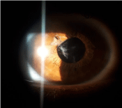
Figure 8: Photography of the right eye 3 months post-operatively.
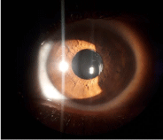
Figure 9: Photography of the left eye 3 months post-operatively.
However, left eye with the best visual acuity has an altered visual field compared to the right eye (Figure 10 and 11).
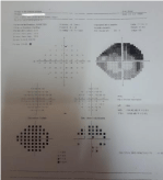
Figure 10: Visual field of the right eye showing significant impairment.
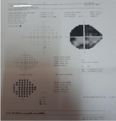
    Figure 11: Visual field of the left eye showing a perimeter alteration giving
way to a tubular vision.
Medical treatment with prostaglandins was commenced 2 months later for ocular pressure between 18mmhg and 20mmhg.
Discussion
Microspherophakia is a generally bilateral disorder, characterized by the presence of a small-diameter lens with an increase in anteroposterior diameter. The equatorial diameter of a microsphere lens measures on average between 6.75 to 7 mm (normal, 9.0mm), and the antero-posterior diameter is larger than normal and exceeds 5.0mm (normal: 3, 4 to 4.5 mm) [4].
Several pathophysiological mechanisms have been suggested: A stop in the development or abnormal insertion of the secondary fibers of the lens, which can be linked to a nutritional deficiency due to defects in tunica vasculosa lentis and which can occur in the fifth and sixth months of embryonic life, the lens normally being spherical at this point.
Zonular fibers can also be rudimentary due to lack of tension and stopping lens development, so they can remain spherical instead of gradually turning into a normal biconvex shape.
Microspherophaqia can present as an isolated, idiopathic, familial anomaly or associated with systemic disorders such as Weill-Marchesani syndrome or Marfan syndrome and rarely with homocystinuria, mandibulofacial dysostosis, Alport syndrome or Klinefelter syndrome [1,3].
Our patient presented no clinical sign suggesting one of these syndromes. His mental state and size were normal. She had no heart, skeletal or muscle abnormalities. In addition, there was no evidence suggesting Alport syndrome in his review. His condition could be of family origin. Genetic counseling could not be done for family conflicts.
Familial microspherophakia is not associated with any systemic defect. Although it is an autosomal recessive disorder, there is one case in the literature with autosomal dominant inheritance [2].
Other ocular features of familial microspherophakia are lenticular myopia, posterior staphyloma, myopic crescent, pupillary ectopia, glaucoma and retinal detachment.
Most patients come to the clinic during adolescence or early childhood [5]. As our patient became symptomatic much later, the diagnosis of microspherophaqia may have been missed and she was treated as angle-closure glaucoma and referred to our training for filtering surgery.
Glaucoma is the most common complication of this disease, which threatens the visual prognosis. Acute angular closure may result from pupillary blockage caused by anterior subluxation of the lens due to zonular hyperlaxity. [6]
Microspherophakia can lead to chronic glaucoma due to recurrent attacks of pupillary blockage, resulting in synechial closure of the angle [3,6].
The management of microspherophakia is still under debate. Medical and laser treatments fail in about 60% of the eyes with this condition. Lens extraction remains the first choice if medical treatment and laser iridotomy fail. Kaushik et al [7] described an adult patient with bilateral angle-closure glaucoma with microspherophakia and who’s intra-ocular pressure (IOP) was successfully controlled by extraction of the lens and anterior vitrectomy.
Kanamori et al. [8] also described a patient with microspherophakia and chronic angle-closure glaucoma who’s IOP was well controlled by goniosynechiolysis and lens extraction.
Although described by some authors (Khokhar et al, 2012) [9] the use of the capsular tension ring is not the treatment of choice because it is most often a small capsular bag. Scleral fixation represents an alternative (Fan et al, 2003) [10] which have been successfully achieved in some patients.
In our case we were able to place the intraocular implant in the capsular bag in both of eyes, this allowed the correction of the high myopia in the spherophakia. However, they are only recommended in patients with an anterior chamber of good depth, with no displacement of the lens and compliance with the annual examinations by the patient.
During follow-up, the authors insist on monitoring the number of endothelial cells, changes in the location of the implant and the development of secondary cataracts (Moshirfar, 2010) [11].
In contrast, Yasar [12] described a patient in whom IOP was not controlled using short-term lens extraction and who subsequently required a trabeculectomy using mitomycin -C for both eyes.
Senthil et al. [13] in their retrospective study noted higher rates of trabeculectomy success (86% at 6 months, 61% at 8 years) in patients with microspherophakia with a long follow-up period. However, they observed significant complications, including a shallow anterior chamber and iridocorneal and iridocrystalline contact, which required surgery. A large part (45%) of the trabeculectomized eyes underwent an extraction of the lens for the control of intra-ocular pressure in their study.
No standard surgical technique has been adopted due to the variability of the laxity of the zonular fibers [14]. Implantation of intraocular lenses is not always possible due to the absence of a stable capsular support, especially after complicated surgery [7,8,14,15]. Also, the implantation of IOL in the capsular bag is questionable due to the post-operative instability of the implant, the unavailability of a small capsular tension ring and the high rate of contraction of the capsular bag. [14,16,17]
The main indications for clear lens extraction in patients with microspherophakia are: corneo-lenticular contact, high unilateral myopia, pupillary blockage and secondary glaucoma refractory to medical treatment [7]. Our patient underwent lens extraction clear due to the rapidly progressive course of glaucomatous neuropathy. However, the main operating difficulty was the implantation of IOL in the small capsular bag and the zonular fragility.
Conclusion
The presence of angle-closure glaucoma associated with high myopia in young subjects should prompt the clinician to diagnose microspherophakia.
The optimal management of glaucoma in microspherophakia is still uncertain. Several factors are responsible for the development of glaucoma in microspherophakia.
Microspherophak patients should be closely monitored to determine the appropriate method for treating their glaucoma.
References
- Johnson VP, Grayson M, Christian JC. Dominant microspherophakia. Arch Ophthalmol. 1971; 85: 534-537.
- Johnson GJ, Bosanquet RC. Spherophakia in a Newfoundland family: 8 years’ experience. Can J Ophthalmol. 1983; 18: 159–164.
- Nelson LB, Maumenee IH. Ectopia lentis. Surv Ophthalmol. 1982; 27: 143– 160.
- Tindall R, Jensvold B. Microspherophakia In: Roy FH, Fraunfelder FW, Fraunfelder FT (eds.) Roy and Fraunfelder’s Current Ocular Th erapy , Sixth Edition. 2008; Chapter 303: 559-560, USA, Elsevier.
- Senthil S, Rao HL, Hoang NT, Jonnadula GB, Addepalli UK, Mandal AK, et al. Glaucoma in microspherophakia: Presenting features and treatment outcomes. J Glaucoma. 2014; 23: 262-267.
- Chan RT, Collin HB. Microspherophakia. Clin Exp Optom. 2002; 85: 294–299.
- Kaushik S, Sachdev N, Pandav SS, Gupta A, Ram J. Bilateral acute angle closure glaucoma as a presentation of isolated microspherophakia in an adult: case report. BMC Ophthalmol. 2006; 6: 29.
- Kanamori A, Nakamura M, Matsui N, Tomimori H, Tanase M, Seya R, et al. Goniosynechialysis with lens aspiration and posterior chamber intraocular lens implantation for glaucoma in spherophakia. J Cataract Refract Surg. 2004; 30: 513–516.
- Khokhar S, Gupta S, Kumar G, Rowe N. Capsular tension segment in a case of microspherophakia. Cont Lens Anterior Eye. 2012; 35: 230-232.
- Fan DS, Young AL, Yu CB, Chiu TY, Chan NR, Lam DS. Isolated microspherophakiawith optic disc colobomata.J Cataract Refract Surg. 2003; 29: 1448-1452.
- Moshirfar M, Meyer JJ, Schliesser JA,Espandar L, Chang JC. Iris-fi xated phakic intra-ocular lens implantation for correction of high myopia in microspherophakia. J Cataract & Refract Surg. 2010; 36: 682-685.
- Yasar T. Lensectomy in the management of glaucoma in spherophakia: is it enough? J Cataract Refract Surg. 2003; 29: 1052–1053.
- Senthil S, Rao HL, Hoang NT, Jonnadula GB, Addepalli UK, Mandal AK, et al. Glaucoma in microspherophakia: presenting features and treatment outcomes. J Glaucoma. 2014; 23:262–267.
- Al-Haddad C, Khatib L. Vitrectorhexis and lens aspiration with posterior chamber intraocular lens implantation in spherophakia. J Cataract Refract Surg. 2012; 38: 1123-1126.
- Wu-Chen WY, Letson RD, Summers CG. Functional and structural outcomes following lensectomy for ectopialentis. J AAPOS. 2005; 9: 353-357.
- Behndig A. Phacoaspiration in spherophakia with corneal touch.J Cataract Refract Surg. 2002; 28: 189–191.
- Dufay-Dupar B, BlumenOhana E, Rodallec T, Nordmann JP. Rare complication in microspherophakia surgery: early capsular contraction. J FrOphtalmol. 2007; 30: e30.