Case Report
Austin J Clin Ophthalmol. 2022; 9(1): 1123.
Endogenous Endophthalmitis - A Rare Complication of Infective Endocarditis: A Case Report
Afaf E¹*, Fatimaezahra H¹, Habiba T¹, Elhassan A² and Berraho A²
¹Ophthalmologist, Morocco
²Professor of Ophthalmology, Morocco
*Corresponding author: Erradi Afaf, Ophthalmologist, Morocco
Received: December 07, 2021; Accepted: February 09, 2022; Published: February 16, 2022
Abstract
Endogenous endophthalmitis (EE) is a severe, acute and diffuse uveitis caused by intraocular haematogenic infection. It affects vulnerable subjects. We report a case of a 42-year-old male with a history of endocarditis. he developed endogenous endophthalmitis in her left eye due to acute endocarditis. Owing to deterioration of the general status, intravitreal injections couldn’t be practiced and therapy consisted only in intravenous antibiotic therapy. We highlight the severity of this disease, associated with unfortunate visual and vital prognosis.
Keywords: Endogenous endophthalmitis; Intra-vitreous injections; Prognosis
Introduction
Endogenous endophthalmitis (EE) is an extremely serious eye condition which threatens the visual prognosis and whose origin can be life threatening. Indeed, it is considered as a secondary localization following the dissemination of an infectious agent by hematogenous route with passage through the blood-retinal barrier. It is a rare pathology representing 2% to 15% of all endophthalmitis and often occurring in debilitated conditions. Its treatment is imperfectly codified. It includes a general anti-infective treatment associated, very often, with intra-vitreous injections (IVT) of anti-infective agents. The aim of our work was to report a case of EE complicating acute endocarditis
Patient and Case
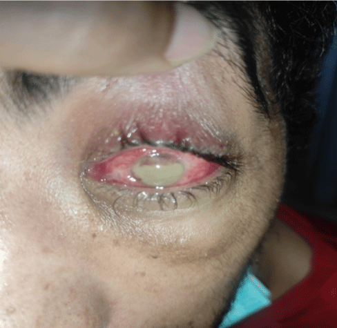
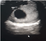
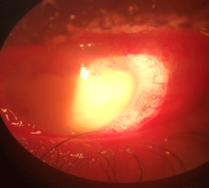
This is a 43-year-old patient with no specific antecedants. History of the disease dates back to 4 days before admission by the installation of a decrease in visual acuity the ophthalmological examination found an anterior non-granulomatous uveitis associated with hypopion and ocular ultrasound is in favor of endophthalmitis general examination reveals fever with intense fatigue and dyspnea blood test and echocoeur returned in favor of infective endocarditis (Figure 1-3).

Figure 1: Hyperhemie conjonctivale, hypopion.

Figure 2: Echographie oculaire en faveur d’une hypereflectivie CA et vitré: endophtalmie unilaterale

Figure 3: Hypopion Chambre anterieure.
Faced with this table, we retained the diagnosis of a bacterial EE and the patient benefited from a triple antibiotic therapy by general route associated with fortified antibiotic eye drops. Topical corticosteroid therapy was introduced secondarily. The very poor general condition of the patient did not allow the practice of either IVT antibiotics or a vitrectomy. The blood cultures came back negative as well as the eye samples. A control ophthalmologic examination of the OG on D4 showed neutralization of the infection with disappearance of secretions and a marked decrease in inflammation in the anterior chamber.
Discussion
Endogenous endophthalmitis is severe, acute, diffuse uveitis caused by blood-borne intraocular infection. It can be uni- or bilateral and preferentially affects the left eye. It is about a pathology which occurs most often on a debilitated ground. According to Jackson et al. ,the main risk factors identified are diabetes mellitus, drug addiction and cancer. A primary genitourinary, dental, hepatic, biliary or pulmonary or cardiac infectious focus is found in 90% of cases
In EAs, the diagnosis with certainty is based on the detection of the infectious agent on microbiological samples which are positive in 80 to 96% of cases. There is a predominance of gram-positive bacteria among bacterial infections. Indeed, blood cultures are positive in 71 to 84% of cases and vitreous culture in 73.9% of cases. Anterior chamber samples can also be contributory. Chiquet et al. described in a review of the literature dating from 2007 those PCRs on bacteriological samples taken in acute postoperative endophthalmitis made it possible to improve the rate of bacterial identification in the aqueous humor (47%) and in the vitreous (68%). In our case, the microbiological analysis was negative.
The curative treatment of EE must be instituted urgently for ocular and general aim. It is a broad-spectrum systemic antibiotic therapy combined with anti-infective IVTs. In our case, this management was not possible because of the altered general condition of the patient. In EE, the place of corticosteroid therapy and vitrectomy is still debated. However, their efficacy has been studied in postoperative endophthalmitis. The visual and general prognosis of EE remains unfortunate even after early and appropriate treatment, and our case is no exception. The death rate varies between 4% and 14.3%, explained by the age and debilitated terrain of the population affected by EE. For this reason, it is necessary to underline the urgency of a general and ocular treatment as soon as the diagnosis is made.
Conclusion
Endogenous endophthalmitis is a rare condition with a poor prognosis. Our case supports the diagnostic and prognostic knowledge available on this subject. We must always think of a more frequent fungal endophthalmitis in the event of EE and seek to eliminate the causes of tumor pseudo-uveitis in the elderly.