Case Report
Austin J Clin Ophthalmol. 2022; 9(1): 1124.
Microbial Keratitis Complicated by Endophthalmitis Hiding Acute Hydrops
Afaf E¹*, Habiba T¹, Fatimazahra H¹, Elhassan A² and Amina B²
¹Ophthalmologist, Morocco
²Professor of Ophthalmology, Morocco
*Corresponding author: Erradi Afaf, Ophthalmologist, Morocco
Received: February 08, 2022; Accepted: March 04, 2022; Published: March 11, 2022
Abstract
Purpose: To report a case of acute hydrops in a 12-year-old child with advanced keratoconus.
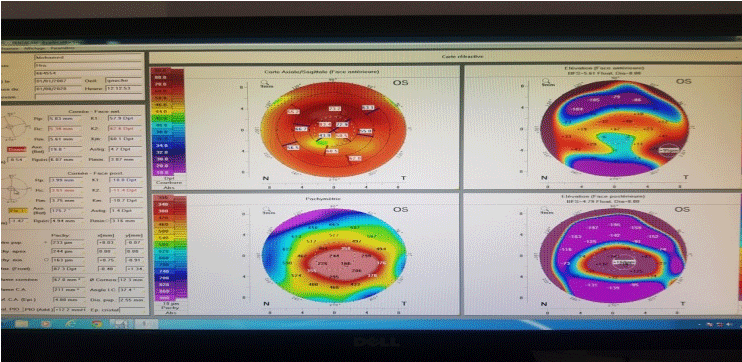
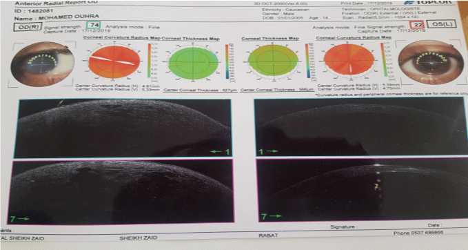
Case Presentation: A twelve-year-old boy diagnosed as having right eye (RE) infectious keratitis, not responding to antimicrobial therapy, was referred to our hospital. The diagnosis of infectious keratitis was established one month prior to his presentation following an episode of acute corneal whitening, pain, and drop in visual acuity. Topical fortified antibiotics followed by topical antiviral therapy were used with no improvement. Slit lamp examination showed significant corneal protrusion with edema surrounding a rupture in Descemet’s membrane in the RE. The diagnosis of acute corneal hydrops from advanced keratoconus was highly suspected and confirmed with corneal topography ans OCT.
Keywords: Hydrops; Corneal topography; Descemet’s membrane
Introduction
Keratoconus (KC) is a non inflammatory ectasia of the cornea. Classically, the onset of KC is during puberty and the condition is progressive until the third or fourth decade of life In fact, KC demonstrates an increased incidence and faster progression at both puberty and pregnancy due to hormonal influences.
Case Presentation
This is the 12-year-old M.O child from and living in ouarzazat.
Background
Personal:
• Poorly monitored eye allergy
• Discovery of a bilateral keratoconus since school age (keratoconus stage 4)
Family: (according to the words of the mother)
• Bilateral Keratoconus in a sister with ODG corneal transplant
• Unilateral keratoconus in a brother
History of disease
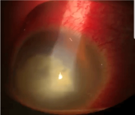
Goes back a week by installation a painful red eye with tearing and photophobia motivating a consultation readressed to our structure for a suspicion of endophthalmitis complicating infectious keratitis (Figure 1-4 and Table 1).

Figure 1: RE: Corneal opacity with stomal.

Figure 2: Infiltration Left Eye.

Figure 3: Corneal Topography.

Figure 4: OCT.
    OD

    OG


    AVSC

    PL+

    2/10F


    Refraction

    Imprenable

    Imprenable


    Annexes

    Saines

    Saines


    Conjonctives

    Hypereamia of Conjonctive

    Normocolored


    Secretions


    CORNEE

    Central Corneal Opacity Measuring 6mm With    Stromal Infiltrate, Fluo

    Paracentral Opacity with Manifeste Ectasia, Vogt    Stries


    CA

    Not Seen

    Normal


    IRIS

    Not Seen

    Normal


    Cristallin

    Not Seen

    Transparent


    VITRE

    N.S

    Transparent


    FO

    N.S

    C/D:3/10 / MBR /VBC / FLAT    RETINA
Table 1: History of Diseases.
Ocular ultrasound
• CA Hyperechogenic
• Crystalline hypoechoic
• Glazed loaded
• Flat retina
Treatment
IV antibiotic therapy: Triaxon 50mg/kg 2*/day.
Fortified eye drops:
• Vancomycin g/h pdt 48hrs then 1g 8*/day.
• Fortum g/h pdt 48hrs then 1g/day.
• Bolus of 40mg of corticosteroids after 48 hours of ATB.
Local treatment: Local antiseptic, hypotonic; hyperosmolar eye drops washing artificial tears.
Discussion
In this case, acute corneal hydrops was the initial presentation of KC in a pediatric patient with a suggestive history of allergic conjunctivitis, eye rubbing, and progressive loss of vision. Corneal hydrops was misdiagnosed as infectious keratitis, and KC was overlooked. This may have been due to the infrequency of KC during childhood but was most likely due to the rarity of occurrence of hydrops in this age group. Acute corneal hydrops is the development of a break in Descemet’s membrane with subsequent marked edema of the corneal stroma and epithelium. The mean age at the onset of corneal hydrops was 39.3 years in one study and 24 years in another. Many risk factors for the development of corneal hydrops in KC were reported and they include childhood diagnosis of KC, male sex, poor corrected visual acuity at the diagnosis of KC, and severe allergic eye disease. In our reported case, besides the early onset of KC and the male sex, allergic eye disease with eye rubbing may have played a major role in the development of corneal hydrops in KC in this young age group. Hence, in our case, allergic eye disease and eye rubbing may have contributed to the development of KC at this early age and eventually lead to the development of acute hydrops. The hypothesis that eye rubbing is the most significant cause of KC is supported by many reports and dates back to 1956 Similar to our case, acute corneal hydrops was the first presentation of KC in the 2 reported cases with both patients diagnosed with allergic conjunctivitis and eye rubbing, further emphasizing the fact that allergic keratoconjunctivitis with eye rubbing may increase the incidence of corneal hydrops in children with KC
Conclusion
In this case, acute hydrops was the initial clinical presentation of advanced KC in a 12-year-old pediatric patient previously misdiagnosed as infectious keratitis. Although it is a relatively rare disease at the age of 12 years, pediatric KC can be rapidly progressive especially in the presence of allergic conjunctivitis and eye rubbing. This entity should always be considered in the differential diagnosis of progressive vision loss and of corneal leukoma in this young age group.