
Research Article
Austin J Clin Ophthalmol. 2022; 9(1): 1125.
Ruptured Sinus of Valsalva Aneurysm from Left Coronary Sinus with Different Causes
Kang R1#, Han M2# and Zhang C1*
¹Department of Ultrasound, The Second Affiliated Hospital of Nanchang University, Nanchang, Jiangxi, China
²Queen Mary School, Nanchang University, Nanchang, Jiangxi, China
#Contributed Equally to this Work and regarded as Co-First Authors
*Corresponding author: Chunquan Zhang, Department of Ultrasound, The Second Affiliated Hospital of Nanchang University, No.1 Minde Road, Nanchang, Jiangxi, 330006, China
Received: February 12, 2022; Accepted: March 08, 2022; Published: March 15, 2022
Abstract
Objective: Analysis of three cases in which the clinical diagnosis was ruptured sinus of Valsalva aneurysm (RSVA) from left coronary sinus, combined with similar cases in the literature, to summarize the etiology of RSVA.
Methods: Retrospective analysis of three cases of RSVA diagnosed in our hospital from 2009 to 2021. Their general condition, examination and treatment were analyzed, and the etiology of the sinus of Valsalva aneurysm (SVA) was summarized in relation to similar cases reported in the literature.
Results: All three cases were diagnosed as RSVA from left coronary sinus with a clear etiology of congenital SVA, Behcet’s disease and infective endocarditis, respectively. They all underwent successful surgical treatment. In the literature, there were 12 cases of SVA from left coronary sinus of definite etiology, 10 of which ruptured.
Conclusions: SVA from left coronary sinus is a rare disease with a complex etiology, and RSVA is one of the most serious complications of SVA. Exploring the etiology of SVA will help to prevent the serious complications of RSVA by targeting the etiology for treatment and timely surgical intervention.
Keywords: Sinus of Valsalva aneurysm; Left sinus; Ruptured; Etiology; Behcet’s disease; Infective endocarditis
Abbreviations
RSVA: Ruptured Sinus of Valsalva Aneurysm; SVA: Sinus of Valsalva Aneurysm; IE: Infective Endocarditis; BD: Behcet’s Disease; BP: Blood Pressure; HR: Heart Rate; RR: Respiratory Rate; ESR: Erythrocyte Sedimentation Rate; B-BNP: B-Type Brain Natriuretic Peptide; ANA: Anti Nuclear Antibody; anti-dsDNA: Antibody, Anti Double Strand DNA Antibody; TTE: Transthoracic Echocardiography; LV: Left Ventricle; LVOT: Left Ventricular Outflow Tract; LA: Left Atrium; CTA: Computed Tomography Angiography; AR: Aortic Regurgitation
Introduction
Sinus of Valsalva Aneurysm (SVA) is an unusual aortic root defect that can be dangerous due to its serious complications; it is defined as dilatation of one or more of the aortic valve sinuses [1]. According to Meier JH et al., SVA commonly arise from the right sinus, (65%~85%), less commonly from noncoronary sinus (10%~30%), and rarely (<5%) from the left sinus [2]. It can be either a congenital or acquired cardiac anomaly [3], and is relatively common in oriental patients4.Congenital SVA is owing to a dilatation generally of a single sinus of Valsalva caused by a separation between the aortic media and the annulus fibrosus, and often with a deficiency of the normal elastic tissue and abnormal development of the bulbus cordis [5]. Congenital SVA is usually seen in patients with Marfan or Ehlers-Danlos syndrome [6]. Acquired SVA is mainly the results of infective endocarditis (IE), syphilis, trauma, Behcet’s disease (BD), atherosclerosis, cystic medionecrosis [7,8].
Although surgical repairs for ruptured sinus of valsalva aneurysm (RSVA) are gradually matured, RSVA is still considered as a special and fetal complication of SVA with severe mortality and morbidit [5]. So it is more revelatory that we focus on the etiology of RSVA from left coronary sinus, for this rare situation is easily neglected and usually fetal. However, there are few cases of RSVA from left coronary sinus and fewer studies on the etiology of SVA at present. Therefore, this paper retrospectively analyzed and reported 3 cases of RSVA from left coronary sinus that occurred in our hospital from 2009 to 2021. 12 cases of SVA from left coronary sinus with a clear cause were also obtained from the literature. And the etiology of SVA was summarized in relation to similar cases reported in the literature.
Our cases reported in this article are remarkable for several reasons: Etiology of SVA, site of rupture, precise preoperative diagnosis and the successful surgical management. By exploring the etiology of SVA will help to prevent the serious complications of RSVA by targeting the etiology for treatment and timely surgical intervention.
Materials and Methods
We retrospectively analyzed three patients with RSVA from 2009 to 2021 IN our hospital and obtained their preoperative, intraoperative, and postoperative conditions. Also, by reviewing the literature, we obtained 12 cases of SVA from left coronary sinus with clear causes [9-20]. We also retrospectively analyzed the etiology of these 12 patients.
Results
See Table 1 and 2.
![]()
    Case 1

    Case 2

    Case 3


    Gender

    Male

    Female

    Male


    Age

    25

    53

    53


    Reasons for admission

    Persistent chest and back pain for 2    years

    Sudden chest tightness and    palpitation for one week

    Chest tightness and shortness of breath    with cough for half a month


    Past medical history

    No special

    Recurrent oral and vulvar ulcers.

    Unexplained fever


    BP/HR/RR

    90/30 mmHg

    160/110 mmHg

    135/95 mmHg


    98 beats/min

    100 beats/min

    100 beats/min


    22 beats/min

    20 beats/min

    21 beats/min


    Chest physical examination

    A 2/6 grade systolic murmur and moderate    diastolic murmur.

    Femoral artery pistol shot sound and    corrigan's pulse.

    A grade 2/6 systolic blowing murmur.


    Abnormal laboratory examination

    ESR was 53mm/h.

    ANA, anti dsDNA were negative.

    B-BNP was 604 pg/ml.


    Electrocardiogram

    Sinus heart rate

    Sinus heart rate

    Prolonged P-R interval.


    Preoperative TTE

    SVA from left coronary sinus ruptured    into LVOT

    SVA from left coronary sinus ruptured into    LV.

    A neoplasm on the aortic valve. SVA from    left coronary ruptured into LA.


    Preoperative CTA

    An aneurysmal expansion of the left    coronary sinus.

    An aneurysm of the left coronary sinus at    the initial segment of the ascending aorta.

    This examination was not done.


    Surgical approach

    Ruptured aortic sinus aneurysm repair

    Congenital aortic sinus aneurysm repair    and aortic valvuloplasty

    Aortic coronary sinus aneurysm repair    and aortic valve replacement


    Surgery detection

    A breaking of approximately 2.0x1.0 cm in    size below the left coronary artery.

    Significant thickening of the aortic wall,    showing changes of aortitis

    A rupture of the left coronary valve of    the aorta was seen, and a neoplasm was seen on it.


    Pathological results/Culture results

    A pseudoaneurysm in LVOT

    Arterial vascular wall fibrosis and    vitreous changes.

    Bacterial cultures of both blood and valve    resulted in Staphylococcus aureus.


    Postoperative TTE

    The saccular structure had disappeared.

    The saccular structure had disappeared.

    The saccular structure had disappeared.    Enlargement of LA and LV.


    Location of rupture into the heart    cavity

    LVOT

    LV

    LA


    Cause of disease

    Congenital SVA

    Behcet's disease

    Infective Endocarditis
Table 1: We got the preoperative, intraoperative and postoperative conditions of these 3 cases.
![]()
    Author

    Age

    Gender

    Causes of Disease

    Location of Rupture into the    Heart Cavity


    Killen DA, et al. [9]

    62

    Male

    Congenital SVA

    Transverse pericardial sinus


    Ryan T, et al. [10]

    33

    Male

    Sepsis with Staphylococcus    aureus after cadaveric renal transplantation

    LA


    Rothbart RM, Chahine RA [11]

    42

    Male

    Infective Endocarditis

    LVOT


    Greiss I, et al. [12]

    56

    Female

    Head-on car accident

    The pericardial space near LA


    Saito T, et al. [13]

    59

    Male

    Infective endocarditis

    LV


    Fazio G, et al. [14]

    30

    Male

    HIV

    Pulmonary artery


    Ryomoto M, et al. [15]

    47

    Male

    Congenital SVA

    LV


    Kawamura A, et al. [16]

    29

    Female

    Takayasu arteritis

    LV


    Bakan S, et al. [17]

    30

    Male

    Behcet's disease

    Unruptured


    Bae K, et al. [18]

    44

    Male

    Infective endocarditis

    LV


    Chamsi-Pasha MA, Lawrie GM [19]

    42

    Female

    Marfan syndrome

    Unruptured


    Talwar S, et al. [20]

    35

    Male

    Congenital SVA

    LVOT


    BP: Blood Pressure; HR: Heart Rate; RR:    Respiratory Rate; ESR: Erythrocyte Sedimentation Rate; B-BNP: B-Type Brain    Natriuretic Peptide; ANA: Anti Nuclear Antibody; anti-dsDNA: Antibody, Anti    Double Strand DNA antibody; TTE: Transthoracic Echocardiography; LV: Left    Ventricle; LVOT: Left Ventricular Outflow Tract; LA: Left Atrium; CTA: Computed    Tomography Angiography.
Table 2: A review of the literature revealed 12 cases of left Valsalva sinus aneurysm with a definite cause. The order in the references corresponds to 9-20.
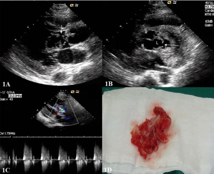
    Figure 1: A: left ventricular long-axis view showed the saccular structure

(SVA) in LVOT and the breach on the side of the ventricular septum; B:

short-axis view of the aorta showed the entrance diameter of the saccular

structure (SVA) is about 6mm; C: the multicolored blood flow signal flows

from the saccular structure to LVOT, and its blood flow velocity was about

4m/s as measured by spectral Doppler; D: ruptured aortic sinus removed

during surgery.
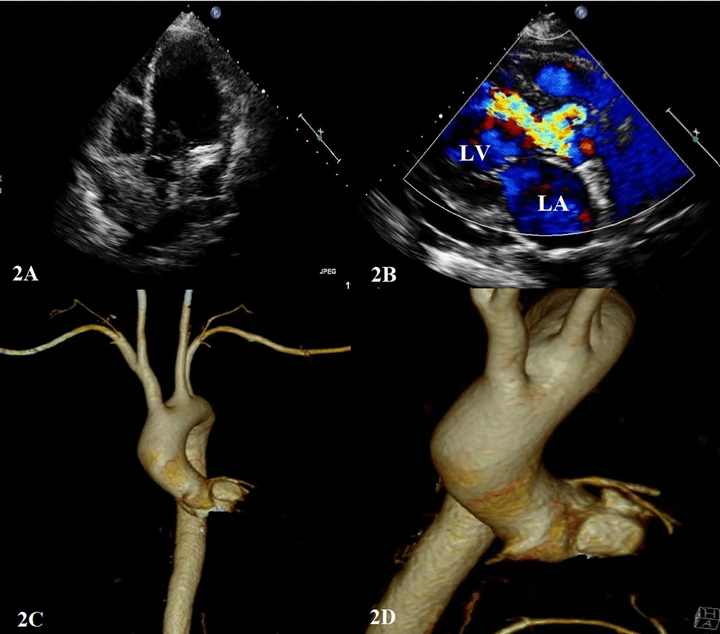
    Figure 2: A: SVA from left coronary sinus rupturing into LV (arrow); B: blood

flow signal from left coronary sinus to LV; C and D: CTA images of patients.
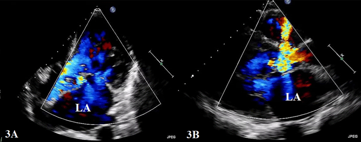
Figure 3: A and B: CDFI shows the blood flow signal from left coronary sinus to LA.
Discussion
SVA is usually thought to be an enlargement or a dilation of one sinus of the aortic root between the aortic valve annulus and the sinotubular ridge [5]. It is an unusual cardiac event with no noticeable symptoms, which poses a challenge for the early diagnosis and treatment of SVA. In the overwhelming majority of SVA cases (90%), the sinus ruptures into the right-sided chambers, and in the other 10%, it ruptures into the left atrium, left ventricle, pulmonary artery, or pericardial cavity [21]. Early diagnosis of SVA is important because surgical treatment is easier to perform at an early stage and facilitates a good prognosis [22]. RSVA is a hazardous complication that can cause AR, left to right shunt following the aneurysm rupture into a cardiac chamber, and acute progressive heart failure [23].
The natural history of SVA is undiscovered; it can be either a congenital or acquired cardiac anomaly [3]. Congenital SVA is usually thought to be the result of incomplete fusion of the two halves of the distal bulbar septum [24-26]. This is the structure that separates the aortic and pulmonic halves of the bulbus cordis, the primary exit tube of the fetal heart. Congenital weakness secondary to an incompletely fused septum predisposes the high-pressure area of the sinuses to aneurysm formation. The left coronary sinus does not arise from the distal bulbar septum, as do the right and noncoronary sinuses [24- 26]. This interprets the infrequency of congenital SVA from the left sinus [26]. Case 1 is considered as a congenital SVA from the left sinus that ruptured into the LVOT. Rupture of a congenital SVA into an adjacent chamber may occur spontaneously, after trauma, after strenuous physical exertion, or from acute bacterial endocarditis.
Acquired SVA are mainly the results of infective endocarditis (IE), syphilis, trauma, Behcet’s disease (BD), atherosclerosis, cystic medionecrosis [7,8], or degenerative diseases, abuse of drugs or alcoholism [27]. In case 2, the aortic wall was seen to be significantly thickened with aortitis changes under surgery. And the patient had a 5-year history of oral and vulvar ulcers, and clinical examination revealed aphthous stomatitis, genital ulceration, and erythema nodosum bilaterally, which could provide clues to differentiate diagnosis. BD is a systemic disorder of recurrent acute inflammation, characterized by major symptoms of oral aphthous ulcers, uveitis, skin lesions and genital ulcers [28]. Involvement of the heart is called Cardio-BD, cardiac manifestations include pericarditis, myocarditis, endocarditis, endomyocardial fibrosis, atrial fibrillation, ventricular arrhythmias, coronary arteritis, acute myocardial infarction, and dilated cardiomyopathy [29]. James et al. proposed that SVA was the main causes of death in patients with BD [30,31]. There is no doubt that surgical treatment is necessary, otherwise it may incur fetal complication. However, treatments that aim directly at the etiology of SVA are the most important.
In case 3, TTE suggested a rupture into LA. Under surgery, a neoplasm was seen on the left coronary valve of the aorta, and the blood and valve culture results were positive for staphylococcus aureus, suggesting that IE might be the cause of this patient. IE appears worldwide, and is defined by infection of a native or prosthetic heart valve, the endocardial surface, or an indwelling cardiac device [32]. Staphylococcus aureus is the most commonly isolated microorganism associated with infective endocarditis in high-income countries and is reported in up to 30% of cases [33,34].
Regardless of manifestation, patients with a persistent unexplained bacteremia should be investigated for infective endocarditis, especially patients with Staphylococcus aureus infection, should be examined with echocardiography [32]. Clinical examination presents alterable signs of disease, with fever (present in about 90% of cases) and a cardiac murmur (in about 85%) being most common. If pathology specimens are accessible (from surgery or autopsy), the diagnosis of IE can be made by histology or positive culture of vegetation or abscess tissue [35].
In summary, discussing the etiology of SVA from the left sinus will help early diagnosis of this rare cardiac anomaly and early treatment of the cause, so as to effectively prevent the serious complication of RSVA.
Conclusion
SVA from left coronary sinus is a very rare disease with multiple etiologies. It can be dangerous due to its serious complications like RSVA. Although surgical repair is gradually matured, it is still considered as a special and fetal complication of SVA. In addition to timely surgical treatment, early treatment for the etiology is also critical. The exploration of etiology of SVA can help early detection of SVA, so that timely surgical treatment can be carried out to prevent serious complication of RSVA.
Financial Support
This work was supported by the (Science and Technology Research Project) from Jiangxi Education Department under Grant (number 190002).
References
- Abohelwa M, Elmassry M, Whisenant T, Thongtan T, Sethi P. Sinus of Valsalva aneurysm presenting with chest pain. Proc (Bayl Univ Med Cent). 2020; 34: 283-285.
- Meier JH, Seward JB, Miller FA, Oh JK, Enriquez-Sarano M. Aneurysms in the left ventricular outflow tract: clinical presentation, causes, and echocardiographic features. J Am Soc Echocardiogr. 1998; 11: 729-745.
- Weinreich M, Yu PJ, Trost B. Sinus of valsalva aneurysms: review of the literature and an update on management. Clin Cardiol. 2015; 38: 185-189.
- Chao D, Wu QY, Tang Y. Ruptured sinus of valsalva aneurysm: a Beijing experience. Annals of Thoracic Surgery. 2002; 74: 1621-1624.
- Ring WS. Congenital Heart Surgery Nomenclature and Database Project: aortic aneurysm, sinus of Valsalva aneury sm, and aortic dissection. Ann Thorac Surg. 2000; 69: S147-163.
- Cao LB, Hannon D, Movahed A. Noncoronary sinus of Valsalva rupture into the right atrium with a coexisting perimembranous ventricular septal defect. World Journal of Clinical Cases. 2013; 1: 146.
- Vural KM, Sener E, Ta?demir O, Bayazit K. Approach to sinus of Valsalva aneurysms: a review of 53 cases. Eur J Cardiothorac Surg. 2001; 20: 71-76.
- Koh KK, Lee KH, Kim SS, Lee SC, Jin SH, Cho SW. Ruptured aneurysm of the sinus of Valsalva in a patient with Beh?et’s disease. Int J Cardiol. 1994; 47: 177-179.
- Killen DA, Wathanacharoen S, Pogson GW. Repair of intrapericardial rupture of left sinus of Valsalva aneurysm. Ann Thorac Surg. 1987; 44: 310-311.
- Ryan T, Markel ML, Waller BF, Armstrong WF, Feigenbaum H. Doppler echocardiographic detection of a ruptured acquired aneurysm of the sinus of Valsalva. Clinica l-morphologic correlations. Chest. 1987; 91: 626-629.
- Rothbart RM, Chahine RA. Left sinus of Valsalva aneurysm with rupture into the left ventricular outflow tract: diagnosis by co lor-encoded Doppler imaging. Am Heart J. 1990; 120: 224-227.
- Greiss I, Ugolini P, Joyal M, Bouchard D, Mercier LA. Ruptured aneurysm of the left sinus of Valsalva discovered 41 years after a decelerational injury. J Am Soc Echocardiogr. 2004; 17: 906-909.
- Saito T, Asano M, Ishida M, et al. Ruptured left coronary sinus of valsalva aneurysm into the left ventricle. Ann Thorac Surg. 2004; 78: 2187.
- Fazio G, Zito R, Dioco DD, et al. Rupture of a left sinus of Valsalva aneurysm into the pulmonary artery. Eur J Echocardiogr. 2006; 7: 230-232.
- Ryomoto M, Mitsuno M, Nishi H, Fukui S, Miyamoto Y, Takanashi S. Surgical repair of a sinus of a Valsalva aneurysm ruptured into the left ventricle. Gen Thorac Cardiovasc Surg. 2009; 57: 426-429.
- Kawamura A, Kato W, Araki Y, Oshima H, Usui A, Ueda Y. Surgical treatment of ruptured aneurysm of the left sinus of Valsalva caused by Takayasu arteritis. Ann Thorac Surg. 2010; 90: e91-92.
- Bakan S, Yamac E, Alis D, Ustabasioglu FE. A Rare Complication of Behcet’s Disease: An Incidentally Detected and Spontaneously Thrombosed Sinus of Valsalva Aneurysm. Ann Thorac Surg. 2016; 101: e171.
- Bae K, Jeon KN, Lee HI, et al. Left sinus of valsalva aneurysm ruptured into left ventricle: A case report of 320-multidetector CT f indings. Medicine (Baltimore). 2017; 96: e7112.
- Chamsi-Pasha MA, Lawrie GM. Aneurysmal left sinus of Valsalva in Marfan’s syndrome. Eur Heart J. 2018; 39: 285.
- Talwar S, Sen Gupta S, Jagia P, et al. Unusual presentation and rupture of left sinus of Valsalva into mitral-aortic intervalvular fibrosa. J Card Surg. 2020; 35: 904-907.
- Liu L, Wan Y, Deng MB. Giant aneurysmal of the left sinus of Valsalva in adults. J Card Surg. 2020; 35: 3145-3147.
- Au WK, Chiu SW, Mok CK, Lee WT, Cheung D, He GW. Repair of ruptured sinus of valsalva aneurysm: determinants of long-term survival. Ann Thorac Surg. 1998; 66: 1604-1610.
- Moustafa S, Mookadam F, Cooper L, et al. Sinus of Valsalva aneurysms--47 years of a single center experience and systematic overview of publis hed reports. Am J Cardiol. 2007; 99: 1159-1164.
- Guo DW, Cheng TO, Lin ML, Gu ZQ. Aneurysm of the sinus of Valsalva: a roentgenologic study of 105 Chinese patients. Am Heart J. 1987; 114: 1169- 1177.
- Jones AM, Langley F. Aortic sinus aneurysms. British heart journal. 1949; 11: 325.
- Yang Y-l, Xie M-x, Cheng TO, Wang X-f, Lü Q, Ghoorah D. Left coronary sinus of Valsalva aneurysm ruptured into the left ventricle: diagnosis by twodimensional and real time three-dimensional echocardiography. International journal of cardiology. 2011; 151: e35-e36.
- Farì G, Pennacchia I, Stigliano E, Oliva A, Carbone A, Arena V. Right sinus of Valsalva aneurysm. Cardiovascular Pathology. 2020; 47: 107209.
- Suzuki N. Behcet’s disease. Clinical and Experimental Medicine. 2004; 4: 10-20.
- Yesudian P, Edirisinghe D, O’Mahony C. Behçet’s disease. International journal of STD & AIDS. 2007; 18: 221-227.
- Zhanwen X, Xingzhou Z, Yaqin L. Aneurysms of the Sinus of Valsalva in a Patient with Behcet’s Disease. Iranian journal of public health. 2014; 43: 372.
- James DG, Thomson A. Recognition of the diverse cardiovascular manifestation in Behcet’s disease. American heart journal. 1982; 103: 457- 458.
- Cahill TJ, Prendergast BD. Infective endocarditis. Lancet. 2016; 387: 882- 893.
- Murdoch DR, Corey GR, Hoen B, et al. Clinical presentation, etiology, and outcome of infective endocarditis in the 21st century: the International Collaboration on Endocarditis-Prospective Cohort Study. Archives of internal medicine. 2009; 169: 463-473.
- Selton-Suty C, Célard M, Le Moing V, et al. Preeminence of Staphylococcus aureus in infective endocarditis: a 1-year population-based survey. Clinical infectious diseases. 2012; 54: 1230-1239.
- Prendergast BD. Diagnostic criteria and problems in infective endocarditis. Heart. 2004; 90: 611-613.