Clinical Image
Austin J Clin Ophthalmol. 2022; 9(2): 1126.
Total Retinal Detachment Following Traditional Couching
Aachak M*, Jeddou I, Boui H, Brarou H, Abdellaoui T, El Asri F, Mouzarii Y, Reda K and Oubaaz A
Department of Ophthalmology, Military Hospital, Mohammed V University, Rabat, Morocco
*Corresponding author: Aachak M, Department of Ophthalmology, Military Hospital, Mohammed V University, Rabat, Morocco
Received: March 07, 2022; Accepted: March 23, 2022; Published: March 30, 2022
Clinical Image
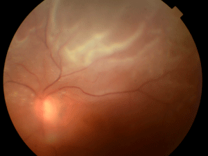
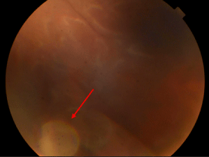
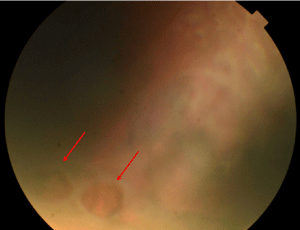
A 65-year-old patient with no notable history, from low socioeconomic status, presented to the ophthalmology clinic for visual impairment of the left eye three months after a traditional couching. Visual acuity was limited to light perception. Slit-lamp examination showed cells and flare in the anterior segment and the vitreous. Fundus examination showed a complete retinal detachment including the fovea with proliferative vitreoretinopathy stage B and two large peripheric retinal tears inferiorly contiguous to the crystalline lens. Surgical treatment was based on pars plana vitrectomy with phacofragmentation of the crystalline lens, pneumatic retinopexy and endophotocoagulation of the retinal tears. Clinical evolution was marked by the improvement of the visual acuity to 20/200 two months after surgery.
Traditional couching of the crystalline lens is the most ancestral documented form of cataract surgery. The clouding lens is dislodged to the vitreous using a sharp instrument, such as a thorn or needle, to pierce the eye with a quick movement at the edge of the cornea or the sclera [1]. Couching continues to be used in developing countries, especially in remote areas. This dangerous technique is done without anaesthesia nor aseptic rules causing irreversible complications such as corneal dystrophy, ocular hypertonia, uveitis, endophthalmitis or retinal detachment as the case of our patient [2,3].
Throughout this work, we are highlighting the danger incurred by patients who are victims of this archaic practice that still exists in several developing countries (Figure 1-3).

    Figure 1: Fundus examination showed a complete retinal detachment

including the fovea.

Figure 2: Crystalline lens in contact with the retina (red Arrow).

Figure 3: Two large peripheric retinal tears inferiorly (red arrow).
References
- A Jait, M Jait. Dislocation of the crystalline lens into the anterior chamber following couching: Case report. Journal Français d’Ophtalmologie. 2017; 40: e327-e328.
- N Belghiti, Alaoui N. Ouarrach, cristallin Empiric treatment of cataract by couching. Journal Français d’Ophtalmologie. 2007; 30: 2S292.
- Mahfouth A Bamashmus. Traditional Arabic technique of couching for cataract treatment in Yemen. Eur J Ophthalmol. 2010; 20: 340-344.