
Case Report
Austin J Clin Ophthalmol. 2022; 9(2): 1127.
Behçet’s Disease Occlusive Vasculitis Progressing to Prepapillary Neovascularization
Ezahra HF*, Habiba T, Afaf E-R, Ahmed B, Louai S, El Hassan A and Amina B
Hôpital des spécialités de Rabat, Morocco
*Corresponding author: Hjira Fatima Ezahra, Hôpital des spécialités de Rabat, Morocco
Received: March 08, 2022; Accepted: March 29, 2022; Published: April 05, 2022
Abstract
53-year-old man who had recurrent oral and genital aphthous, who consulted urgently for bilateral uveitis that is currently active on the right eye with central and peripheral occlusive vasculitis.
The patient was diagnosed with behet’s disease in front of the inflammatory syndrome, ocular involvement and bipolar aphtosis.
The management was early based on bolus of methylprednisolone and immunosuppressant, argon laser of ischemic areas.
Unfortunately, the evolution was unfavorable with the appearance of prepapillary neovessels and persistence of the macular edema having required several intravitreal injections of anti-VEGF and urgent sessions of PRP with argon laser.
Keywords: Behçet’s disease; Occlusive vasculitis; Prepapillary neovascularization
Case Presentation
53-year-old man who had recurrent oral and genital aphthous, who consulted urgently for brutal bilateral decrease in visual acuity associated with eye redness and pain.
The visual acuity in the right eye: 3/10
In the left eye: 1/10
Examination of the anterior segment found pupillary seclusion on the left and normal on the right eye (Figure 1) [1].
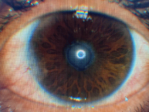
Figure 1: Pupillary seclusion in the left eye.
The posterior segment found in the left eye a pigmented hyalitis and active hyalitis on the right eye.
The optic fundus in the left eye was inaccessible due to seclusion [2,3].
The optic fundus shows in the right eye central retinal vein occlusion with macular edema, retinal periphery shows vascular sheathing and areas of ischemia [4].
The papillary excavation was pathological and intraocular pressure (IOP) was high.
The patient was put on hypotonic topical therapy (Figure 2-4).
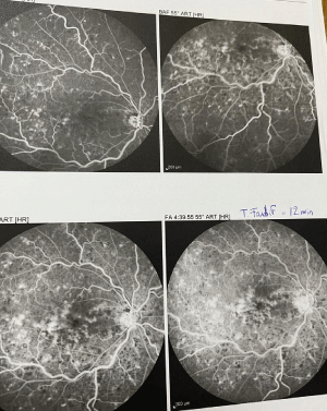
Figure 2: Ocular angiography: Central retinal vein occlusion in the right eye.
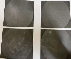
Figure 3: Peripheral vasculitis with ischemic areas in the right eye.
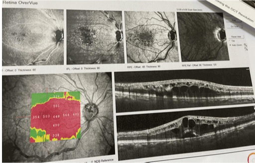
Figure 4: OCT: Macular edema on the right eye.
The patient received a corticosteroid bolus for 3 days with an oral relay.
An internal medicine opinion was carried out and the diagnosis of Behçet’s disease was retained and the patient was put on an immunosuppressive drug [5].
The patient also received two sessions of argon laser photocoagulation in the ischemic areas [6].
Face with the persistence of the macular edema, 3 intravitreal injections of bevacizumab was performed (Figure 5).
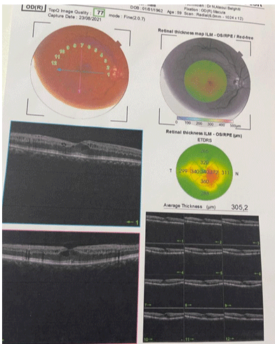
Figure 5: OCT of the right eye after 3 IVT of bevacizumab.
Despite all the therapies, the evolution was marked by the appearance of prepapillary neovessels (Figure 6).
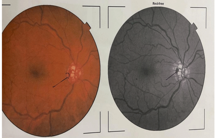
Figure 6: Prepapillary neovessels on the right eye.
So an urgent panretinal photocoagulation was performed.
After a month of PRP, the neovessels began to regress.
Discussion
Retinal vaculitis is a sight-threatening inflammatory eye condition that involves the retinal vessels. Detection of retinal vasculitis is made clinically, and confirmed with the help of fundus fluorescein angiography. Active vascular disease is characterized by exudates around retinal vessels resulting in white sheathing or cuffing of the affected vessels.
Retinal vasculitis affecting predominantly the veins (phlebitis) has been described in association with Behçet’s disease.
Fluorescein angiographic studies in Behcet’s disease show that the major retinal changes occur on the venous side with leakage from both small and large veins. Leakage is initially in the peripheral vessels but also involves the central vessels to produce macular oedema.
Patients with ischemic retinal vasculitis represent a major management problem. It is important to identify the presence of retinal ischemia in patients with retinal vasculitis because panretinal laser photocoagulation should be considered when angiographic evidence of widespread retinal nonperfusion is present, and before (or shortly after) the development of neovascularization.
The periphlebitis may cause nonperfusion of a substantial portion of the retina that may lead to proliferative vascular retinopathy with sequelae such as recurrent vitreous hemorrhage, traction retinal detachment, rubeosis iridis, and neovascular glaucoma
Conclusion
Ischemic retinal vasculitis is an important complication of Behçet’s disease, requiring emergency pan-retinal photocoagulation to avoid the development of preretinal and prepapillary new vessels which may be complicated by intravitreal hemorrhage, tractional retinal detachment or neovascular glaucoma.
References
- Abu El-Asrar AM, Herbort CP, Tabbara KF. Retinal vasculitis. Ocul Immunol Inflamm. 2005; 13: 415-433.
- Ahmed M. Abu El-Asrar, Carl P. Herbort and Khalid F. Tabbara. Differential Diagnosis of Retinal Vasculitis. Middle East Afr J Ophthalmol. 2009; 16: 202- 218.
- Perez VL, Chavala SH, Ahmed M, Chu D, Zafirakis P, Baltatzis S, et al. Ocular manifestations and concepts of systemic vasculitides. Surv Ophthalmol. 2004; 49: 399-418.
- International Study Group for Behçet’s Disease. Criteria for diagnosis of Behçet’s disease. Lancet. 1990; 335: 1078-1080.
- Sakane T, Takeno M. Novel approaches to Behçet’s disease. Exp Opin Invest Drugs. 2000; 9: 1993-2005.
- Okada AA. Drug therapy in Behçet’s disease. Ocul Immunol Inflamm. 2000; 8: 85-91.