
Clinical Image
Austin J Clin Ophthalmol. 2022; 9(2): 1129.
Posterior Scleritis Simulating a Vogt-Koyanagi-Harada Syndrome
Aachak M*, Brarou H, Boui H, Abdellaoui T, El Asri F, Mouzarii Y, Reda K and Oubaaz A
Department of Ophthalmology, Military Hospital, Mohammed V University, Rabat, Morocco
*Corresponding author: Meryem Aachak, Department of Ophthalmology, Military Hospital, Mohammed V University, Rabat, Morocco
Received: April 05, 2022; Accepted: April 27, 2022; Published: May 04, 2022
Clinical Image
A 35 years old patient presented with complaints of diminution of vision in the right eye with ocular pain without any redness nor other clinical signs. Ophthalmic examination revealed a best corrected VA of counting fingers in the right eye. Slit lamp examination of the RE was normal. Fundus evaluation revealed a grade II papillary edema with macular retinal folds (Figure 1).
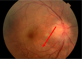
    Figure 1: Fundus evaluation showed a grade II papillary edema with macular

retinal folds (red arrow).
Retinal fluorescein angiography showed a choroïdal filling delay extending to the arteriel phase and a filling delay of a serious retinal detachment located in the macular and the temporal superior area (Figure 2) well shown in the OCT spectral domain (Figure 3).

Figure 2: Retinal fluorescein angiography of the right eye showed a choroïdal filling delay extending to the arteriel phase and late optic disc hyperfluorescence.
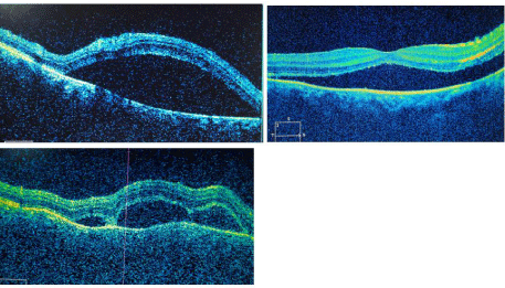
    Figure 3: OCTs of the right eye showing serous retinal detachment located in

the macular and the temporal superior area.
Numerous testings came back negative; brain angiography MRI, microbiological analysis of the cerebrospinal fluid, HSV, CMV, EBV, HIV and hepatitis viruses serologies.
Before this clinical presentation a Vogt-Koyanagi-Harada disease diagnosis was suspected. However, the biological testings, the analysis of the cerebro spinal fluid, the cutaneous and ORL clinical exams were all normal.
B scan ultrasonography revealed increased scleral tickening of 3.02mm with a T sign confirming the diagnosis of the posterior scleritis (Figure 4).
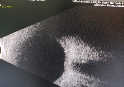
    Figure 4: B scan ultrasonography (red eye) revealed increased Scleral

thickening of 3.02mm with T sign.
Pulse steroid therapy was initiated with an injection of 1-gram intravenous methylprednisolone for 3 days followed by oral prednisolone 60mg once daily tapered gradually. The patient noticed an improvement in vision acuity of 8/10 associated with a decreasing of the serous retinal detachment bubbles (Figure 5 and 6) and of the papillary edema.
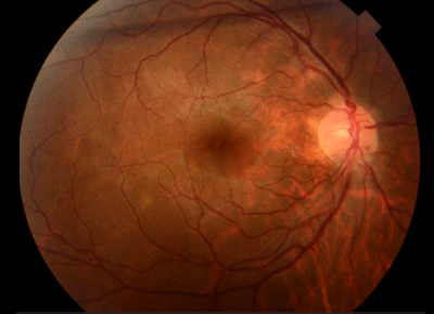
    Figure 5: Fundus evaluation 2 weeks after initiating corticosteroid therapy

showing regression of papillary edema with macular retinal folds.
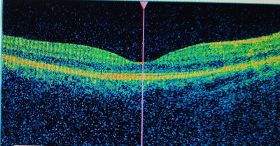
Figure 6: Macular OCT of the right eye after corticosteroid therapy.
The clinical signs of posterior scleritis include typically increase in ocular pain during eye movement with sometimes a decrease in visual acuity. The diagnosis is revealed on fundus evaluation through retinal folds or choroidal detachment, exsudative retinal detachment, papillary edema or choroidal effusions. Ocular hypertension and orbitary signs (ophthalmoplegia, diplopia, proptosis) are possible [1].
Posterior scleritis is a disease with a strong feminine predominance and can occur at any stage of life [2].
B scan ultrasonography is the key exam for the diagnosis of the posterior scleritis. It shows an increased thickness of the posterior coats (>2.0mm) and the presence of fluid in the sub-Tenon’s space responsible for the T sign. Brain MRI shows an enhancement of the posterior sclera [3].
References
- Sandra Vermeirsch, Ilaria Testi, Carlos Pavesio. Choroidal involvement in non-infectious posterior scleritis. Journal of Ophthalmic Inflammation and Infection. 2021; 11: 41.
- E Hérona, T Bourcier. Sclérites et épisclérites Scleritis and episcleritis. Journal français d’ophtalmologie. 2017.
- Lydia Montolio-Chiva. Sclérite postérieure bilatérale révélant une artérite à cellules géantes. Revue du Rhumatisme. 2021; 88: 394-395.