Clinical Image
Austin J Clin Ophthalmol. 2022; 9(2): 1130.
Both Central Retinal Venous Occlusion and Cilioretinal Artery Occlusion Secondary to Hyperhomocysteinemia. A Case Report
Aachak M*, Brarou H, Jeddou I, Boui H, Abdellaoui T, El Asri F, Mouzarii Y, Reda K and Oubaaz A
Department of Ophthalmology, Mohammed V University, Morocco
*Corresponding author: Meryem Aachak, Department of Ophthalmology, Military Hospital, Mohammed V University, Rabat, Morocco
Received: May 06, 2022; Accepted: June 01, 2022; Published: June 08, 2022
Clinical Image
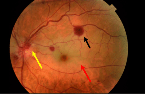
A 50 YO female patient with no medical history presented to the ophthalmologic emergency department for an acute decreasing of visual acuity in the left eye without any redness nor ocular pain. Ophthalmic examination revealed a best corrected VA of ‘finger counting’ in the LE. The LE slit lamp examination was normal. Fundus evaluation showed a retinal white edema in the cilioretinal artery territory associated with pre retinal hemorrhage. The foveola was spared (Figure 1).

    Figure 1: Fundus photography of the left eye showed a retinal white edema

in the cilioretinal artery territory (red arrow) associated with pre retinal

hemorrhage (black arrow) and peripapillary flame-shaped retinal hemorrhage

(yellow arrow).
Clinical examination of the RE was normal and a best corrected VA of 20/20.
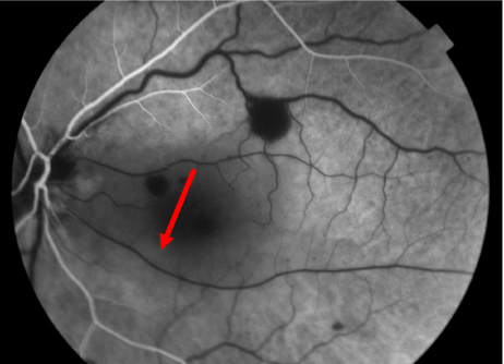
Retinal fluorescein angiography showed a cilioretinal artery perfusion delay in the LE with a mask effect related to re retinal hemorrhage. OCT demonstrated a tickening of the inner retinal layers. An initial testing (VS, CRP, fasting blood sugar, lipid profile, ultrasound of supra aortic trunks, ETT) came back negative.
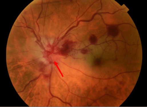
The evolution was marked by the appearance of a papillary edema associated with peri papillary flame-shaped hemorrhages (Figure 2).

    Figure 2: Fundus photography 3 days after first examination showed

papillary edema (red arrow) and worsening of peri papillary flame-shaped

hemorrhages.
It is a mixed occlusion of both the central retinal vein and the cilioretinal artery. Blood screening showed an hyperhomocysteinemia of 19 umol/l.
Homocysteine is a non-proteinogenic a-amino acid. It is biosynthesized from methionine [1].
Hyperhomocysteinemia is a medical condition characterized by an abnormally high level of homocysteine in the blood, conventionally described as above 15 μmol/L. Elevated homocysteine is a known risk factor for cardiovascular disease as well as thrombosis [2].
Cilioretinal artery occlusion is a distinct clinical entity since the cilioretinal artery derive from the posterior ciliary artery instead of the central retinal artery (Figure 3).

    Figure 3: Early-stage retinal fluorescein angiography showed a cilioretinal

artery perfusion delay in the left eye (red arrow) with a mask effect related to

re retinal hemorrhage.
Three types of cilioretinal artery occlusion ‘CRAO’are described in the litterature; isolated Non-arteritic CRAO, arteritic CRAO and non arteritic CRAO associatd with a central retinal vein occlusion [3].
References
- Jean-Philippe Galanaud. Hyperhomocystéinémie et thromboses artérielles. Maladies Artérielles. 2016; 505-507.
- Capuano V, Sellam A. Occlusion mixte simultanée d’une artère ciliorétinienne et de la veine hémisphérique supérieure : quel est le rôle de l’hyperhomocystéinémie. Journal Français d’Ophtalmologie. 2016; 39: 279- 281.
- Pierru A, Héron E, Girmens JF, Paques M. Vitré-Rétine : Occlusions veineuses rétiniennes. EMC Ophtalmol. 2015.