Case Report
Austin J Clin Ophthalmol. 2022; 9(3): 1134.
Descemet’s Membrane Detachment Following Corneal Suture Removal: Case Report
Belidi HE*, Saoiabi Y, Boumehdi I and Cherkaoui O
Department of Ophthalmology, Specialty Hospital, Rabat, Morocco
*Corresponding author: H El Belidi Department of Ophthalmology, Specialty Hospital, Rabat, Morocco
Received: November 07, 2022; Accepted: December 22, 2022; Published: December 28, 2022
Abstract
Introduction: Descemet’s Membrane Detachment (DMD) is the separation of the descemet’s membrane from the overlying corneal stroma. It is a rare and a potential vision-threatening complication of cataract surgery, with incidence rates being reported at 0.044–0.5% after phacoemulsification and 2.5% after extra capsular extraction. In these cases, DMD is mostly seen during surgery or in the early post-operative period and it is associated to surgical technique, surgical equipment or genetic factors. It is one of the most serious complications of cataract surgery, leading to irreversible corneal decompensation. Small DMD may resolve spontaneously, but most large detachments require interventions such as pneumodescemetopexy.
Case Report: We report a 70-year-old female patient, without any underlying disease, presented with the complaint of decreased vision in her pseudophakic right eye after a 15-weeks silent post-extra capsular cataract extraction period and two weeks after corneal suture removal. On slit-lamp examination, massive corneal edema was noticed on the temporal periphery with the involvement of the visual axis. Anterior segment optical coherence tomography revealed the presence of DMD in the superotemporal quadrant. To provide reattachment of DMD, we performed an anterior chamber tamponade with air. No complication associated with descemetopexy was noticed during recovery. Total Descemet’s membrane reattachment was achieved.
Discussion: Among all intraocular surgeries, DMD is most commonly described after cataract surgery. It generally occurs in early- postoperative period and late-onset DMD have been reported less frequently. It presents as localized or diffuse corneal edema. Anterior segment optical coherence tomography examination can be clearly used for observation of the position and the range of DMD. There are several ways to manage DMD: medical treatment, pneumodescemetopexy, penetrating keratotplasty and endothelial keratoplasty. To the best of our knowledge, this is the first reported case of DMD after corneal suture removal.
Conclusion: DMD after cataract surgery is associated with a variety of factors. Anterior segment optical coherence tomography examination can be used to find clear detachment of the descemet’s membrane. The position of detachment and surgical incision were found to be closely related. The location and the scope of detachment can be used to guide clinical treatments and improve prognosis of patients.
Keywords: Descemet membrane detachment; Anterior segment optical coherence tomography; Descemet membrane; Descemetopexy; Cataract surgery
Introduction
Descemet’s membrane is a thick basement membrane, measuring 5–10 μm in thickness, It is built bytwo different layers, an anterior layer formed by proteoglycans and collagen lamellae, and a posterior layer,adjacent to the endothelium, produced by the endothelial cells [1]. It contributes in maintaining the corneal transparency along with the endothelium. Descemet’s Membrane Detachment (DMD) is the separation of the descemet’s membrane from the overlying corneal stroma. It is a rare and a potential vision-threatening complication of cataract surgery.
DMD has also been reported after various other procedures as iridectomy, vitrectomy, cyclodialysis cleft creation, holmium laser sclerostomy, viscocanalostomy, penetrating keratoplasty and trabeculectomy [2].
The incidence of DMD after cataract surgery varies according to the surgical technique. It is a rare complication with incidence rates being reported at 0.044–0.5% after phacoemulsification and 2.5% after extra capsular extraction [1]. In these cases, DMD is usually noticed during surgery or in the early post-operative period associated with genetic factors and surgical technique. Late-onset DMD after cataract surgery is rarely reported [3].
DMD is one of the most serious complications of cataract surgery, leading to irreversible corneal decompensation. The natural history of DMD goes from spontaneous resolution to chronic detachment.
Case Report
We report a 70-year-old female, without any underlying disease. She had a long-term follow-up in our department for cataracts of the bilateral eyes: dense (Grade 4 in The Oxford Clinical Cataract Classification and Grading System) nuclear cataract was observed in her right eye and moderately dense in her left eye with pseudoexfoliation syndrome. The intraocular pressure was 17mmhg in both eyes. We performed a routine extra capsular cataract surgery for the right eye and the procedure was uneventful.
Following the cataract surgery, the cornea was transparent and the Best Corrected Visual Acuity (BCVA) after7 days of suture removal was 0.1 LogMAR.
The patient presented with the complaint of decreased vision in her pseudophakic right eye after 15-weeks silent period post-extra capsular cataract extraction and two weeks after a corneal suture removal. BCVA was decreased to 0.7 LogMAR in the right eye.
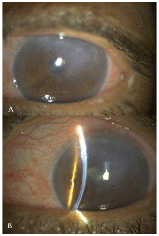
On slit-lamp examination, massive corneal edema was noticed on the temporal periphery with the involvement of the visual axis (Figure 1). Intraocular Pressures (IOP) were 14.0 mmHg in the right eye and 16.5 mmHg in the left eye. No other abnormality was observed in the slit lamp and ultrasound examination was normal.

    Figure 1: (A): A slit-lamp diffuse photograph showing stromal edema.

(B): A slit-lamp slit photograph showing a planar DMD with

stromal edema. DMD: Descemet Membrane Detachment.
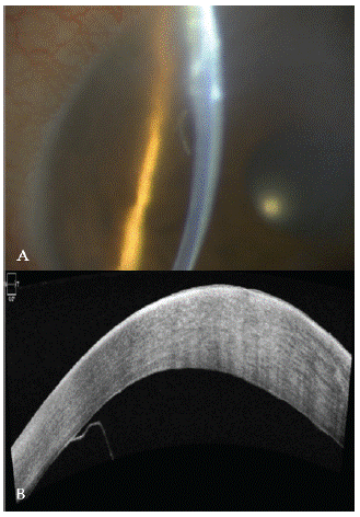
An Anterior Segment Optical Coherence Tomography (ASOCT) was performed and carefully examinated for the presence of stromal clefts. A rhegmatogenous DMD was revealed with the highest point of detachment being at the superotemporal quadrant without rolled scroll (Figure 2).

    Figure 2: (A) The slit beam showing DMD. (B): Anterior segment

optical coherence tomogram showing a planar DMD.
To provide reattachment of DMD, we decided to perform an anterior chamber tamponade with air, under a topical an-esthesia: an anterior chamber paracentesis was performed at 3 O’clock via the clear cornea near the limbus. After expressing out some aqueous humor by depressing gently the posterior lip of the paracentesis, the anterior chamber was filled with sterilized air. The air was allowed to completely fill the anterior chamber for five minutes, and then it was partially removed to avoid pupillary block. The paracentesis wound was left suture less. The patient intraocular pressure at one and two hours after surgery were normal at 14-17 mmHg respectively. He was advised to maintain a left cheek to pillow position. Antibiotics and topical corticosteroids and topical hypertonic saline were administered. One day after the procedure, the edema was lightened, and the cornea had regained much of its clarity. The patient was then recommended to lean into a left lateral position.
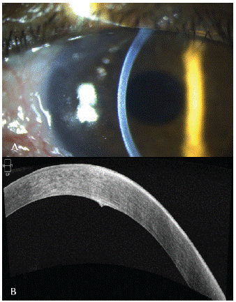
No complication associated with descemetopexy was noticed during recovery. Total Descemet’s membrane detachment reattachment was achieved (Figure 3). The BCVA increased gradually to 0.1 LogMAR, but left a small Descement’s membrane scar. During the 6-month follow-up period, No recurrent DMD was observed.

    Figure 3: (A): photography; (B): AS OCT: result at one week postoperatively.

The edema has totally regressed, the membrane is perfectly

reapplied.
Discussion
DMD was first described by Samuels in 1928 during iridectomy [4]. In 1964, Scheie reported this complication after cataract surgery [5].
DMD is a rare complication following cataract surgery. According to the literature, the incidence rates being reported at 0.044–0.5% after phacoemulsification surgery and 2.5% after extra capsular extraction [6-7]. According to Monroe, this percentage is underestimate because of the large number of DMD that are only detectable by gonioscopy. In his study, Monroe estimates the prevalence of localized DMD is 43% [8]. DMD after uncomplicated cataract surgery is even more unusual and may be the consequence not only of the surgery itself but also of an underlying anatomic abnormality [9]. According to Ti and Chee, the main risk factors of DMD are older age, nuclear sclerosis grade > 4 cataract on LOCS III scale, and preexisting endothelial disease. DMD generally occurs at the main clear corneal incision (87.5%) [10].
Endothelial diseases including Fuchs dystrophy have been documented to be highly correlated with DMD after cataract surgery. Patients with pseudoexfoliation were found to have focal accretion of the pseudoexfoliation material onto or within DM and thickness irregularity of DM. Because of these reasons, DM can detach during cataract surgery [11]. Our case had the risk factor of older age, pseudoexfoliation syndrome and dense cataract.
Late-onset DMD following uneventful cataract surgery has been reported in only seven cases in the literature, and as per our knowledge our case is the first reported one of delayedonset descemet’s membrane detachment after corneal suture removal following uncomplicated cataract surgery.
The management of DMD depends on various factors such as the duration of watchful observation, the area of the detachment, and the degree of anteroposterior separation from the stroma [12]. Due to the unknown course of DMD, there is no consensus regarding the treatment of the disease.
There are several ways to deal with DMD. It is crucial to determine early in the course whether to manage the case with medication or to intervene surgically. The sites, height, extent of DMD, as well as the presence of any scrolled edges are few important parameters that must to be considered while making this decision [2].
The Mackool and Holtz classified DMD into two groups: planar (when there is 1mm or less separation of the DM from the stroma) and non-planar (DMD exceed 1 mm of separation) [12]. In another classification, Samarawickrama et al differentiate peripheral detachments from those reaching the center of the cornea [13].
DMD treatment can be medical, with topical steroids and hyperosmotic agents, if the detachment is small, planar, with nonscrolled edges and non- vision threatening [14-15]. The advantage of this approach is to avoid surgery, to reduce the risk of infection and further damage to the corneal endothelium. Medical treatment seems to be adequate in many cases and may be an appropriate initial therapy. Small detachments are frequent and reattach spontaneously a few days after the surgery. However, an extended corneal edema and a delay of treatment can lead to an irreversible corneal opacification [16]. Surgical intervention as the primary treatment for DMD is indicated for cases with nonplanar DMD, length of DMD >2 mm, scrolled edge, or involving the central cornea. Furthermore, all cases of DMD that are not resolved after conservative therapy request surgical intervention [17].
Descemetopexy has become the gold standard treatment for the management of DMD with success rates reported to be 90– 95%. Tamponading agents successfully used include 100% air, perfluoropropane (12–14% C3F8) and sulphur hexafluoride (14– 20% SF6). Air is usually preferred for many reasons, including: rapid absorption, lower cost, and less risk of pupillary or endothelial toxicity [18-19].
Long-standing gases SF6 and C3F8 were used for cases of failing reattachment with air or of prolonged DMD. Repeated injections with air or other gases are sometimes required to reposit the DMD [19-20]. Descemetopexy with viscoelastic agents has also been reported. Due to the high risk of increasing the IOP, this method has been used only in recalcitrant DMD despite tamponading with gases [21-22].
One study reported the use of Nd: YAG laser to manage a late-onset fluid-filled DMD after cataract surgery [23].
Various authors have described the use of transcorneal suture of DMD with variable success. It is usually combined with descemetopexy [24-25].
If descemetopexy fails to reattach Descemet’s membrane, further endothelial keratotplasty or penetrating keratoplasty are suggested for visual rehabilitation [26-28].
Conclusion
DMD is a rare and a potential vision-threatening complication of any intraocular surgery particularly cataract surgery. The use of ASOCT can help in early diagnosis. The management of DMD varies from case to case, different treatment options are available but the optimal treatment depends on the type and extent of DMD. Prompt diagnosis and timely management determine the prognosis.
Disclosure of Interest
The authors declare that they have no competing interest.
References
- Zhang X, Jhanji V, Chen H. Tractional Descemet’s membrane detachment after ocular alkali burns: case reports and review of literature. BMC Ophthalmol. 2018; 18: 256.
- Singhal D, Sahay P, Goel S, Asif MI, Maharana PK, Sharma N. Descemet membrane detachment. Surv Ophthalmol. 2020; 65: 279- 293.
- Kocak Altintas AG, Ilhan C. Successful treatment of late onset post-phacoemulsification Descemet’s membrane detachment. Ther Adv Ophthalmol. 2019; 11: 2515841419853691.
- Samuels B. Detachment of Descemet’s Membrane. Trans Am Ophthalmol Soc. 1928; 26: 427-37.
- SCHEIE HG. STRIPPING OF DESCEMET’S MEMBRANE IN CATARACT EXTRACTION. Trans Am Ophthalmol Soc. 1964; 62: 140-52.
- Dowlut SM, Brunet M. Décollement de la membrane de Descemet dans la chirurgie de la cataracte [Detachment of Descemet’s membrane in cataract surgery]. Can J Ophthalmol. 1980; 15: 122-4.
- Emery JM, Wilhelmus KA, Rosenberg S. Complications of phacoemulsification. Ophthalmology. 1978; 85: 141-50.
- Monroe WM. Gonioscopy after cataract extraction. South Med J. 1971; 64: 1122-4.
- Das M, Begum Shaik M, Radhakrishnan N, Prajna VN. Descemet Membrane Suturing for Large Descemet Membrane Detachment After Cataract Surgery. Cornea. 2020; 39: 52-55.
- Couch SM, Baratz KH. Delayed, bilateral Descemet’s membrane detachments with spontaneous resolution: implications for nonsurgical treatment. Cornea. 2009; 28: 1160–3.
- Naumann GOH, Schlotzer-Schrehardt U. Keratopathy in pseudoexfolia- tion syndrome as a cause of corneal endothelial decompensation. Ophthalmology. 2000; 107: 1111-1124
- Mackool RJ, Holtz SJ. Descemet membrane detachment. Arch Ophthalmol. 1977; 95: 459-63
- Mulhern M, Barry P, Condon P. A case of Descemet’s membrane detachment during phacoemulsification surgery. Br J Ophthalmol. 1996; 80: 185-6.
- Kumar DA, Agarwal A, Sivanganam S, Chandrasekar R. Height-, extent-, length-, and pupil-based (HELP) algorithm to manage post-phacoemulsification Descemet membrane detachment. J Cataract Refract Surg 2015; 41: 1945-53.
- Kim IS, Shin JC, Im CY, Kim EK. Three cases of Descemet’s membrane detachment after cataract surgery. Yonsei Med J. 2005; 46: 719-23.
- Marcon AS, Rapuano CJ, Jones MR, Laibson PR, Cohen EJ. Descemet’s membrane detachment after cataract surgery: management and outcome. Ophthalmology. 2002; 109: 2325-30.
- Weng Y, Ren YP, Zhang L, Huang XD, Shen-Tu XC. An alternative technique for Descemet’s membrane detachment following phacoemulsification: case report and review of literature. BMC Ophthalmol. 2017; 17: 109.
- Chaurasia S, Ramappa M, Garg P. Outcomes of air descemetopexy for Descemet membrane detachment after cataract surgery. J Cataract Refract Surg. 2012; 38: 1134-9.
- Jain R, Murthy SI, Basu S, Ali MH, Sangwan VS. Anatomic and visual outcomes of descemetopexy in post-cataract surgery descemet’s membrane detachment. Ophthalmology. 2013; 120: 1366–72.
- Ti SE, Chee SP, Tan DT, Yang YN, Shuang SL. Descemet membrane detachment after phacoemulsification surgery: risk factors and success of air bubble tamponade. Cornea. 2013; 32: 454-9.
- Sonmez K, Ozcan PY, Altintas AG. Surgical repair of scrolled descemet’s membrane detachment with intracameral injection of 1.8% sodiumhyaluronate. Int Ophthalmol. 2011; 31: 421–3.
- Donzis PB, Karcioglu ZA, Insler MS. Sodium hyaluronate (Healon) in the surgical repair of Descemet’s membrane detachment. Ophthalmic Surg. 1986; 17: 735-7.
- Rathi H, Venugopal A, Rengappa R. Case of Late-Onset Fluid-Filled Descemet Membrane Detachment After Cataract Surgery and Its Management Using the Nd: YAG Laser. Cornea. 2016; 35: 897-9.
- Amaral CE, Palay DA. Technique for repair of Descemet membrane detachment. Am J Ophthalmol. 1999; 127: 88-90.
- Jeng BH, Meisler DM. A combined technique for surgical repair of Descemet’s membrane detachments. Ophthalmic Surg Lasers Imaging. 2006; 37: 291-7.
- Zhou S-Y, Wang C-X, Cai X-Y, Liu Y-Z. Anterior seg- ment OCT-based diagnosis and management of Descemet’s membrane detachment. Ophthalmol J Int Ophtalmol Int J Oph- thalmol Z Augenheilkd. 2012; 227: 215-22.
- Kim JJ, Kim HK. Descemet membrane stripping endothe- lial keratoplasty for Descemet membrane detachment following phacoemulsification. Can J Ophthalmol J Can Ophtalmol. 2015; 50: 73-6.
- Samarawickrama C, Beltz J, Chan E. Descemet’s mem- brane detachments post cataract surgery: a management paradigm. Int J Ophthalmol. 2016; 9: 1839-42.