Case Report
Austin J Clin Pathol. 2014;1(2): 1006.
Distinguishing Primary Pulmonary Squamous Cell Carcinoma from Metastatic Squamous Cell Carcinoma of the Cervix: Utility of p16
Essel Marie B de Leon*, Jaishree Jagirdar and Nicole Riddle
Department of Pathology, University of Texas, USA
*Corresponding author: Essel Marie B de Leon, Department of Pathology, University of Texas, Health Science Center, Mail Code 7750, 7703 Floyd Curl Drive, San Antonio, Texas, 78229-3900, USA
Received: April 10, 2014; Accepted: May 02, 2014; Published: May 06, 2014
Keywords
Squamous cell carcinoma of the cervix; Lung carcinoma; p16
Abbreviations
CT– Computed Tomography; H&E – Hematoxylin and Eosin; HPV– Human Papilloma Virus; ISH – In–Situ Hybridization; SCC – Squamous Cell Carcinoma.
Background
Pulmonary metastases from squamous cell carcinoma (SCC) of the cervix are uncommon. It is difficult to distinguish SCC of different primary sites based on histologic features, and without appropriate molecular markers and comparative mutational profiling their metastatic nature can only be implied [1]. P16 immunostain is a wellknown excellent surrogate marker for human papilloma virus (HPV) infection in SCC of the cervix with strong expression in more than 95% of cases [2]. Recently, several studies have assessed the utility of p16 expression as a surrogate marker for HPV infection in noncervical primary sites, including lung, oropharynx, nasopharynx, head and neck, esophagus and skin. This letter is to discuss the diagnostic pitfall in squamous cell carcinoma in the lung in a patient with a previous history of cervical squamous cell carcinoma.
Figure 1: The original cervical carcinoma of the cervix (A) showed a poorly differentiated squamous cell carcinoma. The pulmonary squamous cell carcinoma (B) has similar cytomorphologic features as the original cervical carcinoma. (H&E, original magnification x200 [A] and [B]).
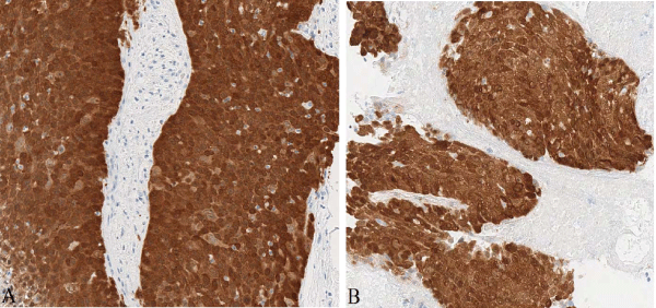
Figure 2: Immunohistochemical staining for p16 showed positive nuclear and cytoplasmic staining in the original cervical SCC (A) and the pulmonary SCC (B). (H&E, original magnification x200 [A] and [B])
Case Presentation
A 40–year–old, female, former smoker, with a history of Stage IIB cervical SCC treated exploratory laparotomy, total abdominal hysterectomy, bilateral salphingo–oophorectomy and chemoradiation presented 7 months later with a one month cough. Chest computed tomography (CT) scan showed a solitary 4.6 cm left lung mass. Transbronchial biopsy was performed and revealed squamous cell carcinoma. The original cervical squamous cell carcinoma and the lung carcinoma have similar morphologic features (Figure1). P16 immunostaining was found in both tumors (Figure 2). HPV (16⁄18⁄6⁄11) genomes by in–situ hybridization (ISH) were not detected.
Conclusion
This case illustrates a potential diagnostic pitfall in a patient with a previous history of cervical squamous cell carcinoma now presenting with solitary lung mass with squamous cell carcinoma. There are still no definitive immunohistochemical markers that candifferentiate between primary pulmonary SCC and cervical SCC metastatic to the lung. It was previously reported that p16 can beused differentiate between primary pulmonary SCC and cervical SCC metastatic to the lung [2]. However a more recent study suggeststhat P16 immunostaining cannot be assumed to signify metastasis from a primary SCC of the cervix since 43% of head and neck SCCand 35% of primary pulmonary SCC can also be p16 positive [3]. Inaddition, Yanagawa et al, found P16 positive immunostaining in 137 (40.8%) of 336 non–small cell carcinoma and five of those patients had a past history of HPV associated SCC of other sites, including three from cervix; and concluded that P16 immunostain is not a surrogate marker for HPV presence in lung cancers [1]. As in this case, P16 immunostaining was found in the cervix and lung tumors. HPV (16⁄18⁄6⁄11) genomes by ISH performed on both cervix and lung tumors were not detected and not useful in this case. It would be supportive of a metastatic cervical SCC if detected in both cervix and lung tumors since HPV DNA by ISH are detected in 20% to 90% depending on geographic locations [4]. This case points out that the distinction between a lung squamous cell cancer being a second primary or a metastasis based on histology, p16 immunostain and HPV DNA by ISH remains difficult and still depends on clinical correlation. The advanced stages of the cervix SCC and former smoking status in this patient are highly suggestive of lung metastasis. However, a primary lung cancer may be considered due to its solitary nature.
References
- Yanagawa N, Wang A, Kohler D, Santos Gda C, Sykes J, Xu J, et al. Human papilloma virus is rare in North American non-small cell lung carcinoma patients. Lung Cancer. 2013; 79: 215-220.
- Wang CW, Wu TI, Yu CH, Wu YC, Teng YH, Chin SY, et al. Usefulness of p16 for Differentiating Primary Pulmonary Squamous Cell Carcinoma From Cervical Squamous Cell Carcinoma Metastatic to the Lung. Am J Clin Pathol. 2009; 131: 715-722.
- Doxtader EE, Katzenstein AA. The relationship between P16 expression and high-risk human paillomavirus infection in squamous cell carcinoma from sites other that uterine cervix: a study of 137 cases. Human Pathology. 2012; 43: 327-332.
- Ostrow R, Manias D, Clark B, Okagaki T, Twiggs L, Faras A. Detection of Human Papillomavirus DNA in Invasive Carcinomas of the Cervix by in Situ Hybridization. Cancer Res. 1987; 47: 649-653.

