Case Report
Austin J Clin Pathol. 2015; 2(2): 1029.
Heterotopic Salivary Gland Tissue in the Rectum of a Patient with Eosinophilic Colitis and Redundant Colon
Ahmed S¹, Lewis M², Matuk R³ and Pisegna J4*
¹Department of Medicine, University of Cedars Sinai Medical Center, USA
²Department of Pathology, University of Cedars Sinai Medical Center, USA
³Department of Gastroenterology, University of Cedars Sinai Medical Center, USA
44Department of Gastroenterology, Digestive Diseases and Hepatology, University of Cedars Sinai Medical Center, USA
*Corresponding author: Joseph Pisegna, Department of Gastroenterology, Digestive Diseases and Hepatology, University of Cedars Sinai Medical Center, Los Angeles, USA
Received: June 22, 2015; Accepted: September 16, 2015;Published: October 03, 2015
Abstract
Although well characterized in the head and neck, heterotopic salivary gland tissue within the sub mucosa of the gastrointestinal tract is a rare finding that has not been clinically well defined. Only 4 reports of heterotopic salivary gland rectal tissue have been previously reported in the literature. We review the previous literature and report on a 65 year old male referred for chronic diarrhea and fecal incontinence who was found with heterotopic salivary gland rectal tissue on colonoscopy. Unique features that have not been previously described within this cohort that were found in this patient include redundant colon and chronic colitis with eosinophilia which suggests a possible causal association.
Keywords: Heterotopic salivary gland; Heterotopic rectal tissue; Salivary gland choristoma
Case Presentation
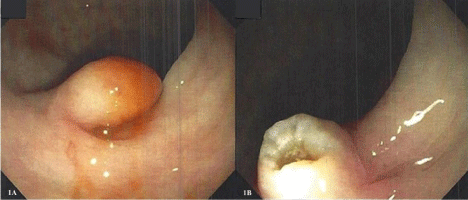
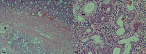
The patient was a 65 year old male who was referred for evaluation of chronic diarrhea and new onset fecal incontinence. The patient noted the onset of fecal incontinence 18 months prior to initial evaluation after a traumatic fall. Fecal incontinence was noted with exacerbations of his cervical neck pain leading to the involuntary passage of non-bloody loose stool. The patient took Lope amide for chronic non-bloody watery diarrhea, Naproxen 500 milligrams by mouth twice daily for chronic musculoskeletal pain and Omeprazole 20 milligrams by mouth daily for gastro esophageal reflux disease. On physical examination the patient pleasant and cooperative in no apparent distress and abdominal exam was benign with a reducible ventral hernia; intact norm active bowel sounds, and was soft nontender to palpation. On rectal exam the patient had no per anal anesthesia, positive anal wink test, normal sphincter tone with ability to contract voluntarily, no per anal masses and brown non-bloody stool in the rectal vault. Electromyography study of the rectum demonstrated normal nerve conduction. Colonoscopy demonstrated redundant colon, a large rectal polyp measuring 2 cm which on gross examination was concerning for sub mucosal diploma versus characinoid lesion (Figure 1), two 5 to 6 mm diminutive colonic ploys, moderate left colon diverticulitis and internal hemorrhoids. Histopathological evaluations of biopsies were significant for ectopic salivary gland tissue in the sub mucosa of the rectum with mixed serous and mutinous glands (Figure 2) as well as poly poidcecal and transverse colonic lesions with mild chronic colitis and tissue eosinophilia. Flexible sigmoidoscopy was performed 4 months after the initial colonoscopy and was unremarkable for any residual rectal lesion. Subsequent endoscopic ultrasound showed a normal 5-layer wall pattern of the rectal wall without per rectal lymphadenopathy or endosonographic abnormalities of the bladder, prostate and seminal vesicles.

    Figure 1:  (a) Endoscopic view of the sub mucosal polyploidy lesion, (b)
Residual lesion following electro cautery snare excision.

    Figure 2:  (a) Heterotopic seromucinous glandular tissue (bracket) within the
rectal sub mucosa (arrowhead). (hematoxylin-eosin stain; 4x magnification),
(b) Mucinous (solid arrow) and serous (clear arrow) acini characteristic of
salivary gland tissue (hematoxylin-eosin stain; 20x magnification).
Discussion
Heterotopic sub mucosal rectal tissue is a rare finding that has been most commonly described as involving gastric mucosa and may present either asymptomatically or with painless rectal bleeding, tenesemus with a discrete ulcer, or even fistula formation to the bladder [1-3]. Rare non-gastric heterotopic rectal tissue has been reported including pancreas [4], prostate [5], respiratory mucosa [6,7] and salivary gland tissue [2,8-10].
Rectal salivary gland heterotopias has been described in four previous cases [2,8-10]. Including the present study, four of five patients were male with an age range from 24 to 65 years old. Two patients presented with rectal bleeding whereas three patients were asymptomatic. Gastric tissue was associated with two of the five patients described. The present patient’s finding of heterotopic tissue was an incidental rectal polyp that did not extend into the adjacent structures which is consistent with previous reports.
Salivary gland tissues are generally restricted to the head and neck where this is potential for malignant transformation [11]. Outside of the rectum within the gastrointestinal tract heterotopic salivary gland tissue has also been reported in the esophagus, jejunum, large intestine and per anal “skin tag” [12-15]. Similar to the characteristics of the patients with rectal salivary gland heterotopias, patients with esophageal, jejuna, large bowel and per anal salivary heterotopias had discrete masses that did not contain any other tissue types and were not associated with malignancy.
Unique features in this patient included redundant colon and chronic colitis with eosinophilia which are not features previously associated with rectal salivary gland heterotopia. Mechanisms leading to heterotopic rectal tissue are unclear however previously have been proposed to be related to failure of developmental descent of the fetal foregut, abnormal differentiation of pluripotent endodermis stem cells or abnormal regeneration of mucosal cells leading to metaphase following inflammatory conditions. The presence of chronic colitis due to a possible allergic trigger in this patient suggests the later mechanism of action.
References
- Limdi JK, Sapundzieski M, Chakravarthy R, George R. Gastric heterotopia in the rectum. Gastrointest Endosc. 2010; 72: 190-191.
- Wolff M. Heterotopic gastric epithelium in the rectum: a report of three new cases with a review of 87 cases of gastric heterotopia in the alimentary canal. Am J Clin Pathol. 1971; 55 : 604- 616.
- Srinivasan R, Loewenstine H, Mayle JE. Sessile polypoid gastric heterotopia of rectum: a report of 2 cases and review of the literature. Arch Pathol Lab Med. 1999; 123: 222-224.
- Yamagishi H, Fukui H, Tomita S, Ichikawa K, Imura J, Ishizuka M, et al. Ectopic gastric mucosa and pancreatic ducts in the rectum. Intern Med. 2011; 50: 1587-1589.
- Dai S, Huang X, Mao W. A novel sub mucosa nodule of the rectum: A case of the ectopic prostatic tissue outside the urinary tract. Pak J Med Sci. 2013; 29: 1453-1455.
- Kawahara K, Mishima H, Nakamura S. Heterotopic respiratory mucosa in the rectum: a first case report. Virchows Arch. 2007; 451: 977-980.
- Ishida M, Iwai M, Yoshida K, Kagotani A, Okabe H. Ectopic respiratory mucosa in the rectum: the second documented case with discussion of its histogenesis. Int J Clin Exp Pathol. 2014; 7: 1819-1822.
- Shindo K, Bacon HE, Holmes EJ. Ectopic gastric mucosa and glandular tissue of a salivary type in the anal canal concomitant with a diverticulum in hemorrhoidal tissue: report of a case. Dis Colon Rectum. 1972; 15: 57-62.
- Weitzner S. Ectopic salivary gland tissue in submucosa of rectum. Dis Colon Rectum. 1983; 26: 814-817.
- Downs-Kelly E, Hoschar AP, Prayson RA. Salivary gland heterotopia in the rectum. Ann Diagn Pathol. 2003; 7: 124-126.
- Cannon DE, Szabo S, Flanary VA. Heterotopic salivary tissue. Am J Otolaryngol. 2012; 33: 493-496.
- Wang C, Chen L, Guo W, Zhu X, Liu Z. Salivary gland choriostoma in the esophagus. Endoscopy. 2014; 46: 658-659.
- Olajide TA, Agodirin SO, Ojewola RW, Akanbi OO, Solaja TO, Odesanya JO, et al. Jejunal choristoma: a very rare cause of abdominal pain in children. Case Rep Surg. 2014: 863- 647.
- Maffini F, Vingiani A, Lepanto D, Fiori G, Viale G. Salivary gland choristoma in large bowl. Endoscopy. 2012; 44: 13-14.
- Evans CS, Goldman RL. Seromucinous (salivary) ectopia of the perianal region. Arch Dermatol. 1987; 123: 1277.