Review Article
Austin J Clin Pathol. 2017; 4(1): 1045.
Spontaneous Splenic Rupture in Malaria due to Plasmodium Ovale: A Case Report
Njoumi N¹*, Naoui H², E¹ brahmi Y¹, Mouhafid F¹, Moujahid M¹, Mimouni B² and Zentar A¹
¹Department of Visceral Surgery, Military Hospital, Mohamed V University, Morocco
²Department of Parasitology, Military Hospital, Mohamed V University, Morocco
*Corresponding author: Noureddine Njoumi, Department of Visceral Surgery, Military Hospital, Hay Ryad 10100, Rabat, Morocco
Received: December 05, 2016; Accepted: January 16, 2017; Published: January 31, 2017
Abstract
Malaria is an endemic parasitic disease caused by five species of Plasmodium in many tropical and subtropical countries. Severe malaria is known to occur with Plasmodium falciparum and Plasmodium vivax. In contrast, Plasmodium ovale is considered as a cause of benign disease. Spontaneous splenic rupture in complicated ovale malaria is extremely rare. Only two cases have been reported in the literature to this day to our knowledge. This is a new case of splenic rupture during an acute malaria infection due to Plasmodium ovale in a 39-year-old man.
Keywords: Malaria; Plasmodium ovale; Splenic rupture
Abbreviations
P: Plasmodium; SRS: Spontaneous Rupture of the Spleen
Introduction
Malaria is the most important tropical parasitic disease in terms of prevalence. Its management often requires medical treatment. Rarely, it may take on a surgical appearance by involvement of the spleen in its chronic form. Spontaneous rupture of a malarial spleen is a rare and potentially lethal complication. Plasmodium (P) falciparum, P vivax, P ovale, P malariae and P knowlesi together account for nearly all human infections with the Plasmodium species [1].
Plasmodium ovale is most commonly associated with benign forms of the disease compared to other parasitic species [2]. We hereby report an uncommon case of spontaneous atraumatic splenic rupture due to Plasmodium ovale malaria that presented with hemoperitoneum.
Observation
A 39-year-old Moroccan male soldier without prior medical history was admitted to the emergency department in September, 2016. He had worked in the Democratic Republic of Congo for six months, and had returned to Morocco four months ago. He had taken anti-malarial chemoprophylaxis made of mefloquine 250 mg once a week during the first three months of his stay in this country. The patient complained of intermittent fever of 40°C, chills, headaches, generalized abdominal pain and severe malaise for five days prior to admission. On clinical examination, the patient was in good general condition, hemodynamically and respiratorily stable, He was febrile at 39.5°C and slightly pale.
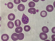
Abdominal palpation revealed diffuse tenderness mainly over the left hypochondrium with painful splenomegaly. Per rectal examination was normal. Patient’s noticeable history of travel to a malaria endemic country warranted investigations for malaria. Microscopic examination of stained peripheral blood smears showed Plasmodium ovale species with an estimated parasitaemia to 1/1000 (Figure 1). The diagnosis of tertian malaria was confirmed in three other thick blood film examinations. Molecular assays have not been performed. Laboratory evaluation showed moderate anemia (9,6 g/dL) thrombocytopenia (77.000/mm3) with elevated C-reactive protein levels of 86,7mg/L. Abdominal sonography revealed a homogeneous splenomegaly with a little peritoneal effusion. The patient was hospitalized in the intensive care unit and after 12 hours, he developed increasing abdominal pain, hypotension (90/60 mmHg) and his hemoglobin level dropped to 8 g/dL.

    Figure 1: Immature schizonts of Plasmodium ovale in giemsa colored thin
smear.
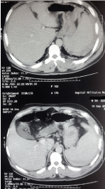
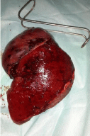
Abdominal CT showed a peri-splenic hematoma with free intraperitoneal fluid (Figure 2). Laparoscopic exploration was performed showing a large subcapsular hematoma of the ruptured spleen with active bleeding, blood clots, enlarged spleen and massive hemoperitoneum. The operation was converted into a laparotomy due to friable splenomegaly with rupture of the splenic capsule, active bleeding, and fatty infiltration. We evacuated approximately 2.5 L of hemoperitoneum. A splenectomy was performed. The spleen measured 20/14/3 cm and weighted 850 g (Figure 3). Per- and Postoperatively, the patient received four packed red blood cell transfusions and four units of fresh frozen plasma. He was administered antimalarial treatment (Artemether/lumefantrine 40/240 mg, 2 tablets twice a day) for three days and antibiotic prophylaxis (amoxicillin 3g a day) for seven days. He was vaccinated against Streptococcus pneumoniae, Neisseria meningitidis and Haemophilus influenzae. The patient had an uneventful postoperative recovery and was discharged on the seventh day on oral daily doses of Phenoxymethylpenicillin (1,000,000 IU twice a day) based prophylaxis. Pathology studies of the specimen showed malarial pigments in the macrophages and congested red pulp of the spleen.

Figure 2: Abdominal CT-scan; splenomegaly and peri-splenic hematoma.

Figure 3: Surgical specimen; enlarged spleen with rupture of splenic capsule.
Discussion
Spontaneous Rupture of the Spleen (SRS) is observed in infectious, neoplastic, hematological and systemic diseases [3]. SRS related to malaria is a rare and potentially fatal complication. Its prevalence appears to be between 1/50 000 and 1/100 000 cases [4]. Plasmodium ovale is the rarest of the five species of Plasmodium infecting humans (3-5%) and mostly associated with the benign form of disease. Reports of severe ovale infection such as splenic rupture remain rare. In contrast, splenic rupture is more common with Plasmodium falciparum and Plasmodium vivax infections [2,5,6].
The spleen plays an important role in fighting malaria, producing antibodies against the parasite. Hyperactive Malarial Syndrome also known as Tropical Splenomegaly Syndrome is the first cause for splenomegaly in endemic regions. This Splenomegaly makes the spleen more fragile and more exposed to rupture [7,8].
Three mechanisms can be involved in the splenic rupture: Episodic increase in intra abdominal pressure on a friable spleen, vascular occlusion due to reticuloendothelial hyperplasia causing a splenic infarction, and brutal splenic congestion [8,9].
Spleen enlargement is considered to be one of the most important clinical signs in the parasitic infection. Clinical presentation of SRS is not specific, delaying and often making diagnosis difficult. Left hypochondrial pain and systemic signs related to circulatory failure occurring during an attack of malaria are the most common presentations of splenic rupture [7,10].
An abdominal ultrasound can detect subcapsular hematomas, perisplenic collections, splenic rupture, and hemoperitoneum. An abdominal CT scan is more accurate and more useful in diagnosis and monitoring [8].
Concerning the treatment, choosing between surgical or conservative treatment depends on the severity of splenic lesions and the hemodynamic repercussions. Laparotomy and splenectomy remains the first-line in management of uncontrolled hemorrhagic shock secondary to splenic rupture. However, conservative treatment is often preferred in patients hemodynamically stable as in the case in traumatic spleen rupture. Antimalarial treatment should always be associated in the management of spontaneous splenic rupture due to malaria parasites [5,7].
Conclusion
Plasmodium ovale, long considered to cause benign disease, is increasingly recognised as a cause of severe malaria. Splenic rupture should be suspected in patients with malaria who present with an acute abdomen.
Consent
Written informed consent was obtained from the patient for publication of this case report and any accompanying images. A copy of the written consent is available for review by the Editor-in-Chief of this journal.
References
- Cook GC, Zumla AI. Malaria in Manson’s Tropical Diseases. London Saunders. 2009: 1201-1300.
- Strydom KA, Ismail F, Frean J. Plasmodium ovale: a case of not-so-benign tertian malaria. 2014; 10: 13-85.
- Fuks D, Browet F, Brevet M, Vidal B, Manaouil D, Regimbeau JM. Spontaneous splenic rupture during a febrile crisis of Plasmodium falciparum malaria. J Chir (Paris). 2005; 142: 403-405.
- Kianmanesh R, Aguirre HI, Enjaume F, Valverde A, Brugiere O, Vacher B, et al. Ruptures non traumatiques de la rate : trois nouveaux cas et revue de la literature. Ann Chir. 2003; 128: 303-309.
- Lemmerer R, Unger M, Voßen M, Forstner C, Jalili A, Starzengruber P, et al. Case report: Spontaneous rupture of spleen in patient with Plasmodium ovale malaria. Wien Klin Wochenschr. 2016; 128: 74-77.
- Mueller I, Zimmerman PA, Reeder JC. Plasmodium malariae and Plasmodium ovale – the ‘bashful’ malaria parasites. Trends Parasitol. 2007; 23: 278–283.
- Osman MF, Elkhidir IM, Rogers SO, Williams M. Non-operative management of malarial splenic rupture: The Khartoum experience and an international review. International Journal of Surgery. 2012; 410-414.
- Correia M, Amonkar D, Audi P, Kudchadkar S. Spontaneous rupture of a malarial spleen - A case report and review of literature. The Internet Journal of Surgery.
- Kapoor U, Chandra A, Kishore K. Spontaneous rupture of spleen with complicated falciparum malaria in a United Nations Peacekeeper: a case report. Med J Armed Forces India. 2013; 69: 288-290.
- Ozsoy MF, Oncul O, Pekkafali Z, Pahsa A, Yenen OS. Splenic complications in malaria: report of two cases from Turkey. J Med Microbiol. 2004; 53: 1255- 1258.