
Case Report
Austin J Clin Pathol. 2021; 8(1): 1072.
A Single Firm Nodule of the Upper Lip: A Case Report
Czarny K¹*, Le Pelletier F² and Ejeil AL³
¹Department of Oral Surgery, University of Paris-Saclay, France
²Department of Oral and ENT Pathology, Sorbonne University, France
³Department of Oral Surgery, University of Paris, France
*Corresponding author: Katarzyna Czarny, Department of Oral Surgery, University of Paris Saclay, France, 165 rue Jean Pierre Timbaud, 92400 Courbevoie, France
Received: July 22, 2021; Accepted: August 09, 2021;Published: August 16, 2021
Abstract
Canalicular Adenoma (CA) is a benign accessory salivary gland neoplasm. It represents less than 1% of salivary gland tumors. It occurs generally on the upper lips in female patients over 50 years of age. We report a case of canalicular adenoma that arose from buccal minor salivary glands of the upper lip without any symptomatology.
Clinically, CA appears as an often single firm mass of 1cm, sparing the main salivary glands which is not pathognomonic of canalicular adenoma. Treatment of choice is enucleation to rule out many differential diagnoses that may present clinically as an asymptomatic nodule. Recurrences are rare.
Histological features are characteristic and usually allow a definitive diagnosis to be made with confidence. However, many differential diagnoses remain to be ruled out.
Keywords: Canalicular adenoma; Minor salivary gland; Benign neoplasia; Upper lip
Abbreviations
CA: Canalicular Adenoma; PA: Pleomorphic Adenoma; PLGA: Polymorphous Low-Grade Adenoma; ACC: Adenoid Cystic Carcinoma; BCA: Basal Cell Adenoma; CK7: Cytokeratin 7; MSG: Minor Salivary Gland
Introduction
Canalicular Adenoma (CA) is a benign neoplasia of salivary glands, which account for less than 1% of salivary gland tumors [1,2]. It occurs almost exclusively in the oral cavity and involves the minor salivary glands of the upper lip mucosa in around 80% of cases [3].
The most frequent clinical presentation is a single nodule, and patients are asymptomatic or complain about discomfort which is not pathognomonic of a specific tumor.
Histological features are nevertheless evocative of the right diagnosis. Canalicular adenoma is characterized by a beading pattern of anastomosing duct/trabecular/papillary-like structures bordered by a single layer of cuboidal to tall columnar epithelial cells, embedded in a loose, not fibrous but sometimes hemorrhagic, and highly vascular connective tissue stroma [3]. However, many differential diagnoses remain to be ruled out such as basal cell adenoma, Pleomorphic Adenoma (PA), Polymorphous Low-Grade Adenoma (PLGA), or Adenoid Cystic Carcinoma (ACC).
Case Presentation
A 80-year-old female patient, treated for hypertension with 10mg of lercanidipine and for hypercholesterolemia with a statin, was referred by her general dentist for a nodule on the left part of her upper lip that appeared 4 months ago. The patient did not report any symptomatology or wound.
The extraoral examination revealed nothing unusual. Bidigital palpation of the upper lip near the left commissure revealed a firm and well circumscribed infracentrimetric nodule within the labial mucosa, without deep adherences.
Clinical diagnoses included mucocele (usually in the lower lip), venous thrombus, sialolithiasis with secondary sialadenitis, lipoma or any other benign tumor of the accessory salivary glands. Surgical excision was decided and performed under local anesthesia. During the surgical procedure, the tumor was well limited allowing easy dissection and excision. After 10 days, complete healing was achieved.
Histological examination of the surgical specimen showed a well-limited and encapsulated tumor without perineural invasion or necrosis (Figure 1 and 2). The stroma was loose with few cells. It was neither chondroid nor hyaline without adipocytes. On all the sections, the same quasi-pathognomonic aspect of cellular cords, sometimes parallels, bordering anastomosing tubes, sometimes distant, forming a “beading” pattern was observed (Figure 3). Cells were cubic-basaloid or cylindrical, in a single epithelial layer, without myoepithelial cells. The cytoplasms were syncytial (without any precise limits); the nuclei were not atypical: neither hyperchromatic nor pleomorphic and without any mitoses (Figure 4). Histological findings were consistent with a canalicular adenoma.
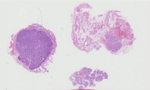
    Figure 1: Histologic picture of well limited tumor encapsulated without
perineural invasion or necrosis. Picture is consistent with a canalicular
adenoma. Hematoxylin and eosin stain, initial magnification 7x.
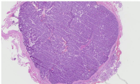
    Figure 2: Histologic picture of well limited tumor, encapsulated, without
perineural invasion. Note the healthy gland lower right. Picture is consistent
with a canalicular adenoma. Hematoxylin and eosin stain, initial magnification
20x.
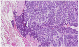
    Figure 3: Histologic picture of hemorrhagic per operative capsular effraction.
Picture is consistent with a canalicular adenoma. Hematoxylin and eosin
stain, initial magnification 80x.
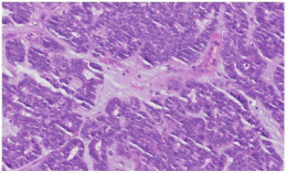
    Figure 4: Histologic picture of cubic-basaloid or cylindrical cells, with absence
of atypia and mitotic activity. Picture is consistent with a canalicular adenoma.
Hematoxylin and eosin stain, initial magnification 200x.
Discussion
Canalicular adenoma is a benign accessory salivary gland tumor that do not affect neither child nor main salivary glands [1-3].
In a literature review, Alberto J. Peraza et al. [3] found 60 articles published from 1973 to 2016, reporting a total of 430 cases. The most common location was the upper lip (66,3%). Clinically, it presented as a nodule (46.5%) in women (64%) older than 66.3 years of age. In 29.5% of patients there were no symptoms. Discomfort was reported in 28.7% of cases.
There are only a few case series, the most important of them reporting on 67 cases [4-7].
Most often, canalicular adenoma is single, but multifocal cases have been reported in the literature, most often 2 simultaneous localizations [8-14].
Other sites that may be affected are the oral mucosa [15], the palate [16] and even the bone [17].
In the present case, the patient had all the most common clinical criteria for canalicular adenoma: an 80-year-old woman with a single asymptomatic nodule of the upper lip. However, in the case of single nodule of the upper lip, other diagnostic hypotheses must be evoked. Only pathology can rule out these diagnoses.
Surgical excision is the treatment of choice and canalicular adenoma only seldom recurs (3% recurrence) [18]. It has been shown in the literature that salivary gland neoplasms may have common histologic features that sometimes make the diagnosis tricky.
Histological analysis should rule out basal cell adenoma (BCA, a benign tumor with a predilection for patients over 65 years of age), pleomorphic adenoma, chondroid syringoma (benign sudoral mixed tumor of the skin located in the dermis or subcutis, polymorphous low-grade adenocarcinoma, or adenoid cystic carcinoma [4,18]. When the histology is not specific enough, immunohistochemistry may help to take a decision (Table 1 and 2).
![]()
    Diagnosis versus Histology [4,18]
    Canalicular Adenoma CA
    Pleomorphic Adenoma PA
    Polymorphous Adenocarcinoma PAC
    Adenoid Cystic Carcinoma ACC
    Basal Cell    Adenoma BCA


    Mostly tubular
    ++++
    ++
    ++
    ++
    ++


    1 layer tubes
    ++++
    0
    0
    0
    0


    Beading
    ++++
    0
    0
    0
    0


    Mixture of pattern
    0
    +
    ++++
    ++
    +


    Hemorrhage
    +++
    0
    0
    0
    0


    Luminal balls
    +++
    0
    0
    0
    0


    Calcifications
    ++
    ++
    ++
    ++
    +


    Myxoid stroma
    +++
    +
    ++
    0
    0


    Fibrous stroma
    ++
    ++
    ++
    +++
    ++


    Peri-neural invasion
    0
    0
    +++
    +++
    0


    Infiltrative borders
    0
    0
    +++
    +++
    0


    Mitoses
    +
    0
    +
    +
    0


    0 = Never; + = Seldom; ++ = Sometimes; +++ =    Often; ++++ = Always.
Table 1: Diagnosis versus histology.
![]()
    Diagnosis versus
    Canalicular Adenoma CA
    Pleomorphic Adenoma PA
    Polymorphous Adenocarcinoma PAC
    Adenoid Cystic Carcinoma ACC
    Basal Cell Adenoma BCA


    P63 Nuclear
    0
    +++
    ++++
    +++
    +++


    PS100
    ++++
    ++++
    +++
    ++++
    ++


    CK5/6
    +++ (balls)
    ++
    ++
    +++
    +++


    CK7
    ++++
    +
    ++++
    ++
    +


    CK8
    +
    +++
    ++
    +
    +


    CK13
    +
    +++
    0
    0
    0


    CK14 luminal and myoepithelial
    +
    +++
    +++
    +++
    ++


    CK19
    +++
    ++
    +
    +
    ++


    α smooth muscle actin    myoepithelial
    0
    +++
    +
    +++
    +++


    Muscle specific actin
    0
    ++
    +
    ++
    ++


    SMmyosin
    0
    +++
    +
    +
    ++


    Calponin myoepithelial
    ++
    +++
    +
    +++
    ++


    SOX 10
    ++++
    ++++
    ++++
    +++
    ++++


    EMA luminal
    0
    ++++
    0
    ++++
    ++++


    DOG 1 luminal
    0
    ++
    +++
    ++
    ++


    CD 117
    +++
    ++
    ++
    +++
    ++


    Vimentin
    +++
    +++
    ++
    ++
    ++


    0 = Never; + = Seldom; ++ = Sometimes; +++ =    Often; ++++ = Always.
Table 2: Diagnosis versus immunochemistry.
Analysis by site or age, although non-specific, can also guide diagnosis (Table 3 and 4).
![]()
    Pathology versus site [18]
    Canalicular Adenoma CA
    Pleomorphic Adenoma PA
    Polymorphous Adenocarcinoma PAC
    Adenoid Cystic Carcinoma ACC
    Basal Cell Adenoma BCA


    Major Salivary Gland
    0
    +++
    +
    +++
    +++


    Minor Salivary Gland
    ++++
    ++
    +++
    ++
    +


    0 = Never; + = Seldom; ++ = Sometimes; +++ =    Often; ++++ = Always.
Table 3: Diagnosis versus site.
![]()
    Pathology versus age [18]
    Canalicular Adenoma CA
    Pleomorphic Adenoma PA
    Polymorphous Adenocarcinoma PAC
    Adenoid Cystic Carcinoma ACC
    Basal Cell Adenoma BCA


    Child
    0
    ++
    +
    0
    +


    Middle-age
    ++
    +++
    ++
    ++
    +++


    Elderly
    +++
    ++
    +++
    +++
    +


    0 = Never; + = Seldom; ++ = Sometimes; +++ =    Often; ++++ = Always.
Table 4: Diagnosis versus age.
Immunohistochemical analysis of canalicular adenoma may show PS100 positivity with strong cytoplasmic and nuclear staining, Cytokeratin 7 (CK7) positivity (it is common to differential diagnoses, in CA it stains all tumor cells underlying that no peripheral cells are present), P63 negativity (no nuclear staining) vimentin and actin smooth muscle negativity [4,18].
Basal cell adenoma tubular type, very rare in minor salivary gland, could be syncytial and with luminal staining by immunohistochemistry CK7 like CA, but BCA would have a hyaline stroma and only BCA would have a distinctive peripheral P63 and smooth muscle actin and vimentin positivity indicative of myoepithelial cells [4,18]. This tumor tends to occur much more commonly in the major salivary glands.
Pleomorphic adenoma is much common in minor salivary gland tumor than CA, it has an epithelial and mesenchymal component in a myxoïd stroma with sometimes cartilage or bone islands and adipocytes in the accessory salivary glands. Furthermore, there would be a continuum in standard histology between the stromal cells and those of the tubes; the appearance would be less monomorphic than for the canalicular adenoma. In PA unlike in CA, Immunohistochemistry CK7 will stain a minority of cells, and only BCA would show nuclear positivity for P63 [4,18].
Polymorphous low-grade adenocarcinoma the second most common malignancy in minor salivary gland, affects like CA mostly elderly women. The cytological morphology is bland, there is a mixture of variable patterns, stroma may be myxoid, peri neural invasion and infiltration of adjacent tissue are frequent [18]. Immunohistochemistry may help to differentiate PA from CA since the former and not the later express P63 in peripheral cells and DOG 1 in luminal ones.
Vimentin results are contradictory. In CA, they are positive according to Thompson (43/52 cases) [4] and Ferreiro (6 cases) [19], but more than 10 negative cases are also described [9,10,20-23]. In PGLA, vimentin is positive according to Furuse (10/10 cases) [22] and de Araujo [23].
Adenoid cystic carcinoma, not so rare in minor salivary gland, affects like CA mostly elderly women. Cytoplasms are scant, Nuclei are angulated with coarse chromatin, the pattern may be tubular, peri-neural invasion is frequent.
Immunohistochemistry may help to differentiate PA from CA since the former and not the later express P63 in peripheral cells, DOG 1 and CD117 in luminal ones [24,25].
Conclusion
In conclusion, keep in mind message is that the canalicular adenoma is a rare benign sometimes multifocal tumor of the MSG in the upper lip of elderly women which should be excised completely to make a confident diagnosis. His histology and immunohistochemistry are rather characteristics, but partial excision may be tricky to rule out the more frequent and recurrent pleomorphic adenoma or malignant tumors.
References
- Yih W-Y, Kratochvil FJ, Stewart JCB. Intraoral Minor Salivary Gland Neoplasms: Review of 213 Cases. Journal of Oral and Maxillofacial Surgery. 2005; 63: 805-810.
- Wang D, Li Y, He H, Liu L, Wu L, He Z. Intraoral minor salivary gland tumors in a Chinese population: a retrospective study on 737 cases. Oral Surg Oral Med Oral Pathol Oral Radiol Endod. 2007; 104: 94-100.
- Peraza AJ, Wright J, Gómez R. Canalicular adenoma: A systematic review. J Craniomaxillofac Surg. 2017; 45: 1754-1758.
- Thompson LDR, Bauer JL, Chiosea S, et al. Canalicular adenoma: a clinicopathologic and immunohistochemical analysis of 67 cases with a review of the literature. Head Neck Pathol. 2015; 9: 181-195.
- Pons Vicente O, Almendros Marqués N, Berini Aytés L, Gay Escoda C. Minor salivary gland tumors: A clinicopathological study of 18 cases. Med Oral Patol Oral Cir Bucal. 2008; 13: E582-E588.
- Levine J, Krutchkoff DJ, Eisenberg E. Monomorphic adenoma of minor salivary glands: a reappraisal and report of nine new cases. J Oral Surg. 1981; 39: 101-107.
- McMillan MD, Smith CJ, Smillie AC. Canalicular adenoma: report of five cases with ultrastructural observations. J Oral Pathol Med. 1993; 22: 368-373.
- Ortega RM, Bufalino A, Almeida LY, et al. Synchronous Polymorphous Adenocarcinoma and Canalicular Adenoma on the Upper Lip: An Unusual Presentation and Immunohistochemical Analysis. Head Neck Pathol. 2018; 12: 145-149.
- Samar ME, Avila RE, Fonseca IB, Anderson W, Fonseca GM, Cantín M. Multifocal canalicular adenoma of the minor labial salivary glands. Int J Clin Exp Pathol. 2014; 7: 8205-8210.
- Siqueira CS, Fernandes KS, Vivas APM, Pinto D dos S, de Sousa SCOM. Clinical and histological features of multifocal canalicular adenomas of the upper lip. Braz Dent J. 2013; 24: 542-546.
- Mansueto G, Falleti J, De Cecio R, Papa F, De Rosa G. Synchronous bilateral multifocal canalicular adenoma: a case report of an unusual finding. Clin Exp Dermatol. 2009; 34: e587-e589.
- Rousseau A, Mock D, Dover DG, Jordan RC. Multiple canalicular adenomas: a case report and review of the literature. Oral Surg Oral Med Oral Pathol Oral Radiol Endod. 1999; 87: 346-350.
- Nelson ZL, Newman L, Loukota RA, Williams DM. Bilateral multifocal canalicular adenomas of buccal minor salivary glands: a case report. Br J Oral Maxillofac Surg. 1995; 33: 299-301.
- Queiroz LMG, da Silveira EJD, Silva Arruda M de L, Ramos CCF. A rare salivary gland neoplasm: multiple canalicular adenoma: A case report. Auris Nasus Larynx. 2004; 31: 189-193.
- Maamouri F, Bellil K, Bellil S, et al. Canalicular adenoma of buccal mucosa. Pathologica. 2007; 99: 69-70.
- Werder P, Altermatt HJ, Zbären P, Bornstein MM. Canalicular adenoma of a minor salivary gland on the palate: a case presentation. Quintessence Int. 2009; 40: 623-626.
- Dayisoylu EH, Pampu AA, Mungan S, Taskesen F. Intra-mandibular canalicular adenoma: report of a rare case. J Pak Med Assoc. 2012; 62: 1239-1241.
- El-Naggar AK, Chan JKC, Grandis JR, Takata T, Slootweg PJ, eds. WHO Classification of Head and Neck Tumours. 4th edition. International Agency for Research on Cancer. 2017.
- Ferreiro JA. Immunohistochemical analysis of salivary gland canalicular adenoma. Oral Surg Oral Med Oral Pathol. 1994; 78: 761-765.
- Machado de Sousa SO, Soares de Araújo N, Corrêa L, Pires Soubhia AM, Cavalcanti de Araújo V. Immunohistochemical aspects of basal cell adenoma and canalicular adenoma of salivary glands. Oral Oncol. 2001; 37: 365-368.
- Matsuzaka K, Murakami S, Shimono M, Inoue T. Canalicular adenoma arising in the upper lip: review of the pathological findings. Bull Tokyo Dent Coll. 2004; 45: 229-233.
- Furuse C, Tucci R, Machado de Sousa SO, Rodarte Carvalho Y, Cavalcanti de Araújo V. Comparative immunoprofile of polymorphous low-grade adenocarcinoma and canalicular adenoma. Ann Diagn Pathol. 2003; 7: 278- 280.
- de Araújo VC, de Sousa SOM, Carvalho YR, de Araújo NS. Application of Immunohistochemistry to the Diagnosis of Salivary Gland Tumors: Applied Immunohistochemistry & Molecular Morphology. 2000; 8: 195-202.
- Khurram SA, Speight PM. Characterization of DOG-1 Expression in Salivary Gland Tumours and Comparison with Myoepithelial Markers. Head Neck Pathol. 2019; 13: 140-148.
- Andrade EP de, Teixeira LN, Montalli VAM, et al. Epithelial membrane antigen and DOG1 expression in minor salivary gland tumours. Ann Diagn Pathol. 2019; 43: 151408.