
Systematic Review
Austin J Clin Pathol. 2022; 9(1): 1078.
Treatment Protocol According to the Meniscal Tears: A Review the Literature for all Comparing Methods
Bernardino S*
Department of Orthopaedic and Trauma Surgery, ASL Bari, Italy
*Corresponding author: Saccomanni Bernardino Department of Orthopaedic snd Trauma Surgery, ASL Bari, Viale Regina Margherita, 70022, Altamura (Bari), Italy
Received: October 19, 2022; Accepted: November 10, 2022; Published: November 17, 2022
Abstract
Purpose: Menisci are fibrocartilage formations that have multiple functional roles in the knee joint. After the better understanding of their function and also the observations of the changes of the knee joint after their removal, such as osteoarthritic changes, instability and changes on the allocation of the weight, a solution of repairing the tear was a demand.
Methods: In our effort to conclude in one treatment protocol according to the meniscal tears we reviewed the literature for all the review articles of comparing methods of several suturing techniques of the meniscal tears.
Results and Conclusions: After reviewing all these articles someone could conclude that simple sutures, mostly horizontal but also vertical have more stability and they are a good and trustable solution for the suturing of a meniscal tears. They demand then very good technique and a lot of surgical time. These very important disadvantages try to solve the various meniscal implants, but with a lower stability so far.
Keywords: Μeniscal tear; Suture
Introduction
Meniscal tears are the most common intra-articular knee injury [7]. Total meniscectomy was for decades the treatment of choice for meniscal tears [7,20]. The findings of the degenerative changes that accompany the removal of the menisci in association with the detailed study of the anatomy and the function of the menisci had as result (also with the improvement of the surgical technique and the available implants) the current practice [7,21,22].
So, for the last two decades it is common knowledge that we always must try to preserve as big as possible functional part of the meniscus [7,22]. The arthroscopic partial meniscectomy has replaced the total meniscectomy that nowadays has been used only when there is no other solution. The maintenance of the complete meniscus is nowadays possible in the 10% of the meniscal tears using the suturing of the tear. It is also acceptable that minor peripheral meniscal tears can be treated conservatively [7,21].
PURPOSE OR HYPOTHESIS
The PURPOSE OR HYPOTHESIS of the study was the literature review of all the studies that compare the techniques of suturing the meniscal tears.
Suturing of the Meniscus
Although the first meniscal suturing has been reported in 1883 by Annandale [7] and Ikeuchi has started arthroscopically suturing of the menisci in 60’s, the progress of different techniques started in 80’s.
Indications of Suturing The Meniscal Tear
The indications of repairing the meniscus or for partial meniscectomy depend on different clinical parameters such as the type of the tear, the geometry, the position, the blood supply, the size, the stability, the presence of other lesions such as the tear of the anterior cruciate ligament [18]. The age is not one of the major parameters, but has relation to the type of the tear (degenerative or not) and the quality of the meniscus. One of the primary parameters is also the wish of the patient because partial meniscectomy has better direct result and easy rehabilitation but suturing has difficult rehabilitation and uncertain result [7,18]. Excellent indication is the recent vertical-longitudinal tears on the red-red zone (lateral 20-30% side of the meniscus) [6,23]. Tears more medial on the red-white zone are relative indications [6,23], but they have been reported good results on old tears and on the degenerative tears [3,6]. Suturing techniques The suturing techniques were developed with the time on an effort for the suturing to be less invasive and with less complications.
a. Open suturing. Initially open suturing technique was used but it had the opportunity to approach only peripheral tears [6,24].
b. Inside-out technique. Afterwards, the arthroscopically assisted suturing was following with an approach from inside to outside that minimizes the disadvantages of the open technique and has the opportunity of approaching all of the tears [6,25] (Figure 1).
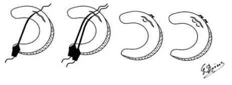
Figure 1: Inside-out suturing.
c. Outside-in technique. Nearly at the same time the arthroscopically assisted suturing was used with an approach from outside to inside that minimizes the danger of neurovascular injuries [6,7,27] (Figure 2).
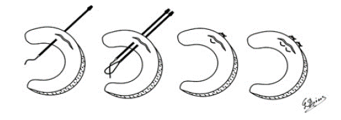
Figure 2: Outside-in suturing.
d. All inside technique. The arthroscopically all inside technique that avoids the open approach of the bursa [6,7].
Suturing Instruments
The instruments that are used nowadays for meniscal suturing are many and their use varies. The mechanical characteristics of the compression (closing the gap of the tear) but also the holding of the pieces of the meniscus until the healing are related to:
a. Their application. This has special meaning for the sutures that have a lot of different techniques. b. Technical characteristics of the material.
c. The bio absorption capacity of the material. The material are non absorbable (such as the sutures), absorbable (sutures and other) and combined.
Review Comparing Studies of Suturing the Meniscal Tears
In the first experimental comparing study of different suturing techniques, Rimmer et al. 19954 compare the failure rate of three meniscal suturing techniques, single horizontal, double vertical and single vertical. Single vertical suture was found to have better mechanical characteristics than the others, better endurance, lower cost and less surgical time. Writers conclude that this is the recommended suture for repairing the meniscal tears. In 1995 Barret et al. [5] present the suturing technique using T-Fix instrumentation (Acufex Microsurgical, Inc, Mansfield, MA). That suturing technique is suggested to central tears of the posterior horn, area very difficult for the common sutures that have to avoid the neurovascular complexes. Vertical tears, bucket handle tears, flapping tears and horizontal tears can be stabilized initially with a single suture and then using the T-Fix suture (Figure 4). In 1999 Song et al. [9] compare the failure rates and the re-tear force in the laboratory between the bio absorbable implant Meniscus Arrow (Bionx, Blue Bell, PA) and three suturing techniques (final knot, horizontal and vertical suture). They conclude that “final knot” suture has comparable failure rates to the new implant (Figure 3). In the same year Miura et al.10 introduce a new technique with a lot of knots using absorbable suture Νο 3-0 that gave very good results in the laboratory on bovine menisci.
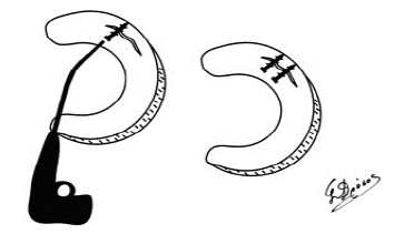
Figure 3: Suturing with arrows.
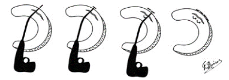
Figure 4: Suturing using Fast-Fix.
The other experimental study that compares different suturing techniques and implants of Barber and Herbert in 20002 compares the endurance of 9 new materials to the traditional single or double suturing techniques with suture and concludes that the best endurance had the double vertical suture. The writers underline that their results are only an indication and not an evidence of what happens clinically.
Later in 2001 Arnoczky et al. [12] on a big experimental study they compare the hydrolysis time of 5 absorbable implants and one suture. The implants they used were: Bionx Meniscus Arrow (Bionx Implants, Inc., Blue Bell, Pennsylvania), Linvatec BioStinger (Linvatec Corp., Largo, Florida), Innovasive Clearfix Screw (Innovasive Devices, Inc., Marlborough, Massachusetts), Surgical Dynamics S.D sorb Staple (Surgical Dynamics, Inc., Norwalk, Connecticut), Mitek Meniscal Repair System (Mitek Products, Inc., A Division of Ethicon, Inc., Westwood, Massachusetts) (Figure 5) and the suture was a vertical 2-0 Polydioxanone. The results they conclude were that in 24 weeks the hydrolysis didn’t affect the power of retention of the implants that have as ingredient the poly L-lactate (such as: Bionx Meniscus Arrow, Linvatec BioStinger, Innovasive Clearfix Screw και Surgical Dynamics S.D sorb Staple that consists of 82% of L-lactate). The implant with the ingredient polydiaxone, Mitek Meniscal Repair System, but also the suture had an important decrease of their endurance in 12 and 24 weeks. Additionally Bionx Meniscus Arrow had an important higher failure rate than all the other implants in 0 and 6 weeks except the vertical 2-0 polydiaxone suture.
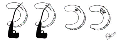
Figure 5: Suturing using Mitek.
In a study in Germany in 2001 Seil et al. [12] compare the sutures in meniscal tears with the application of circular force. They conclude that the initial force of the suture depends on the material of the suture. So, they suggest the use of PDS 0 and PDS 1 for better stability and less possibility of gapping.
Also Germans Tingart et al. 13 (2001) in a literature review asking the question “sutures or arrows for meniscal tears” and after having studied 10 studies that relate to that subject and have been published between 1996 and 2000, they conclude that: the percentage of meniscal tears healing after suturing is between 75 and 100%. The advantages of the arrows comparing to simple sutures are: less surgical time, easy surgical technique and less danger of damage of the neurovascular tissues. Also the failure rates are lower than sutures on experimental studies and similar on clinical studies. There have also been referred complications such as the infection of foreign body, lesions in articular cartilage and migration of the arrow. They suggest that maybe the combination of sutures and arrow is the better solution for meniscal tears, but randomized prospective studies are needed to verify that.
In an other study from Britain Walsh et al. [14] (2001) compare experimentally the endurance of four all inside techniques: Meniscal Arrow, Bionx Implants Inc, Meniscal Staple, Surgical Dynamics Inc, horizontal and vertical sutures. The results of the experiments showed that the classification depending on the endurance beginning with the most stable is: horizontal suture, vertical suture, meniscal arrow and meniscal staple that had ineffective holding. In 2001 also from a comparing study of Becker et al. [15] excludes the result that different meniscal implants have less endurance than simple sutures. They compare 6 different implants: Meniscus Arrow (Bionx Tampere Finland), Dart (Arthrex Naples FL), Stinger (Linvatec Largo FL), Meniscal Screw (Innovasive Marlborough MA), T-Fix (Acufex Manfield MA), Fastener (Mitek Westwood MA) with the simple horizontal 2/0 Ethibond (Ethicon Norderstedt Germany) suture. They suggest that meniscal implants should be used very close to each other for better holding and also that combination with sutures has a better result according to the stability. In 2002 Bellemans et al. [16] after an extended study they compare several implants of different sizes with simple sutures. They conclude that the stability depends on the size of the implant. So, 13 and 16 mm Bionix Arrow and T-fix Device have similar stability to the horizontal and vertical sutures. Opposite, 10 mm Bionix Arrow, S.D. Sorb Stapler and 12 mm Arthrex Meniscal Dart have very low stability.
In 2003 in a review paper for the meniscal surgery Sgaglione et al. [18] on the paragraph that refers to the surgical repair of menisci, is referred to all the known at that time implants classify them to first and second generation and without comparing them they conclude to some results according to their general use. Their queries start that all the implants make less the surgical time but have many specification to their technical implantation and they need very good technique that results to many mistakes. Also, there is a questioning about their stability according to the traditional suturing techniques. Also they think that the combination of sutures and implants maybe increase the stability. Finally they refer that in complicated tears and tears with decreased vascularity is suggested the use of traditional simple sutures for better holding.
Finally in 2005 Haas et al. [17] on a prospective study they compare the results of FasT-Fix (Smith & Nephew Adnover MA) to the traditional suturing techniques and they conclude that the results are similar. Such as the other implants the use on the anterior horn tear is very difficult. Those tears anyway are very rarely alone, often they are accompanied by bucket handle tears that extend posterior.
Discussion
With the use of the arthroscope meniscal suturing is easier. The techniques that are in use today are: all inside, outside in and inside out. The techniques that don’t need additional incisions are very attractive. Only then the technique factors that are important are the biomechanical features of the implants [2] and the factors that affect the progress of the healing of the meniscal tear such as blood supply, size of the tear, type of the tear, concomitant ACL reconstruction and rehabilitation program.
According to the bio absorbability of the implants the opinions differ. The initial opinion that the use of non absorbable sutures is preferable, because the use of absorbable ones has as a result their absorption before the healing of the tear6, is still supported [8]. Then, recent studies support that the use of non absorbable sutures causes more histological destruction to the meniscus and the around tissues [11].
After the study of all the studies that refer to the techniques on the meniscal suturing concludes someone that simple horizontal suturing, but also vertical ones have better stability and they are a good and reliable solution of suturing of a meniscal tear. They need then a very good technique and more surgical time. Those important disadvantages try to deal with the different meniscal implants but with lower rates of stability so far.
References
- Rauschmann M, Deb R, Thomann K, et al Die Geschichte der Meniskuschirurgie. Orthopade. 2000; 29: 1044-54.
- Barber F, Herbert M. Meniscal repair devices. Arthroscopy. 2000; 16: 613-8.
- Hamberg P, Gillquist J, Lysholm J. Suture of new and old peripheral meniscus tears. JBJS. 1983; 65: 193-7.
- Rimmer M, Nawana N, Keene G, Pearcy MJ. Failure strength of different meniscal suturing techniques. Arthroscopy. 1995; 11: 146-50.
- Barrett G, Richardson K, Koenig V. Technical note. T-Fix endoscopic meniscal repair: Technique and approach to different types of tears. Arthroscopy. 1995; 11: 245-51.
- Jakob R, Staubli H, Zuber K, Esser M. The arthroscopic meniscal repair. Techniques and clinical experience. Am J Sports Med. 1988; 16: 137-42.
- Morgan C, Wojtys E, Casscells C, Casscells SW. Arthroscopic meniscal repair evaluated by second-look arthroscopy. Am J Sports Med. 1991; 19: 632-8.
- Barrett G, Richardson K, Ruff C, Jones A. The effect of suture type on meniscus repair. A clinical analysis. Am J Knee Surg. 1997; 10: 2-9.
- Song E, Lee K: Biomechanical test comparing the load to failure of the biodegradable meniscus arrow versus meniscal suture. Arthroscopy. 1999; 15: 726-32.
- Miura H, Kawamura H, Arima J.: A new, all-inside technique for meniscus repair. Arthroscopy. 1999; 15: 453-5.
- Yasunaga T, Kimura M, Kikuchi S.: Histologic change of the meniscus and cartilage tissue after meniscal suture. Clin Orth Rel Res. 2001; 387: 232-40.
- Seil R, Rupp S, Jurecka C, Rein R, Kohn D. Der Einfluss verschiedener Nahtstarken auf das Verhalten von Meniskusnahten unter zyklischer Zugbelastung. Unfallchirurg. 2001; 104: 392-8.
- Tingart M, Hoeher J, Bouillon B, et al : Meniskusrefixierung: Faden oder Anker? Unfallchirurg. 2001; 104: 507-12.
- Walsh PS, Evans LS, O’ Doherty MD, Barlow IW. Failure strengths of suture vs biodegradable arrow and staple for meniscal repair: an in vitro study. The Knee. 2001; 8: 151-6.
- Becker R, Schroeder M, Staerke C, Urbach D, Nebelung W. Biomechanical investigations of different meniscal repair implants in comparison with horizontal sutures on human meniscus. Arthroscopy. 2001; 17: 439-44.
- Bellemans J, Vandenneucker H, Labey L, Van Audekercke, R. Fixation strength of meniscal repair devices. The Knee. 2002; 9: 11-4.
- Haas A, Schepsis A, Hornstein J, Edgar CM. Meniscal repair using the FasTFix all inside meniscal repair device. Arthroscopy. 2005; 21: 167-75.
- Sgaglione N, Steadman JR, Shaffer B, Miller MD, Fu FH. Current concepts in meniscus surgery: Resection to replacement. Arthroscopy. 2003; 19: 161-88.
- Boyd K, et al. Meniscus preservation; rationale, repair techniques and results. The Knee. 1983; 10: 1-10.
- Larson RL. Physical examination of rotatory instability. Clin Orth Rel Res. 1983; 172: 38-44.
- Belzer JP, Cannon WD. Meniscus tears: Treatment in the stable and unstable knee. J Am Acad Orthop Surg. 1993; 1: 41-7.
- Rath E, Richmond JC. The menisci: basic science and advances in treatment. Br J Sports Med. 2000; 34: 252-7.
- DeHaven KE. Peripheral meniscus repair. An alternative to meniscectomy. Orthop Trans. 1981; 5: 399-400.
- Cassidy RE, Shaffer AJ. Repair of peripheral meniscus tears. A preliminary report. Am J Sports Med. 1981; 9: 209-14.
- DeHaven KE, Black KP, Griffiths HJ. Open meniscus repair. Technique and two to nine years results. Am J Sports Med. 1989; 17: 788-95.
- Henning CE, Lynch MA, Clark JR. Vascularity for healing of meniscus repairs. Arthroscopy. 1987; 3: 13-8.
- Morgan CD, Casscells SW. Arthroscopic meniscus repair: a safe approach to the posterior horns. Arthroscopy. 1986; 2: 3-12.