
Case Report
Clin Oncol Res. 2018; 1(1): 1005.
Granular Cell Tumor of the Tongue: A Case Report
Koutsias G¹, Chatzifotiou E¹, Tsompanidou C², Pavlidis P³, Zagelidou E4 and Anestakis D¹*
¹Department of Histopathology, Laboratory of Forensic Medicine and Toxicology, Aristotle University of Thessaloniki, Greece
²Department of Pathology, General Hospital of Thessaloniki ‘’Agios Dimitrios’’, Greece
³Laboratory of Forensic and Toxicology, Democritus University of Thrace, Greece
4Forensic Medical Service of Thessaloniki, Ministry of Justice, Transparency, and Human Rights, Greece
*Corresponding author: Anestakis D, Department of Histopathology, Laboratory of Forensic Medicine and Toxicology, Aristotle University of Thessaloniki, Greece
Received: March 26, 2018; Accepted: May 29, 2018; Published: June 05, 2018
Abstract
Granular Cell Tumor (GCT) is a relative uncommon benign neoplasm and also a non-epithelial tumor or a tumor which is able to develop a pseudoepitheliomatous hyperplasia of the covering epithelium, as particular specialists believe [1,2]. The etiology of Granular Cell Tumor is still unknown, but some scientists support that there is a connection between Granular Cell Tumor and Schwann cell, a special form of a glial cell, as a probable cause of it. Plus, Mesenchymal cells and striated muscles are some other possible reasons that cause Granular Cell Tumor, which is characterized by accumulation of plump cells with abundant granular cytoplasm [3]. Furthermore, we cannot admit Granular Cell Tumor’s diagnostic difficulties, as its histopathological features can be confused with a well-distinguished oral squamous cell carcinoma [2]. About 45 to 65% of all cases occur in the head and neck region and of these 70% located in the oral cavity. The mean average of discovery in Granular Cell Tumor is the fourth to six decade of life, but this is not certain, as the entity could be found in any age. The middle age could depicted as the age peak of the disease, nevertheless the manifestation time varies widely [1,4]. We report a case of 52-years old female with a granular cell tumor of the tongue. There is a need for further investigation because this lesion of the tongue could be easily confused with a well-distinguished oral squamous cell carcinoma, so there is a big possibility to misdiagnose.
Keywords: Granular cell tumor; Immunohistochemistry; Tongue; Abrikossoff; Carcinoma; Neoplasm
Introduction
Granular Cell Tumor (GCT), also known as “granular cell myoblastoma” or “Abrikossoff’s tumor” was first described by Abrikossoff, who was a pathologist, in 1926. At first, GCT was originally thought to be a muscle tumor and that is why it was named granular cell myoblastoma [5].
The belief that GCT has neural origin has been more widely accepted [6]. Although, reports suggesting a potential muscular, histiocytic, fibroblastic or pericytic origin can be discovered in the literature [5,6].
As a matter of course, GCT lesions are restricted to the submucosa and present as a benign, small, asymptomatic, polypoid firm lesions most ordinarily found fortuitously on endoscopy or colonoscopy evaluation for hemorrhoids and fissures [7]. Nevertheless, the 50% of intraoral GCT lesions present pseudoepitheliomatous hyperplasia (PEH) connected with mucosal epithelium [2].
Most of the cases are asymptomatic, but there are 1-2% of the cases that are malignant [7]. It would be remiss of us if we did not mention that there is a comparison between histological findings in benign and malignant lesions. Hence, Malignant cells have the ability to rarely demonstrate cellular necrosis, enlarged nucleoli, cell elongation, pleomorphism and mitotic activity [7,8].
Commonly GCT’s histopathological aspects comprise polygonal cells with small nuclei and copious eosinophilic granular cytoplasm. The tissue size is usually 3mm or less, painless, a non-ulcerated nodule and presents as a solitary lesion [2,7]. The Granular Cell Tumor has a mean average of discovery in the fourth to six decade of life, butthis is not absolute as the entity could be found in any age. In particular, there is a greater female than male predilection of 1:5:1 [1,4,7]. The colour of this specific tumor is usually pink, but may also have a yellow hue.
Case Presentation
We report a case of 52-years old female with agranular cell tumor of the tongue. The patient visited “Agios Dimitrios” General Hospital in Greece complaining of a swelling on the dorsum of the tongue. The patient reported a history of painfully swelling of her tongue, admitting the same time that this particular pain was increasing as the time was passing by. On intraoral examination revealed a single firm, well circumscribed, non-tender lesion, measuring 0.8 x 0.6 cm on the dorsal of the tongue. Written informed consent was obtained from the patient for publication of this Case Report and any accompanying images.
Surgical excision of the lesion was done, under local anesthesia, and the Granular cell tumor was histopathologically defined. Images from this examination are shown in Figure 1 (pseudoepitheliomatous hyperplasia, acidophilic granular cytoplasm x100), Figure 2 (x200) and Figure 3 (S-100(+). X40). Also, in Figure 4 we can distinguish the lesion clinically before and after the excision. The histological examination was crucial in adjudicating biological aspects of the lesion for the final diagnosis as it showed a well-defined, not encapsulated nodule in a focal area of submucosal connective tissue.
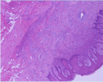
    Figure 1:  The histopathology report was as follows: “pseudoepitheliomatous
hyperplasia of the stratified squamous epithelium beneath which are large
polyhedral cells with acidophilic granular cytoplasm and pyknotic nuclei.
(X100).
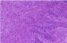
Figure 2: Granular cell tumour of tongue (X200).
There was pseudoepitheliomatous hyperplasia of the stratified squamous epithelium beneath which there were large polyhedral cells with eosinophilic granular cytoplasm and pyknotic nuclei, positive to S-100.
Results
The histological examination with the immunohistochemical findings confirmed the definite diagnosis of granular cell tumor of the tongue (Figure 2). Specifically, this examination demonstrated large polyhedral cells with acidophilic granular cytoplasm which were beneath the stratified squamous epithelium with pseudoepitheliomatous hyperplasia (Figure 1). These cells, with eosinophilic granular cytoplasm and pyknotic nuclei were positive to protein S-100 (Figure 3). After the surgical excision of the lesion, patient’s pain was decreasing gradually as she declared (Figure 4b).
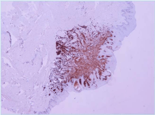
Figure 3: Granular celltumour of tongue S-100 (+)(X40).
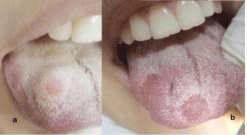
    Figure 4:  a) Clinical picture of the lesion before excision; (b) Clinical picture
of the lesional site, 3 weeks after excision.
Discussion
As we mentioned above, GCT is generally asymptomatic and due to its immunohistochemically similarity with nerve tissue, it is described as neural based tumor in nowadays. GCT is a rare soft tissue tumor, which is mostly localized in the head-neck region usually, lesions are small (0.5-3 cm) and solitary. Moreover, GCT can take an uncapsulated form or it can presents as a pseudo-invasive lesion [11], so we have to be extremely careful in the diagnosis because in the overlying lingual epithelium alternative degrees of pseudohyperplasia are often seen, which can mimic squamous cell carcinoma [5]. In this point, we have to mention that if a biopsy is performed, it should be deep enough to include underlying infiltrating cells [12].
The primary and first choice, both for diagnosis and treatment, is the surgical excisional biopsy of the tumor, which is very therapeutic in most of cases. As it is recommended, removal of the lesion should be wide enough, so we can grant oncological radicality, despite the terminal diagnosis [12]. Referring to our previous point, GCT’s histopathological aspects comprise polygonal cells with small nuclei and copious eosinophilic granular cytoplasm. An additional corollary is S100-reactivity, that demonstrates a neural origin of the tumor. Albeit, there are tumors like dermatomyofibroma, cutaneous leiomyosarcoma and benign fibrous histiocytoma, which are S100- negative in opposition to the classic Granular Cell Tumor [5].
Significantly, protein S-100 is used to identify LCs. Langerhans’ Cells (LCs) is cells with dendritic morphology located in the squamous epithelia of the epidermis and mucosa. This specific kind of cells has the ability to present antigens to CD4+ T cells and because of this fact, LCs are momentous modulators of immune response against antigens found within the epithelial tissue [9,10]. Thus, protein S-100 was first isolated from the bovine brain and then was identified in glial and Schwann cells of the nervous system ,as well as in epidermal LCs, myoepithelial cells of the salivary and mammary glands and in many other cells of various organs [9].
Furthermore, we should mention the role of c-erbB-2 oncoprotein in the immunohistochemical results of different carcinomas, as GCT consists a sector of them. The connection between the overexpression of the c-erbB-2 gene and the tumorigenesis was first reported by Schechler in 1982 in rats. From than onward, many researches came to the surface reporting the function of this oncoprotein in plenty human malignant neoplasms [10].
Conclusion
In summary, a case of granular cell tumor of the tongue is reported in a 52-years old female patient. The recognition of the unknown neoplasm might help the surgeon for the correctly diagnosis and surgical treatment of these tumors. As GCT constitutes a rare lesion, there is a big possibility to misdiagnose. For that reason, we moved to further investigations because this lesion of the tongue could be confused with a well-distinguished oral squamous cell carcinoma. A careful histological examination is the key to achieve the right diagnosis. Due to this uncommonness of GCT, patients should be informed about its metachronicity and also they should often revisit the hospital, because a close follow-up is very important in any case and especially in tumor cases.
References
- Recep Bedir, Rukiye Yilmaz, Ibrahim Sehitoglu, Abdulkadir Ozgur. Coexistence of Granular Cell Tumor with Squamous Cell Carcinoma on the Tongue: A Case Report. Iranian Journal of Otorhinolaryngology. 2015; 27: 69-74.
- Jean Carlos Barbosa Ferreira, Angélica Ferreira Oton-Leite, Rafaela Guidi, Elismauro Francisco Mendonça. Granular cell tumor mimicking a squamous cell carcinoma of the tongue: a case report. BMC Research Notes. 2017.
- Nagaraj PB, Ongole R, Bhujanga-Rao BR. Granular cell tumor of the tongue in a 6-years-old girl- a case report. Med Oral Patol Oral Cir Bucal. 2006; 11: E162-E164.
- John Spencer M. Daniels. Granular cell tumour of tongue: A case report. The Saudi Dental Journal. 2009; 21: 75–78.
- Barbieri M, Musizzano Y, Boggio M, Carcuscia C. Granular cell tumour of the tongue in a 14-years-old boy: case report. Acta Otorhinolaryngol Ital. 2011; 31: 186-189.
- Ashwini, Naveen Shankar V, Praveena V. Oral Abrikossoff Tumor: A Report of Uncommon Presentation. J Cancer Sci Ther. 2011; 3: 233-234.
- Emily F Kelly, Alan A Stein, Xiaoling Charlene Ma, Jose Yeguez. A rare case of perianal granular cell tumor: case report and literature review. Journal of Surgical Case Reports. 2017; 2017.
- Cohen MG, Greenwald ML, Garbus JE, Zager JS. Granular cell tumor--a unique neoplasm of the internal anal sphincter: report of a case. Dis Colon Rectum. 2000; 43: 1444-1446.
- Turusov VS. [Protein S-100 in the histological diagnosis of tumors]. Arkh Patol. 1990; 52: 71-78.
- Albuquerque RL, Jr, Miguel MC, Costa AL, Souza LB. Correlation of c-erbB-2 and S-100 expression with the malignancy grading and anatomical site in oral squamous cell carcinoma. Int J Exp Pathol. 2003; 84: 259–265.
- Giuliani M, Lajolo C, Pagnoni M, Boari A, Zannoni GF. Granular cell tumor of the tongue (Abrikossoff's tumor). A case report and review of the literature. Minerva Stomatol. 2004; 53: 465-469.
- Leboulanger N, Rouillon I, Papon JF, Josset P, Roger G, Garabédian EN. Childhood granular cell tumors: two case reports. Int J Pediatr Otorhinolaryngol. 2008; 72: 279-283.