
Case Report
Austin Crit Care J. 2014;1(1): 2.
Adenovirus Pneumonia in Immunocompetent Adult
Saad Khan and Kashif Hussain*
West Virginia University Hospital, USA
*Corresponding author: Kashif Hussain, West Virginia University Hospital, USA
Received: July 31, 2014; Accepted: September 05, 2014; Published: September 08, 2014
Abstract
Pneumonia in immune competent adults is rare and has been described in literature as a much more common entity in immune compromised individuals. It presents as a nonspecific viral illness which can cause severe respiratory distress and lead to respiratory failure. Early detection is essential and diagnosis should be considered in patients not responding to antimicrobial therapy. More than 80 % of the adenovirus infection occurs in children less than 4 years old. Immune compromised individuals are more susceptible to this infection. We report a case of severe adenovirus pneumonia in a young healthy immune competent adult who developed respiratory distress leading to respiratory failure.
More studies need to be conducted to understand the pathophysiology and triggering factors leading to severe adenovirus infections in individuals Adenovirus.
Introduction
Adenovirus is non enveloped lytic DNA virus and was first isolated in 1950 in adenoid tissue- derived cell cultures. Adenovirus is ubiquitous in human and animal populations, survives long periods outside a host, and is endemic throughout the year. More than 80 % of the adenovirus infection occurs in children less than 4 years old. Immunocompromised individuals are more susceptible to this infection. We report a case of severe adenovirus pneumonia in a young healthy immunocompetent adult who developed respiratory distress leading to respiratory failure.
Case Presentation
32 year old white male with no significant past medical history presented with complains of dyspnea, productive cough and chest pressure. He stated that his symptoms felt like his usual "Bronchitis" that he gets every year. No preceding pharyngitis or conjunctivitis symptoms. His symptoms progressively worsened over one week. He felt feverish but did not check his temperature.
He has also had anorexia, chills, nausea, vomiting, diarrhea and abdominal pain. He described his abdominal pain as a dull pain that worsened with his coughing. He denied any hemoptysis, weight loss, rash, and dysuria. Patient is a known smoker and smoked half a pack a day for the last 21 years and drank less than 4 beers a week. He had been unemployed for quite some time and lived with girlfriend and her two children. He denied any IV drug use or any other substance abuse. He had been tested for HIV and was negative. No history of sexually transmitted disease. He did admit to exposure to dog and a rabbit both of them outside the house. He recently did have fresh water exposure along with soil, landscaping and dust exposure.
He went to an outside hospital for an evaluation and was found to have a WBC count of 3.4 with 1 band and 80% PMNs and platelet count of 94,000. He also had hyponatremia with Na of 123 and acute renal insufficiency with CR of 1.4. He was hypoxic with pO2 49 on ABG. CXR at OSF was concerning for Right lower lobe Pneumonia and was started on broad spectrum antimicrobial therapy.
At the time of transfer to our facility his Temp was 100.4 , BP 100/66mm/hg, Pulse 114 beats/ min, resp rate of 32 breaths/ min and oxygen saturation of 93 % on 4 liters. His Arterial blood gas on arrival showed 7.45/ 26/49/21.5 with his WBC count of 3.7 with 72% PMNs and Platelet count of 72,000. CXR on admission to our hospital showed extensive consolidation in the right lower lobe and left mid lung zone. He was continued on broad spectrum antimicrobial therapy but hypoxemia and dyspnea continued to get worse.
He was started on noninvasive positive pressure ventilation but he continued to be in respiratory distress prompting admission to MICU. There he was intubated and was placed on mechanical ventilation. He was continued on broad spectrum antimicrobial therapy. His blood cultures stayed negative.
He continued to spike fever and underwent a CT scan of his chest which showed the presence of ground glass opacities in the bilateral upper lungs, left side greater than right and large consolidation in the right mid-to-lower lung zones. Work up including urine legionella, Urine Strep pneumonia, and HIV test were negative. His other blood work up including Ehrlichia, Ana plasma ,Influenza, Para influenza, human meta pneumovirus, Mycoplasma IGM antibody, Leptospira antibody , Pneumococcal antibody, Histoplasma antibody, CMV by PCR , Aspergillus galactomannan all were found to be negative.
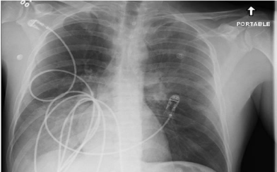
Figure 1: Right lower lobe and left mid lung infiltrates.
Figure 2: Bilateral ground glass opacities with right lung consolidations.
He then underwent a Bronchoscopy with BAL and Adenovirus DNA quantitative PCR grew >2000000 copies/ ml. Specific type of Adeno virus was type 7. All other cultures from the BAL sample remained negative. He only required supportive care treatment for his Adenovirus Pneumonia. His IGG and IGA levels were also found to be low and he was treated with one time dose of IVIG with repeat check of IGG level in 14 days. Patient completed total of 10 days of antimicrobial therapy. Patient was extubated 7 days after he presented to the MICU and was transferred to the medicine floor. His antimicrobial therapy was stopped. At Discharge his WBC count was 12.9 with Platelet count of 842. His PO2 had improved to 86 on his arterial blood gas. His CXR on discharge did show marked improvement and he did not require oxygen on ambulation at the time of discharge.
Discussion
Adenoviruses cause approximately 5% of upper respiratory tract infections and 10% of pneumonias [1]. Most commonly; upper respiratory tract disease presents as mild pharyngitis or tracheitis accompanied by coryza .The common serotypes that cause these syndromes are adenovirus 1, 2, 5, and 6 and occasionally 3 and 7. Other systemic manifestations, including fever, malaise, headache, myalgia, and abdominal pain, are common. Exudative tonsillitis and cervical adenopathy may be present and can be clinically indistinguishable from group a streptococcal infection. Among adults, adenoviral disease typically occurs in epidemics involving military recruits and, occasionally, hospitalized or institutionalized civilians. In Immunocompromised persons, dissemination and/ or severe respiratory failure develop in 10 to 30% of cases. Patients with adenovirus infection generally present with a nonspecific viral illness. Laboratory findings generally show lymhopenia, leukopenia, and thrombocytopenia. Radiological evidence shows the presence of patchy ground glass opacities bilaterally [2].
Adenovirus can be detected in the affected sites including nasopharyngeal aspirates/swabs, washings, BAL, Virus specific direct or indirect immunoflourescent stains, conventional or shell viral cultures or PCR. Viral cultures by conventional techniques are the gold standard but could be insensitive for certain samples (eg, blood) and may take up to 21 days to detect the cytopathic effect. PCR of Adeno virus DNA in plasma, urine, or infected sites may establish the diagnosis and is highly sensitive for disseminated disease. Quantification of the viral load using real-time PCR is a useful marker to assess response to therapy.
Treatment of adenovirus generally includes supportive care and no antiviral has been shown to alter the course of the disease process. In review of literature Cidofovir and Immunoglobulin therapy has been used in patients with liver and bone marrow transplant [3]. Prospective, randomized trials are needed to elucidate indications for therapy in both symptomatic and asymptomatic patients with Adenovirus infections.
Literature review does show that adenovirus pneumonia in immunocompetent adults did become a concern after Ryan et al published death in 2 military recruits in 2001[4].
Case report from Japan described an immunocompetent 42-yearold man with mild adenovirus pneumonia following pharyngitis and conjunctivitis [5]. Diagnosis was established on the basis of chest radiologic findings, detection of adenovirus type 7 DNA by PCR assay in material obtained from (BAL), and a greater than fourfold rise in adenovirus-specific antibody titers during the course of illness. The patient's self-limiting symptoms improved within 2 weeks, and chest radiologic findings improved within PCR assay of material obtained by BAL was useful for the rapid diagnosis of adenovirus pneumonia.
Rapid diagnosis of adenovirus pneumonia using PCR can help initiate adaptation of adequate infection control measures and avoidance of unnecessary use of antimicrobial therapy for bacterial infection and usage of antiviral therapy if needed for severe disease process.
References
- Barker JH, Luby JP, Sean Dalley A, Bartek WM, Burns DK, Erdman DD. Fatal type 3 adenoviral pneumonia in immunocompetent adult identical twins. 2003; 37: e142-146.
- Chong S, Lee KS, Kim TS, Chung MJ, Chung MP, Han J. Adenovirus pneumonia in adults: radiographic and high-resolution CT findings in five patients. AJR Am J Roentgenol. 2006; 186:1288-1293.
- Ribaud P, Scieux C, Fraymuth F, Morinet F, Gluckman E. Successful treatment of adenovirus disease with intravenous cidofivir in an unrelated stem cell transplant recipient. Clin Infect Dis. 1999; 28: 690-691.
- From the Centers for Disease Control and Prevention Two fatal cases of adenovirus related illness in previously healthy young adults. J A M A. 2001; 286: 782-783.
- Naoya Hijikata, Noboru Takayanagi. Adenovirus pneumonia in an immunocompetent adult. J Infect Chemotherapy. 2012; 18: 780-785.