Case Report
J Dent App. 2014;1(3): 33-36.
Immediate Loading of a Single Implant Retained Mandibular Complete Denture Utilising a Magnetic Attachment: A Case Report
Sethi T1*, Kheur M1, Harianawala H1, Jambhekar S2, Kantharia N1, Kheur S3 and Sandhu R1
1Dept of Prosthodontics, M.A. Rangoonwala Dental College and Research Centre,Maharashtra University of Heath Sciences, Pune, India
2I.T.I. Scholar, University of Connecticut, U.S.A and Lecturer, Terna Dental College, Nerul, Navi Mumbai,India
3Dept Of Oral Pathology, D.Y.Patil Dental College, Pimpri, Pune, India
*Corresponding author: Dr. Tania Sethi, Department of Prosthodontics, M. A. Rangoonwala Dental College and Research Centre, 808, Road no 8 , Sind Society, Aundh, Pune - 411007, Maharashtra, India
Received: June 25, 2014; Accepted: Aug 02, 2014; Published: Aug 04, 2014
Abstract
Implant retained overdentures have significantly improved patient satisfaction and prosthetic outcomes in edentulous patients. There is consensus that two implants, splinted or unsplinted in the interforamina region are adequate to retain/support an overdenture. However, single implants retained overdentures are gaining popularity in recent times. The literature has reported on single implant retained overdentures using a delayed loading protocol and utilising Locator or O ring attachments. However, data on overdentures retained by single immediately loaded implants is limited.
This case report describes a simple and viable treatment protocol of immediately loaded, single implant-retained mandibular overdenture, using a magnetic attachment. The protocol allows the magnetic attachment to be used universally with any implant system. Patients reported an immediate and significant improvement in quality of life and better oral function as assessed by a Visual Analogue Scale.
Keywords: Single implant; Midline; Overdentures, Geriatric
Introduction
The use of two implants to retain a mandibular overdenture is considered the standard of care for treatment of an edentulous patient [1,2]. Recent reports in the literature have indicated that the single implant retained mandibular overdenture is also becoming a popular treatment option for edentulous patients [3-7]. Literature has reported on single implant retained mandibular overdentures using O Ring or Locator attachments and delayed loading [3-9]. Recent studies have shown that single implant retained overdentures in the mandible could achieve clinical outcomes similar to those of multiple implants [6,10].
Immediate loading of dental implants in the edentulous mandible has been scientifically and clinically validated with an implant survival rate of 96%-100% and prosthetic survival rate from 88.3%-100% [11].
Magnetic attachments have been successfully used to retain overdentures [12]. These magnetic attachments are known to reduce lateral stresses on the surrounding bone, are convenient for the patient to handle, and offer long term durability [12,13]. They are also relatively easier to maintain.
Single implant retained overdentures have advantages over two implant retained overdentures. Two implant retained overdentures require the implants to be parallel to each other, be equidistant from the midline, at the same level and failure of one may lead to unequal stresses on the other. These are avoided in case of a single implant retained overdenture.
However, data on overdentures retained by single immediately loaded implants is limited.
This case report describes a simple and viable treatment protocol of immediately loaded, single implant-retained mandibular overdenture, using a magnetic attachment. The protocol allows the magnetic attachment to be used universally with any implant system.
The patient treated by this protocol reported satisfactory oral function as assessed by a Visual Analogue Scale.
Case Report
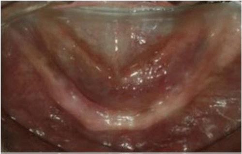
A 62 year female patient reported to the Department of Prosthodontics and Implantology with the complaint of an ill fitting mandibular denture. The patient lost her teeth to periodontal disease and was edentulous for 5 years. She reported having had three sets of complete dentures being fabricated in this time span not being satisfactory with any. Detailed evaluation of the dentures revealed retentive and stable maxillary dentures, however the mandibular denture lacked retention and stability (Figure 1).
Figure 1: Preoperative situation.
A variety of treatment options were offered to the patient which included a conventional mandibular denture, mandibular overdenture retained by two implants and a single implant retained mandibular overdenture with a conventional maxillary denture.
A Cone Beam Computed Tomography scan was performed prior to finalising the treatment. Based on clinical, radiologic findings and patients financial limitations and expectations, an immediately loaded single implant retained mandibular overdenture with a conventional maxillary denture was selected for the patient. Lingualised occlusion with bilateral balance was utilised to reduce lateral forces [14].
Presurgical medical evaluation and initial radiographic examination using an Orthopantomograph was carried out. A conventional complete denture was fabricated. The mandibular denture was duplicated in clear acrylic resin (Acryln `R`, Asian Acrylates, India) to fabricate a dual purpose stent-initially as a radiographic stent and later as surgical guide. Radiographic markers were placed in a duplicated mandibular denture and a Cone Beam Computed Tomography (i-CAT, Imaging sciences International, LLC, PA, U.S.A) was performed. The horizontal (occlusal) and cross-sectional (sagittal) slices of the mandible made at the midline at the level of the ridge crest were examined carefully to rule out the presence of the sublingual artery perforating the lingual cortex. Additionally, the cross sectional images ruled out presence of lingual undercut or large incisive canal. The scans revealed a height and width of 11mm and 6.9mm respectively at the midline. Based on these findings it was decided to place 4.6 X9.5 implant (Biohorizon, Birmingham, AL, U.S.A).
The patient was administered 1000mg Amoxicillin one hour prior to surgery as a loading dose.
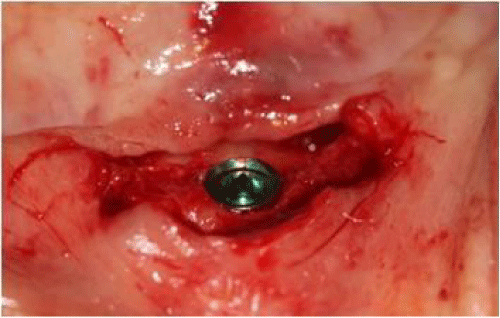
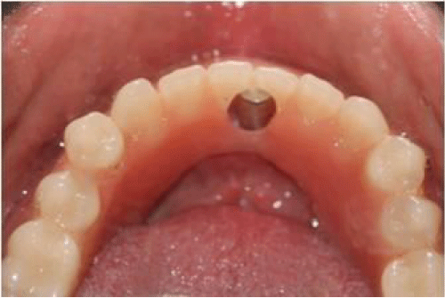
An implant measuring 4.6 mm in diameter and9.5mm in length was placed in the midline using a conventional surgical protocol. Adequate primary stability was obtained on placement (Figure 2).
Figure 2: Implant placed in the midline of the mandible.
This was then confirmed using an implant stability tester (Osstell AB, Gotebörg,Sweden). As Implant Stability Quotient value obtained was 66, it was decided to go ahead with immediate loading of the implant.
An implant level impression was made (Impregum, 3M Deutschland Gmbh, Neuss, Germany), the laboratory analogue attached and the impression was poured in die stone (Ultrarock ,Kalabhai Karson Pvt.Ltd ,India) . Healing abutment (4.6 mm, Standard height healing abutment, BioHorizons, Birmingham, AL, U.S.A) was placed and the patient was dismissed.
The patient was prescribed Amoxicillin 500 mg and anti-inflammatories (Ibuprofen-Paracetamol) twice a day for 3 days. The patient was instructed to eat a soft diet for one week. Chlorhexidine 0.12% mouthwash was prescribed to be used 3 times a day starting 1 day post of surgery until 10 days post operative.
A stock straight abutment was selected (Laser-Lok Simple Solution,Bio Horizons, U.S.A). The abutment was milled down to 3mm in height.
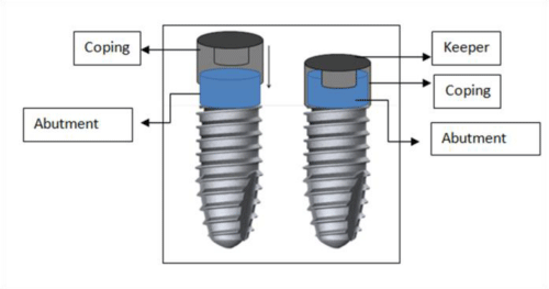
A wax pattern of a coping incorporating the keeper (Magfit DX, Aichi Steel Corporation, Japan) on the superior surface was fabricated onto the abutment. The assembly was cast in Nickel- Chromium alloy (Wiron 99, Bego, Lincoln RI, U.S.A). The coping had an inner extension that fit into the inner diameter of the abutment in order to improve its retention (Figure 3).
Figure 3: Design of the coping with the magnetic attachment.
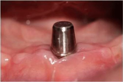
The abutment was placed and torqued into the implant at 30Ncm torque the next day. The access hole was blocked using a temporary cement (Fermit, IvoclarVivadent AG, Schaan, Liechtenstein) .The coping was cemented onto the abutment with dual cure resin cement (Multilink Speed, IvolcarVivadent, Schaan, Liechtenstein) (Figure 4). An abutment replica technique was used to minimize the amount of cement used for cementation [15]. After cleaning the excess cement, a radiograph was taken to ensure no cement remnant was left behind.
Figure 4: Abutment and coping in situ.
After cementation of the coping, the denture was relieved in the area of the coping, creating adequate space for the attachment assembly (Figure 5). Due to the resorbed nature of the ridge, the adequate inter-arch distance allowed incorporation of a greater amount of acrylic at the midline area of the denture and permitted the use of a conventional heat cured acrylic denture base without any metal substructure. Also, acrylic dentures are easy to fabricate, adjust, reline and have lower costs.
Figure 5: Denture trimmed to provide space for attachment assembly.
The magnet (Magfit DX, Aichi Steel Corporation, Japan) was placed insitu over the keeper.
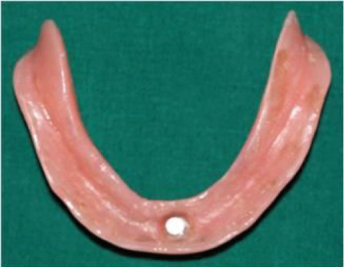
Rubber dam was used to protect tissues and prevent sutures from getting incorporated into acrylic resin. The magnet was picked up into the denture using self cure acrylic resin (Figure 6).
Figure 6: Intaglio surface of denture housing the magnet.
The excess acrylic resin was trimmed and the denture was polished. The area around the magnet was relieved so as to prevent the acrylic from contacting the coping thereby minimising lateral forces onto the implant.
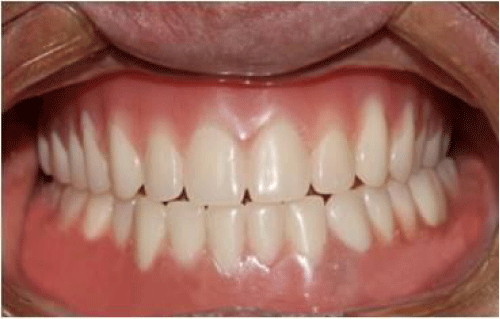
The denture was delivered and denture maintenance, oral hygiene instructions to avoid/prevent plaque accumulation on the coping surface were given. Patient was also briefed about the recall visits (Figure 7).
Figure 7: Dentures in situ.
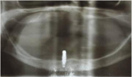
The follow up protocol included routine checkups at 1 week for suture removal and occlusal adjustments and thereafter at 3 weeks, 1 month and every 3 months. Hygiene maintenance was reinforced at every appointment. Intraoral radiographs were taken to assess the bone level around the implants at every stage and after 3 and then 6 month recall visits. A radiograph at 1 year follow-up is presented (Figure 8). The bone loss measured radio graphically after one year was 0.2 mm.
Figure 8: Radiographic evaluation after 1 year.
Objective evaluation of the treatment was done using a Visual Analogue Scale (VAS) to assess retention, masticatory efficiency, phonetics and over all comfort. A maximum score of 10 was assigned to each parameter. The VAS scores were recorded twice- once, prior to surgery while wearing conventional dentures and the other one month following use of the implant supported overdenture. The total score before treatment was 25/40 and after treatment was 36/40. A marked improvement was noted in masticatory efficiency, retention, speech and overall comfort with the use of the single implant retained overdenture.
Discussion
The use of a single implant to support an overdenture was first documented by Cordioli [6]. Gradually, keeping the principles of minimal surgery in mind, as well taking economic considerations into account, the use of a single implant to retain a mandibular denture has gained popularity and now has been shown to be a viable treatment option. Some studies have documented the use of the same method using O Ring and Locator attachments [3-9]. Liddelow et al. [3,4] in their studies used an immediate loading protocol for implants with a ball attachment. They concluded that for mucosa borne overdentures, this treatment is a safe, reliable and cost effective. In the author's experience, the ball abutment requires frequent maintenance and replacement of components. Plaque accumulation and soft tissue problems are also frequent. Grover et al. [16] used an early loading protocol for placement of a single implant with a magnet supported overdenture with conventional and shortened dental arches. They concluded that single implant supported magnet retained mandibular overdentures significantly improve the Oral Health- Related Quality of life of completely edentulous patients.
One of the prime requirements of successful immediate loading of implants is minimal transfer of lateral forces onto the fixture causing micro movement. Magnets have been known to impart less lateral forces and at the same time positively influence osseointegration of implants [13,17]. This was the principle followed for the treatment of this patient. The authors have treated multiple patients with this protocol and have found it to be a successful option.
This method uses an implant abutment with a customised coping that harbours the keeper. The placement of the coping and picking up of the magnet in self cure acrylic resin can be done chairside and does not require additional laboratory time.
Conclusion
For the case described, an immediately loaded, single implant retained mandibular overdenture is a feasible option for an edentulous patient who could not tolerate conventional dentures. This treatment protocol has a potential to be used for patients who cannot wear conventional mandibular dentures and who cannot afford multiple implant therapy. It is a simple and economical protocol that allows a greater number of edentulous patients to benefit from an implant-retained prosthesis.
References
- Feine JS, Carlsson GE, Awad MA, Chehade A, Duncan WJ, et al. The McGill consensus statement on overdentures. Mandibular two-implant overdentures as first choice standard of care for edentulous patients. Gerodontology. 2002; 19: 3-4.
- Thomason JM, Feine J, Exley C, Moynihan P, Müller F, Naert I et al. Mandibular two implant-supported overdentures as the first choice standard of care for edentulous patients--the York Consensus Statement. Br Dent J. 2009; 207: 185-186.
- Liddelow GJ, Henry PJ. A prospective study of immediately loaded single implant-retained mandibular overdentures: Preliminary one-year results. J Prosthet Dent. 2007; 97: S126-S137.
- Liddelow G, Henry P. The immediately loaded single implant-retained mandibular overdenture: a 36-month prospective study. Int J Prosthodont. 2010; 23: 13-21.
- Cheng T, Ma L, Liu XL, Sun GF, He XJ, et al. Use of a single implant to retain mandibular overdenture: A preliminary clinical trial of 13 cases. Journal of Dental Sciences. 2012; 7: 261-266.
- Cordioli G, Majzoub Z, Castagna S. Mandibular overdentures anchored to single implants: A five-year prospective Study. J Prosthet Dent. 1997; 78: 159-165.
- Gerald Krennmair, Christian Ulm. The Symphyseal Single-tooth Implant for Anchorage of a Mandibular Complete Denture in Geriatric Patients: A Clinical Report. Int J Oral Maxillofac Implants. 2001; 16: 98-104.
- Alsabeeha N, Payne A, De Silva RK, Thomson WM. Mandibular single-implant overdentures: preliminary results of a randomized control trial on early loading with different implant diameters and attachment systems. Clin Oral Impl Res. 2011; 22: 330-337.
- Alsabeeha N, Payne A, De Silva RK, Swain MV. Mandibular single-implant overdentures: a review with surgical and prosthodontic perspectives of a novel approach. Clin Oral Impl Res. 2009; 20: 356-365.
- Walton JN, Glick N, Macentee MI. A randomized clinical trial comparing patient satisfaction and prosthetic outcomes with mandibular overdentures retained by one or two implants. Int J Prosthodont. 2009; 22: 331-339.
- Gallucci GO, Morton D, Weber HP. Loading Protocols for Dental Implants in Edentulous Patients. Int J Oral Maxillofac Implants. 2009; 24(Suppl): 132-146.
- Ceruti P, Bryant SR, Lee JH, Mac Entee M. Magnet-Retained Implant-Supported Overdentures: Review and 1-Year Clinical Report. J Can Dent Assoc. 2010; 76: 52-57.
- Yang TC, Maeda Y, Gonda T, Kotecha. Attachment systems for implant overdenture: influence of implant inclination on retentive and lateral forces. Clin Oral Impl Res. 2011; 22: 1315-1319.
- Jambhekar S, Kheur M, Kothavade M, Dugal R. Occlusion and Occlusal Considerations in Implantology. IJDA. 2010; 2: 125-130.
- Jambhekar S, Matani J, Sethi T, Kheur MG. Reduction of excess cement during cementation of implant -retained crowns: A clinical tip. J Dent Implant. 2013; 3: 168-1671.
- Grover M, Vaidyanathan AK, Veeravalli PT. OHRQoL, masticatory performance and crestal bone loss with single-implant, magnet-retained mandibular overdentures with conventional and shortened dental arch. Clin Oral Implants Res. 2013; 00: 1-7.
- Maeda Y, Horisha M, Yagi K. Biomechanical rationale for a single implant-retained mandibular overdenture: an in vitro study. Clin Oral Impl Res. 2008; 19: 271-75.
