Research Article
J Dent App. 2014;1(4): 55-60.
Analysis of Average Horizontal and Vertical Mandibular Growth Components Relative to the Horizontal Plane as Determined from Cross-sectional Cephalometric Normal Standards
Richard G. Standerwick*
Associate Clinical Professor, UBC Faculty of Dentistry, Dept. of Orthodontics, Vancouver, BC, Canada
*Corresponding author: Dr. Richard G. Standerwick, 20159 88th Ave, Suite E207, Langley, BC, Canada V1M
Received: June 26, 2014; Accepted: September 02, 2014; Published: September 04, 2014
Abstract
Objective: To provide some insight into annual horizontal and vertical components of mandibular growth.
Material and Methods: The averaged growth data for the ages of 8 to 16 were garnered from an Atlas of craniofacial growth: Cephalometric standards from the university school growth study, the University of Michigan and organized into horizontal and vertical growth components utilizing trigonometry.
Results: Considering the limitations of the data, during puberty it would seem the average horizontal growth vector of the mandible is approximately 1.5mm per year for females and 2mm/yr for males. The average vertical component of growth during puberty is approximately 2.8mm/yr for males and 1.5 mm/yr for females.
Conclusion: The amount of horizontal growth may be overestimated and vertical growth underestimated with current use of growth along anatomic planes rather than relative to the horizontal or Frankfort plane.
Introduction
Diagnosis and treatment often rely on the amount of craniofacial growth available, however it is difficult to find a description of annual horizontal (ventral) and vertical (caudad) components of normal mandibular growth relative to the Frankfort horizontal (FH) on a search of orthodontic literature. Mandibular growth in length is often described utilizing the distance between condyle associated landmarks and mandibular symphysis associated landmarks eg condylion-gnathion (Co-GN), condylion-pogonion (Co-Pg), articulare-pogonion (Ar-Pg), and articulare-gnathion (Ar-Gn) [1-4]; however these measured changes are angled relative to the Frankfort Horizontal /horizontal plane. For this reason, the measured change does not adequately reflect clinically observed facial development [5] nor the ventral change resulting in decreased facial convexity.
According to Proffit [6,7] mandibular ramus height growth (condylion-gonion) is 1 to 2 mm per year and mandibular body (gonion-pogonion) growth is 2 to 3 mm per year based on the growth standards utilized in this study [8], and Ricketts reported 1.5mm/yr of mandibular growth measured from Co to symphysis [9]. Burstone and Marcotte [10] observed Ar-Pg annual growth and growth increments for female ages 10 to 14 which displayed annual growth increments of 2mm, 3.5mm, 1.9mm and 1.4mm, and for male ages 12 to 16 which displayed annual growth increments of 2.2mm, 2.8mm, 4.7mm and 2.3mm. Female mandibular length as Ar-Pg has been observed to display 4.29mm of growth for 18months prior to menarche and then a subsequent decrease in growth rate to 2.84mm for 18 months post-menarche [3], which is approximately 2.4mm a year over the 36 months of pubertal growth. Mandibular length measured as Co-Gn displayed growth of 4.31mm per year measured over a 5 to 24 month observation period [1], 2.5mm/year over 5 years from 91/2 years of age to 14½ [2], and approximately 2mm per year for an untreated control group [11,12]. Mandibular length measured as Ar-Gn displayed a 3.26 ± 1.47mm/yr for males and 2.90 ± 1.32 mm/yr of incremental growth [4] and 1.33mm/yr as observe for an untreated control group [12]. Bjork described 2-5mm of condylar growth per year [13], and Buschang [14] described incremental condylar growth with an approximate average of 2.8mm/yr for males and 2.2mm/yr for females, however this may not directly translate into the desired growth direction at pogonion [15].
Growth vector analysis has been previously described utilizing a constructed reference of a perpendicular line to the sella turcica-nasion line (SN), SN-7 or FH which originates from sella, however growth periods presented often do not allow an annual assessment of growth without additional assumption [5,15-18].
The purpose of this study was to provide an estimate for the average amount of horizontal and vertical circumpubertal mandibular growth using cross-sectional data from normal growth standards relative to the horizontal plane approximating the Frankfort horizontal (FH) [8]. Potential application includes orthodontic diagnosis and treatment planning when considering normal growth and the potential for orthopedic or dento-alveolar modification.
Material and Methods
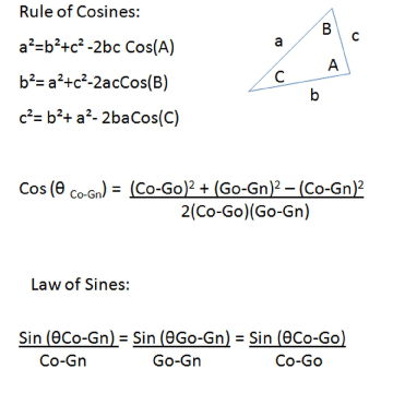
The sample for this study was obtained from an Atlas of Craniofacial Growth: Cephalometric Standards from the University School Growth Study, the University of Michigan and consisted of 83 Caucasian individuals (47 male, 36 female). Growth standard measurements for condylion-gonion (Co-Go), condylion-gnathion (Co-Gn), gonion-gnathion (Go-Gn), and sella-nasion-gonion-gnathion (SN-GoGn) (Table 1) were obtained for the ages of 8 to 16 years of age and were chosen as they were the only available landmark measurements which would relate the condyle, gonion and mandibular symphysis; Co, Go, Gn. 6A measurement for the gonial angle was not available; therefore, the cosine rule was used to establish the angle of Co-Go-Gn (θCo-Gn) (Figure 1). The Law of Sines was then used to calculate the remaining angles θGo-Gn and θCo-Go (Figure 1).
Figure 1: Shown are the equations representative of the Rule of Cosines and the relationship of the angles to their respective to the sides of a triangle. A rule of Cosines equation is adapted to solve for C(θCo-Gn) and then the Law of Sines is shown adapted to solve for θGo-Gn and θCo-Go.
    A    Average Age related length of Co-Gn (mm)
    Average    Age related length of Go-Gn (mm)
    Average    Age related length of Co-Go (mm)
    Average    Age related angle of SN-GoGn (mm)
    Growth    Increments for Co-Gn (mm)


    Co-Gn
    Sex

    Go-Gn
    Sex

    Co-Go
    Sex

    SN-GoGn
    Sex


    AGE
    M
    F
    AGE
    M
    F
    AGE
    M
    F
    AGE
    M
    F
    AGE
    M
    F


    6
    103.0
    100.5
    6
    65.4
    65.6
    6
    48.7
    46.5
    6
    34.8
    35.8
    6    to 7
    2.3
    2.8


    7
    105.3
    103.3
    7
    68.2
    67.3
    7
    49.1
    47.7
    7
    35.6
    36.3
    7    to 8
    3.9
    3.0


    8
    109.2
    106.3
    8
    70.5
    69.8
    8
    51.3
    49.1
    8
    34.7
    34.9
    8    to 9
    2.5
    2.0


    9
    111.7
    108.3
    9
    72.4
    70.9
    9
    52.8
    50.8
    9
    34.2
    35.0
    9    to 10
    2.8
    3.0


    10
    114.5
    111.3
    10
    74.4
    73.4
    10
    54.0
    51.5
    10
    34.4
    35.0
    10    to 11
    3.1
    2.1


    11
    117.6
    113.4
    11
    76.6
    75.1
    11
    55.8
    52.4
    11
    34.3
    34.6
    11    to 12
    2.1
    2.3


    12
    119.7
    115.7
    12
    77.9
    75.9
    12
    57.2
    54.6
    12
    33.5
    34.0
    12    to 13
    3.4
    2.1


    13
    123.1
    117.8
    13
    79.9
    77.8
    13
    59.4
    55.1
    13
    32.9
    34.2
    13    to 14
    3.4
    2.1


    14
    126.5
    119.9
    14
    82.4
    78.9
    14
    61.6
    56.8
    14
    32.9
    33.5
    14    to 15
    2.2
    2.1


    15
    128.7
    122.0
    15
    84.0
    80.0
    15
    62.7
    58.9
    15
    32.9
    32.1
    15    to 16
    4.9
    1.6


    16
    133.6
    123.6
    16
    86.3
    81.0
    16
    66.1
    60.5
    16
    32.6
    31.1
Table 1: Average age related length of Co-Gn, Go-Gn, Co-Go in millimeters and angulation of SN-GoGn in degrees. (from An Atlas of craniofacial growth: Cephalometric standards from the University school growth study, the University of Michigan1). Also shown are growth increments for Co-Gn, Co-Go, Go-Gn.
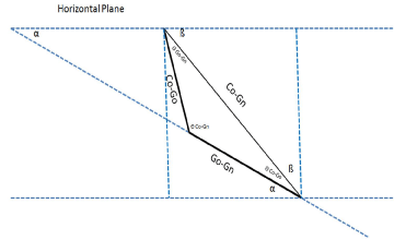
The sella-nasion line minus 7 degrees (SN-7) was utilized to represent the horizontal plane as it approximates the FH (16, 17, 19, 20) (Figure 2). The SN-GoGn value [8] was adjusted by 7 degrees to compensate for the 7° difference between the SN-GoGn and SN-7- GoGn.
Figure 2: The orientation on an arbitrary co-ordinate system and conceptualization used to relate the Frankfort Horizontal/Horizontal plane to the mandibular anatomic planes (Co-Go, Go-Gn, Co-Gn). The Frankort Horizontal/ Horizontal plane represents the x-axis and the y-axis is drawn perpendicular to the Horizontal plane at the position representing condylion (Co).
Because SN-7 is known, the angle between Go-Gn and a horizontal constructed by an arbitrary Cartesian co-ordinate system (α) could be extrapolated (Figure 2). Adding the angles θCo-Go and α (Figure 2) will allow one to determine the x- component (horizontal) and y-components (vertical) of growth (Figure 3).
Figure 3: The formulae used to solve for the X and Y component of growth. Derived from Cosθ= adjacent /hypotenuse and Sinθ= opposite/hypotenuse.
The ages at puberty were considered separately. The ages for puberty were assumed to display a 2 year sexual dimorphism [21], and were separated using growth increments that represented the ages during puberty; 10-13 years of age for females and 12 to 15 for males [17,22,23].
The sample is represented by an SNA, SNB, MPA, and ANB consistent with a mesoprosopic/mesocephalic craniofacial type (normal growth standards) [8].
Results
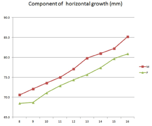
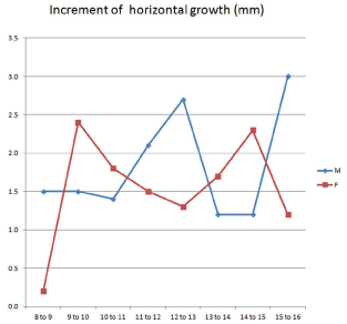
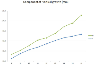
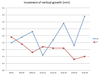
The calculated x and y components based on averaged growth data relative to age are shown in Table 3 and 5, and Figures 4-7. It would seem that the horizontal component of growth during puberty (12 to 16 for males, 10-14 for females) is approximately 1.5mm per year for females and 2mm/yr for males on average. The vertical component of growth during puberty is approximately 2.8mm/yr for males and 1.5 mm/yr for females.
Figure 4: The age related growth component along the x-axis (in millimeters; adapted from Riolo [8]). Male (M) and female (F) values are shown.
Figure 5: The increment of yearly growth along the x-axis (in millimeters). Male (M) and female (F) values are shown.
Figure 6: The age related growth component along the y-axis (in millimeters; adapted from Riolo [8]). Male (M) and female (F) values are shown.
Figure 7: The increment of yearly growth along the y-axis (in millimeters). Male (M) and female (F) values are shown.
    Cosine    rule to determine θ(Co-Gn) in degrees(°)
    Law    of Sines to determine θ(Go-Gn) and     θ(Co-Go) in degrees(°)


    Sex
    θ    (Go-Gn)
    Sex
    θ(Co-Go)
    Sex


    Age
    M
    F
    Age
    M
    F
    Age
    M
    F


    8
    126.7
    125.9
    8
    31.2
    32.1
    8
    22.1
    22.0


    9
    125.6
    124.9
    9
    31.8
    32.5
    9
    22.6
    22.6


    10
    125.4
    125.1
    10
    32.0
    32.7
    10
    22.6
    22.3


    11
    124.6
    124.6
    11
    32.4
    33.0
    11
    23.0
    22.4


    12
    124.0
    124.1
    12
    32.7
    32.9
    12
    23.4
    23.0


    13
    123.5
    124.0
    13
    32.8
    33.2
    13
    23.7
    22.8


    14
    122.3
    123.3
    14
    33.4
    33.4
    14
    24.4
    23.3


    15
    122.0
    122.2
    15
    33.6
    33.7
    15
    24.4
    24.1


    16
    121.9
    121.1
    16
    33.3
    34.1
    16
    24.8
    24.8
Table 2: Average age related angulation change of θCo-Gn as determined by using the Cosine Rule and θ(Go-Gn) as determined by the Law of Sines (in degrees).
    Component    of growth along the X-axis (mm)
    Increment    of Growth Per Year along the X-axis (mm)


    Sex
    Sex


    Age
    M
    F
    Age
    M
    F


    8
    70.6
    68.5
    8    to 9
    1.5
    0.2


    9
    72.1
    68.7
    9    to 10
    1.5
    2.4


    10
    73.6
    71.1
    10    to 11
    1.4
    1.8


    11
    75.0
    72.9
    11    to 12
    2.1
    1.5


    12
    77.1
    74.4
    12    to 13
    2.7
    1.3


    13
    79.8
    75.7
    13    to 14
    1.2
    1.7


    14
    81.0
    77.4
    14    to 15
    1.2
    2.3


    15
    82.2
    79.7
    15    to 16
    3.0
    1.2


    16
    85.2
    80.9
    Ttl
    14.6
    12.4


    Puberty
    8.1
    6.3


    2    mm/yr at Puberty
    1.5    mm/yr at Puberty
Table 3: The age related growth component along the x-axis and the increment of yearly growth along the x-axis (in millimeters; assumed growth direction is ventral).
    Component    of growth along the Y-axis (mm)
    Increment    of Growth Per Year along the Y-axis (mm)


    Sex
    Sex


    Age
    M
    F
    Age
    M
    F


    8
    83.3
    81.3
    8    to 9
    2.0
    2.4


    9
    85.3
    83.7
    9    to 10
    2.4
    1.9


    10
    87.7
    85.6
    10    to 11
    2.8
    1.3


    11
    90.5
    86.9
    11    to 12
    1.1
    1.7


    12
    91.6
    88.6
    12    to 13
    2.2
    1.6


    13
    93.8
    90.2
    13    to 14
    3.4
    1.6


    14
    97.2
    91.6
    14    to 15
    1.8
    0.8


    15
    99.0
    92.4
    15    to 16
    3.9
    1.0


    16
    102.9
    93.4
    Ttl
    19.6
    12.3


    Puberty
    11.3
    6.2


    2.8    mm/yr at Puberty
    1.55    mm/yr at Puberty
Table 4: The age related growth component along the y-axis and the increment of yearly growth along the y-axis (in millimeters; assumed growth direction is caudad).
    Ratio    of x/y


    Sex


    Age
    M
    F


    8 to 9
    0.8
    0.1


    9 to 10
    0.6
    1.3


    10 to 11
    0.5
    1.4


    11 to 12
    1.9
    0.9


    12 to 13
    1.2
    0.8


    13 to 14
    0.4
    1.1


    14 to 15
    0.7
    2.9


    15 to 16
    0.8
    1.2
Table 5: Ratio of the increments of horizontal growth relative to vertical growth.
Discussion
Growth of the mandible is complex as there are horizontal, vertical, transverse and rotational components of mandibular growth for which the affects cannot be easily separated. Yet, it is still useful to have normal values for the horizontal and vertical components of growth to aid in diagnosis and treatment planning. This study demonstrates normal growth of a mesoprosopic/ mesocephalic nature (normal growth standards) with limitations of the unknown change in condyle-fossa position and the lack of a reported gonial angle which required mathematical determination rather than direct measurement. In future studies, the calculation of the gonial angle would be avoidable if simple direct radiographic measurement of Co-Go-Gn is possible. The average growth effect presented in this study means there will be situations where growth will be greater and situations where growth will be less than described. The use of the terms horizontal and vertical to describe the relative growth change are more consistent terms for use with the presented data [17,24], however the assumption of growth direction should consider that sella-turcica may be displaced cephalad during growth [25-29] and that the asymmetrically growing spheno-occipital synchondrosis [27] bisects the anterior cranial base and mandibular condyle [28,29]. A caudad and ventral growth vector of the mandible and mandibular condyles could be reasonably assumed through application of observations from superimposition on the posterior cranial base [25,26,28-31] and the occurrence of airorhyncy (posterior and upper portions of the face rotate dorsally relative to the posterior cranial base by extension of the anterior cranial base relative to the posterior cranial base) [32-34] as growth progresses from a central axis based on the preservation of neural structures (eg. brain stem) and airway [31,32,35,36]. This is not to say the glenoid fossae positions are not altered between different craniofacial types [37-39], but rather considers that the development process may require further investigation. Other studies have attempted to describe the horizontal nature of mandibular growth: Johnston's pitchfork analysis, McNamara's nasion perpendicular.
For this study, the calculated average amount of horizontal growth seemed slightly less than commonly assumed magnitudes (common assumption of growth along Go-Gn seems to over-estimate) and the average amount of calculated vertical growth seemed slightly greater than commonly assumed (common assumption of growth along Co- Go seems to under-estimate).
It is notable that the ratio of horizontal relative to vertical growth is not consistently equal to 1 (equal horizontal and vertical growth components). This may reflect the fluctuating horizontal/vertical pattern of mandibular growth described by Bergersen [28], Bjork [18] and Downs [29].
Growth vector analysis of hyperdivergent/ leptoprosopic and hypodivergent/euryprosopic individuals could be considered in the future relative to a "stable" reference point while preserving sexual dimorphism [26]. Further analysis of growth standard samples to determine horizontal and vertical components of mandibular growth may improve our understanding, diagnosis, treatment planning and would corroborate the available information.
Conclusion
Based on normal craniofacial growth standards:
- Average horizontal growth of the mandible during puberty as measured at pogonion appears to be 2mm/ year for males and 1.5mm/year for females.
- Average vertical growth of the mandible during puberty as measured at pogonion appears to be 2.8mm/year for males and 1.5mm/year for females.
The amount of horizontal growth may be overestimated and vertical growth underestimated with current use of growth along anatomic planes rather than relative to the Frankfort Horizontal/ horizontal plane.
References
- Gomes AS, Lima EM. Mandibular growth during adolescence. Angle Orthod. 2006; 76: 786-790.
- Mitani H, Sato K. Comparison of mandibular growth with other variables during puberty. Angle Orthod. 1992; 62: 217-222.
- Tofani MI. Mandibular growth at puberty. Am J Orthod. 1972; 62: 176-195.
- Lewis AB, Roche AF, Wagner B. Growth of the mandible during pubescence. Angle Orthod. 1982; 52: 325-342.
- Mannchen R. A critical evaluation of the pitchfork analysis. Eur J Orthod. 2001; 23: 1-14.
- Proffit WR, Fields HW. Contemporary Orthodontics 3e. St. Louis: Mosby; 2000. p. 99.
- Proffit WR, Fields HW, Sarver DM. Contemporary Orthodontics 5e. St. Louis: Elsevier; 2012. p. 97.
- Riolo ML, University of Michigan. Center for Human Growth and Development., United States. Public Health Service. An Atlas of craniofacial growth : cephalometric standards from the University school growth study, the University of Michigan. Ann Arbor: Center for Human Growth and Development, University of Michigan; 1974. 379 p. p.
- Ricketts RM. Planning treatment on the basis of facial pattern and an estimate of its growth. Angle Orthod. 1957; 27: 14-37.
- Burstone CJ, Marcotte MR. Problem Solving in Orthodontics: Goal-Oriented Treatment Strategies. Carol Stream: Quintessence Publishing Co., Inc; 2000. pp237, 40 p.
- Marschner JF, Harris JE. Mandibular growth and class II treatment. Angle Orthod. 1966; 36: 89-93.
- Chen JY, Will LA, Niederman R. Analysis of efficacy of functional appliances on mandibular growth. Am J Orthod Dentofacial Orthop. 2002; 122: 470-476.
- Bjork A. Variations in the growth pattern of the human mandible: longitudinal radiographic study by the implant method. J Dent Res. 1963; 2: 400-411.
- Buschang PH, Santos-Pinto A, Demirjian A. Incremental growth charts for condylar growth between 6 and 16 years of age. Eur J Orthod. 1999; 21: 167-173.
- Bjork A, Skieller V. Facial development and tooth eruption. An implant study at the age of puberty. Am J Orthod. 1972; 62: 339-383.
- Buschang PH, Martins J. Childhood and adolescent changes of skeletal relationships. Angle Orthod. 1998; 68: 199-206.
- Buschang PH, Santos-Pinto A. Condylar growth and glenoid fossa displacement during childhood and adolescence. Am J Orthod Dentofacial Orthop. 1998; 113: 437-442.
- Bjork A, Skieller V. Normal and abnormal growth of the mandible. A synthesis of longitudinal cephalometric implant studies over a period of 25 years. Eur J Orthod. 1983; 5: 1-46.
- Burstone CJ, James RB, Legan H, Murphy GA, Norton LA. Cephalometrics for orthognathic surgery. J Oral Surg. 1978; 36: 269-277.
- Legan HL, Burstone CJ. Soft tissue cephalometric analysis for orthognathic surgery. J Oral Surg. 1980; 38: 744-751.
- Iuliano-Burns S, Hopper J, Seeman E. The age of puberty determines sexual dimorphism in bone structure: a male/female co-twin control study. J Clin Endocrinol Metab. 2009; 94: 1638-1643.
- Lowrey GH. Growth and development of children. 8th ed. Chicago: Year Book Medical Publishers; 1986. xi, 507 p. p.
- Bordini B, Rosenfield RL. Normal pubertal development: part II: clinical aspects of puberty. Pediatr Review. 32: 281-292.
- Agronin KJ, Kokich VG. Displacement of the glenoid fossa: a cephalometric evaluation of growth during treatment. Am J Orthod Dentofacial Orthop. 1987; 91: 42-48.
- Standerwick R, Roberts E, Hartsfield J, Jr., Babler W, Kanomi R. Cephalometric superimposition on the occipital condyles as a longitudinal growth assessment reference: I-point and I-curve. Anat Rec (Hoboken). 2008; 291: 1603-1610.
- Standerwick RG, Roberts EW, Hartsfield JK, Jr., Babler WJ, Katona TR. Comparison of the Bolton Standards to longitudinal cephalograms superimposed on the occipital condyle (I-point). J Orthod. 2009; 36: 23-35.
- Melsen B. The postnatal growth of the cranial base in Macaca rhesus analyzed by the implant method. Tandlaegebladet. 1971; 75: 1320-1329.
- Coben SE. The spheno-occipital synchondrosis: the missing link between the profession's concept of craniofacial growth and orthodontic treatment. Am J Orthod Dentofacial Orthop. 1998; 114: 709-712.
- Coben SE. Growth Concepts. Angle Orthod. 1961; 31: 194-201.
- Moss ML. Ontogenetic aspects of cranio-facial growth. [1st ed. Moyers RE, Krogman WM, editors. Oxford, New York,: Pergamon Press; 1971. ix, 360 p. p.
- Frankel R. The applicability of the occipital reference base in cephalometrics. Am J Orthod. 1980; 77: 379-395.
- Lieberman DE, Ross CF, Ravosa MJ. The primate cranial base: ontogeny, function, and integration. Am J Phys Anthropol. 2000; Suppl 31: 117-169.
- McCarthy RC, Lieberman DE. Posterior maxillary (PM) plane and anterior cranial architecture in primates. Anat Rec. 2001; 264: 247-260.
- Ross CF, Ravosa MJ. Basicranial flexion, relative brain size, and facial kyphosis in nonhuman primates. Am J Phys Anthropol. 1993; 91: 305-324.
- Vital JM, Senegas J. Anatomical bases of the study of the constraints to which the cervical spine is subject in the sagittal plane. A study of the center of gravity of the head. Surg Radiol Anat. 1986; 8: 169-173.
- Bjork A. Cranial base development: A follow-up x-ray study of the individual variation in growth occurring between the ages of 12 and 20 years and its relation to brain case and face development. Am J Orthod. 1955; 41: 198-225.
- Pirttiniemi P, Kantomaa T, Ronning O. Relation of the glenoid fossa to craniofacial morphology, studied on dry human skulls. Acta Odontol Scand. 1990; 48: 359-364.
- Ingervall B. Relation between height of the articular tubercle of the temporomandibular joint and facial morphology. Angle Orthod. 1974; 44: 15-24.
- Kantomaa T. The relation between mandibular configuration and the shape of the glenoid fossa in the human. Eur J Orthod. 1989; 11: 77-81.
