1Department of Periodontology, Kocatepe University, Turkey
2Department of Periodontology, Erciyes University, Turkey
3Department of Periodontology, Erciyes University, Turkey
4Department of Prosthodontics, Erciyes University, Turkey
5Department of Oral and Maxillofacial Surgery, Kocatepe University, Turkey
*Corresponding author: Cakmak O, Department of Periodontology, Kocatepe University, Guvenevler Street, Inonu Boulevard, 03030, Afyon, Turkey
Received: September 10, 2014; Accepted: September 20, 2014; Published: September 22, 2014
Citation: Cakmak O, Alkan BA, Tasdemir Z, Kilinc HI and Atalay Y. Free Gingival Grafting Before Implant Placement In A Geriatric Patient : A Case Report. J Dent App. 2014;1(5): 91-94. ISSN:2381-9049
Geriatric patients generally suffer from edentulism or partial edentulism, and those without a dental prosthesis can experience siginificant health issues, mostly due to inadequate nutrition. There are various techniques for rehabilitation of total or partial edentulism. Implant-supported overdentures have become an effective treatment alternative for edentulous patient. In this case report, a free gingival graft technique was used to increase the width of keratinized tissue before implant placement to improve long-term implant-supported prosthesis prognosis in a geriatric patient.
Keywords: Gingiva; Dental implantation; Dental Implants; Maintenance; Dental Prosthesis; Implant-Supported
In years of 2000, there were approximately 600 million geriatrics in the world, and that number is increasing due to advances in medical science [1]. In recent years, geriatrics have become more susceptible to chronic illnesses such as cancer, diabetes mellitus, and oral diseases [2]. Among oral diseases, periodontal disease is the most prevalent among geriatrics [3]. There may be several possible causes of this. First, a decrease in hand flexibility of geriatric patients often causes a decline in oral hygiene , which can contribute to periodontal disease. Second, they often experience a compromised immune system, which can also contribute to periodontal disease [4]. Oral status directly affects general health. Geriatric patients generally have edentulism or partial edentulism, and those without a dental prosthesis can experience siginificant health issues, mostly due to inadequate nutrition [5].
There are various techniques for rehabilitation of total or partial edentulism. Implant-supported overdentures have become an effective treatment alternative for edentulous patient [6]. The success and maintenance of implant rehabilitation depends on many factors. The amount of keratinized tissue (KT) around implants may be important for peri-implant tissue health. A recent systematic review concluded that lack of adequate KT around dental implants is associated with more plaque accumulation, tissue inflammation, mucosal recession, and attachment loss [7]. A complete absence of KT, especially with non-optimal oral hygiene status, may negatively influence the long-term maintenance of restored teeth and/or dental implants.
Various methods have been described for increasing KT width around the implants [8-11]. In the presence of both shallow vestibules and inadequate KT, free gingival graft (FGG) can be performed successfully [12]. This procedure can be done either before, after, or at the time of implant placement [13].
In this case report, a FGG technique was used to increase the width of KT before implant placement to improve long-term prognosis of the implant-supported overdenture in a geriatric patient.
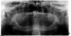
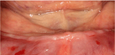
A 73-year-old female patient who was edentulous for approximately 20 years received a total prosthesis at Erciyes University, Faculty of Dentistry, Department of Prosthodontics. She did not have any medical problems and was not taking any medication. Two implants with ball attachments supporting the overdenture were planned, and the patient was referred to the Department of Periodontology. After a clinical examination, a lack of KT, a shallow vestibular sulcus, and alveolar bone loss on the mandibular anterior region were determined (Figure 1 and 2). A preoperative cone-beam computed tomography (CBCT) scan was conducted to examine the bone before deciding on periodontal plastic surgery. The CBCT images revealed that bone length and size were suitable for implants. A bilaterally FGG was planned to provide KT prior to implant and prosthetic rehabilitation. A signed informed consent form was obtained after the patient was provided with detailed information describing the nature and duration of the surgical and prosthetic procedures.
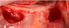
All surgical procedures were performed by the same clinician (OC) under local anesthesia. The patient was instructed to rinse with 0.12% chlorhexidine mouthwash prior to surgery. Following the administration of local anesthesia to recipient and donor sites, the recipient sites were prepared in the canine regions (Figure 3). Tissue tags were removed and bleeding was controlled using sterile gauze dampened with saline. The desired graft size was harvested from the palate without rugae and divided into two pieces. A prefabricated acrylic stent was placed to protect the donor site. Subsequently, grafts were sutured to the recipient sites with non-resorbable and resorbable 4-0 round sutures (Dogsan, Yalincak, Trabzon, Turkey) (Figure 4a and 4b). Firm pressure was applied over the grafted areas with a saline-soaked gauge to enhance close approximation. The patient was prescribed an antibiotic (amoxicicilin/clavulanate 1000 mg, two times daily for one week), analgesic (diclofenac, 50 mg every six hours), and mouthwash (0.2% chlorhexidine-digluconate, twice a day for two weeks). The sutures were removed 10 days later (Figure 5a and 5b) and follow-up visits were carried out every two weeks during the two months (Figure 6a and 6b). The healing period was uneventful. The patient was allowed to wear a temporary total prosthesis after four weeks. After two months, an adequate amount of non-moveable KT and deep vestibular sulcus were observed at the canine regions with no signs of inflammation, and the patient was scheduled for implant surgery (Figure 7).
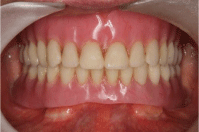
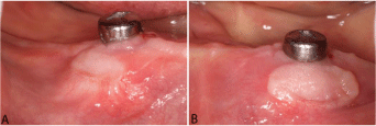
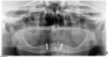
Implant placement was performed by routine procedures. After local anesthesia was administered, a crestal incision over the canine areas were made and the mucoperiosteal flap was raised. The osteotomies were prepared using the manufacturer’s protocol and evaluated with a probe to ensure that all bony walls were intact. Then, two implants (4.1x12mm, Dentium®, Implantium, Seoul, Korea) were placed in the canine regions (Figure 8). The implants were placed using a two-stage surgical procedure. The flap was repositioned and sutured with 3.0 non-resorbable round sutures (Dogsan, Yalincak, Trabzon, Turkey) (Figure 9). The same medications and mouthwash were prescribed, and the sutures were removed after seven days. Healing was uneventful (Figure 10). Aproximately 3 months after implant placement, a panoramic radiograph was taken and reviewed, which showed that the implants were osseointegrated and completely healthy (Figure 12). After gingiva formers were placed (Figure 11a and 11b), oral hygiene instructions were provided and the patient was referred to the Department of Prosthodontics for permanent dentures. The treatment was finalized by providing a maxillary total prosthesis and a mandibular implant-supported overdenture. Patient was followed for 18 months without any problem (Figure 13 and 14).
This clinical case report presents the treatment of a geriatric patient with atrophic mandible, shallow vestibular depth, and inadequate KT. The patient’s chief complaint was the lack of retention of her mandibular complete denture. A FGG and two implants with ball attachments supporting the overdenture were performed, respectively.
It is known that the implant to bone and the KT interface are weaker than that of natural teeth [10]. Altthough the bacteria that causes peri-implantitis and peri-implant mucositis is similar to the bacteria that causes periodontal disease, the magnitude of destruction in dental implants may be more devastating [10]. Thus, soft tissue surrounding implants may play an important factor in disease initiation and progression. There are varying opinions regarding the influence of KT surrounding dental implants. Wennstrom and Derks have claimed that, in good oral hygiene conditions, the marginal gingiva around dental implants is clinically healthy, even when there is an absence of KT [14]. However, Bouri et al. [15], reported a relationship between implant survival rate and KT width. A recent systematic review concluded that lack of adequate KT around dental implants is associated with more plaque accumulation, tissue inflammation, mucosal recession, and attachment loss [7]. Indeed, proper oral hygiene practices are very difficult to achieve around dental implants without the protection of KT, especially in geriatric patients who are not able to maintain adequate oral hygiene. In addition, a shallow vestibule may cause food retention, impaired quality of oral hygiene procedures, and mobility of the peri-implant mucosa [16]. Therefore, to enhance long-term peri-implant health and prognosis of the prosthesis, FGGs in conjunction with vestibuloplasty was performed before implant placement in this case report.
Various surgical methods, such as an apically positioned flaps, laterally positioned flaps, FGGs, or connective tissue grafts, can be applied to maximize the width of KT [11]. To our knowledge, the most indicated procedure to increase the width of KT and depth of the vestibular sulcus seems to be FGGs, which is a predictable and reliable surgical procedure. However, this procedure may cause some problems for the patient, such as the need for a donor site, two surgical sites with a possibility of pain after surgery, and a longer treatment period [17]. Moreover, wound healing may be another risk factor for geriatric patients. However, in this case study, the healing period was uneventful and the patient’s compliance was excellent. An acceptable amount of non-moveable KT and deep vestibular sulcus was observed at the canine regions with no signs of inflammation after periodontal plastic surgery.
A FGG procedure may be performed before, after, or at the time of implant placement [13]. The grafting procedure was carried out before the implant surgery to facilitate tissue manipulation at the time of implant placement by increasing the width of KT around the implants. A wider zone of KT may provide easier flap reflection and suturing and a decreased surgical time. This situation may be important in the post-operative period for geriatric patients because of the possibility of compromised healing. However, obtained KT may be moveable if a FGG operation is performed after implant placement.
It has been reported that ball attachments are less costly, less technique sensitive, and easier to clean than bars [18]. In addition, the probability of gingival hyperplasia reportedly is more easily reduced with ball attachments [19]. Thus, ball attachments were used in this case report.
Using FGGs is a safe and predictable method to increase KT, thus allowing for better oral hygiene for the patient. Maintaining proper oral hygene is especially important for geriatric patients beause of the possibility of decreased hand flexibility. Mucogingival surgery resulted in an appropriate environment for proper oral hygiene procedures for the geriatric patient. However, the patient was informed that the long-term prognosis of the prosthesis would depend on the supporting periodontal therapy. The prognosis was highly favorable and no significant soft or hard tissue changes were observed during the follow-up period. It was observed that the results were maintained and the patient was satisfied by the esthetic and functional outcomes of her overdenture after 18 months.
The authors report no conflicts of interest related to this case report.
Panoramic radiograph before treatment.
Preoperative clinical situation: Absence of adequate KT, decreased vestibular depth and severely resorbed ridge.
Recipient sites were prepared in the mandibular canine regions.
Free gingival graft was sutured: right mandibular canine region.
Free gingival graft was sutured: left mandibular canine region.

Ten days after surgery after suture removal: right mandibular canine region.
Ten days af ter surgery after suture removal: left mandibular canine region.
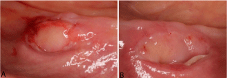
Twenty-one days after surgery: right mandibular canine region.
Twenty-one days after surgery: left mandibular canine region.
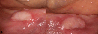
Two-month postoperative healing of the FGG. Note presence of enough KT and deep vestibular sulcus.
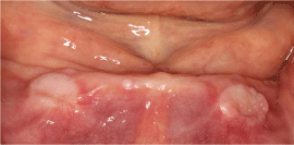
Implants were placed at the mandibular canine regions.
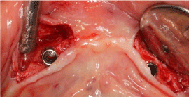
Primary and tension-free closure of flaps.
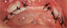
One month after implant surgery.
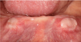
Three months after implant surgery: right mandibular canine region.
Three months after implant surgery: left mandibular canine region.
Panoramic radiograph: three months after implant surgery.
Stable peri-implant soft tissue, 16 months after implant placement.
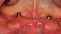
The final intraoral view at 18 months.
