1Department of Dental Implants, U.O.C. Maxillofacial Surgery and Odontostomatology, Fondazione IRCCS Cà Granda, University of Milan, Italy
*Corresponding author: Poli PP, Department of Dental Implants, U.O.C. Maxillofacial Surgery and Odontostomatology, Fondazione IRCCS Cà Granda, University of Milan, Via Commenda 10, 20122 Milan, Italy
Received: September 15, 2014; Accepted: October 21, 2014; Published: October 23, 2014
Citation: Beretta M, Poli PP, Bassi G and Maiorana C. Vertical and Horizontal Guided Bone Regeneration around Endosseous Dental Implants: An 8-Year Follow-Up Clinical Case-Report. J Dent App. 2014;1(6): 100-104. ISSN:2381-9049
Guided bone regeneration (GBR) technique was developed to increase the bone volume in deficient implant sites. The aim of the present case-report was to evaluate the outcome up to an 8-year follow-up, of a mandibular bilateral alveolar ridge augmentation by means of horizontal and vertical GBR, in order to obtain a proper amount of bone around dental implants placed simultaneously with the alveolar ridge reconstruction. In both surgical sites, a 1:1 mixture of demineralized bovine bone mineral (DBBM) and autologous bone was used in association with a bioabsorbable collagen membrane or with a non-resorbable titanium reinforced e-PTFE membrane when horizontal or vertical regeneration were respectively needed. Clinical and radiological assessments were conducted once a year during the follow-up period up to 8 years. Results were comparable with those reported in the current available literature. A higher marginal bone resorption was radiographically observed after the first year of functional loading, whereas at the following examinations all the implants showed no further crestal bone loss.
Keywords: Barrier membranes; Bone substitutes; Dental implants; Guided Bone Regeneration
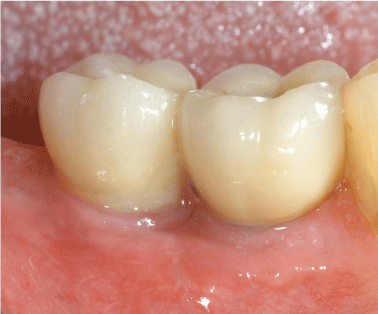
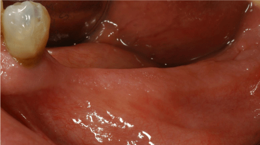
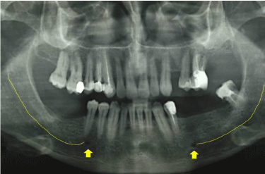
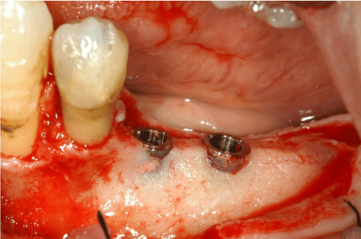
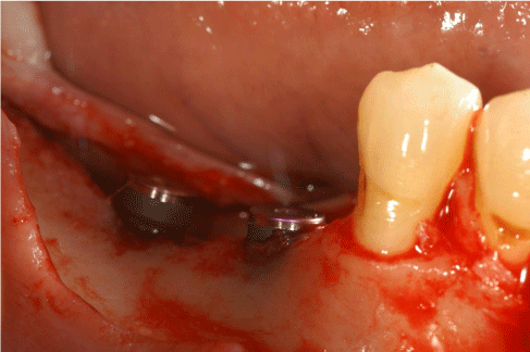
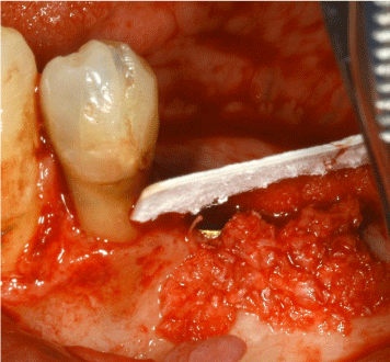
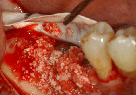
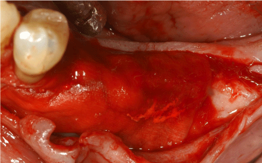
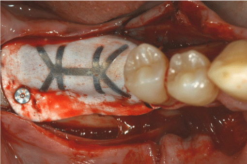
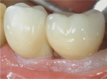
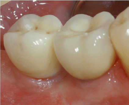
A 62 year-old male patient came to our attention presenting a posterior bilateral mandibular partial edentulism, requiring bone regeneration procedures due to an alveolar ridge resorption, for implant placement purposes. Pre-operative clinical (Figures 1 and 2) and radiological examinations (Figure 3) were used to assess the morphology of the alveolar ridge and to identify the inferior alveolar nerve anatomical course. Presurgical medication consisted of a 0.2% chlorhexidine digluconate mouth rinse and an extraoral scrub with a povidone–iodine solution. A sedative premedication was orally administered before the surgery. Local anaesthesia consisted of 4% articaine with epinephrine 1:100,000 infiltrations. For each mandibular side, a midcrestal full-thickness incision was made, and vertical releasing incisions were used to allow for a wide flap basis as well as sufficient access to the defective ridge area. Buccal and lingual flaps were reflected, the bone ridge was examined and any soft tissues remaining on the crest were debrided with a surgical curette. Depending on the amount of the residual crestal bone height and width, a simultaneous approach was chosen for implant placement. Dental implants were placed in a prosthetically ideal position simultaneously with the regenerative technique. Two Camlog® Screwline (Camlog Biotechnologies, Basel, Switzerland) with 3.8 mm and 4.3 mm diameters and 11 mm length were placed at the bone level on the left side (Figure 4), whereas two Camlog® Screwline with 4.3 mm diameter and 9 mm length were placed on the right side and were left to protrude 3-4 mm from the top of the bone surface, due to an insufficient vertical height (Figure 5). The cortical bone plate was perforated with a round bur in order to expose the medullary spaces and stimulate bleeding, allowing access of the cells from the bone and bone marrow to the area of regeneration. Subsequently a 1:1 ratio mixture of demineralized bovine bone mineral (DBBM) (Bio-Oss, Geistlich AG, Wolhusen, Switzerland) and autogenous bone chips harvested from the retromolar region was placed in the defect area around the implants on both sides. On the left a horizontal regeneration was performed, with a bioabsorbable collagen membrane (Bio-Gide, Geistlich AG, Wolhusen, Switzerland) adapted onto the crestal bone protecting the graft (Figure 6), while on the right side a vertical reconstruction was carried out through a titanium-reinforced e-PTFE Gore-Tex membrane (W. L. Gore & Associates, Inc., Flagstaff, AZ, USA) fixed on the buccal and lingual aspect with cortical pins over the defect (Figure 7). The membranes were contoured and trimmed in order to extend 3 to 4 mm laterally from the bone margin of the defects (Figure 8 and 9). This allowed undisturbed bone regeneration without the interference of competing soft tissue. A horizontal periosteal-releasing incision was made in both sides to allow for an adequate flap mobilization so that a tension-free primary wound closure could be obtained by means of horizontal mattress and single interrupted stitches. The patient underwent an antibiotic prophylactic treatment starting 1 day before surgery and then twice daily for 1 week (Amoxicillin/Clavulanate, Augmentin, GlaxoSmithKline, Brentford, UK). After the surgery, an anti-inflammatory agent (Ketoprofen, Orudis, Aventis Pharma, Origgio, Varese, Italy) was prescribed for 1 week. The patient was instructed to rinse twice daily with a 0.2% chlorhexidine solution and to refrain from mechanical plaque removal in the surgical area for 1 week. Sutures were removed 10–14 days after surgery. After a healing period of 6 months, all implants achieved successful integration. A partial thickness flap was made on the left side to uncover the implants and to place the healing abutments. A full thickness flap was contrariwise performed on the right side to remove the e-PTFE membrane before screwing the healing abutments. Temporary crowns were placed after a 3 weeks healing period and were left in place for 6 months to modify and condition the soft tissue shape and contour. At the end of the restoration, four zirconia abutment and metal free zirconia dental crowns were placed to restore the missing teeth (Figures 10 and 11). After the final prosthetic restoration, patient was registered in a maintenance program consisting of recalls for professional oral hygiene and clinical evaluation every 6 months. A radiological follow-up evaluation with periapical X-rays and orthopantomographs was conducted once a year.
All implants were placed with an adequate primary stability and the healing proceeded without neither major complications nor mucosal dehiscences and exposures of the membranes. Upon raising the flaps for re-entry surgery, in both sides the regenerated area appeared as mineralized bone tissue, and implants were clinically stable. An integration of the DBBM particles into the newly formed bone was observed. The bone volume following regeneration was satisfactory on the buccal and lingual aspect of the ridge. The radiographic appearance demonstrated an increased osseous healing over the bone defect during the 6 months healing period. No remnants of the collagen membrane could be detected at the re-entry surgery on the left side. A thick layer of periosteum-like tissue was found on top of the newly formed bone, possible remnants of the partly resorbed membrane. At e-PTFE membrane removal and abutment connection on the right side, the membrane was found in proper position and well integrated with the underlying tissue. A regenerated tissue, clinically similar to bone, was observed surrounding the implant heads. A thin soft tissue layer was present between the membrane and the regenerated bone-like tissue. Six months after the placement, all implants were well integrated as demonstrated by the clinical measurements regarding the soft tissue and by radiographic analysis regarding the bone, and were subsequently prosthetically loaded. After a 8 years follow-up, implants showed a survival and success rate of 100% according to Albrektsson et al. criteria, namely immobility, absence of peri-implant radiolucency, and marginal bone loss not exceeding 1.5 mm after the first year of loading and up to 0.2 mm yearly. An implant was considered survived if it was still physically in the mouth during the clinical evaluation [1]. A higher marginal resorption was radiographically observed after the first year of functional loading, whereas at the following examinations, all the implants showed no further crestal bone loss (Figure 12). At the last recall, the patient referred satisfactory function of the implant-supported prostheses, with no clinical signs of peri-implant tissue inflammation and suppuration (Figures 13 and 14).
In the present case-report, barrier membranes were used to physically protect the graft isolating the bone defect from the overlying soft tissue, obtaining an adequate amount of bone circumferentially around the exposed threads simultaneously with implants insertion.
The use of articaine deserves some words to be spent. Indeed, evidence suggests that it is the local anaesthetic agent that best diffuses within soft and hard tissues. Furthermore, the quicker onset and shorter elimination time associated with no relevant side effects or gross toxicity make this molecule the drug of choice in dentistry when local anaesthesia is needed [2,3].
The results pointed out that the combination of DBBM and autologous bone, grafted under a collagen or an e-PTFE membrane, may be respectively used when horizontal and vertical bone augmentation was respectively needed. As a matter of fact, in both surgical techniques, the survival and success rate were 100% over an 8-year follow-up. Non-resorbable membranes were frequently applied to have a total control over the resorption rate [4], however main drawbacks were an extensive surgical site needed for membrane removal and frequent soft tissue dehiscences and membrane exposures allowing for bacterial contamination and infection of the area intended for regeneration, jeopardizing the osteogenesis [5,6]. In literature, several advantages of collagen, including the haemostasis, the weak immunogenicity, the easy manipulation, the presence of a direct effect on bone formation [7] and the capacity to augment the width of the tissue [8], have been cited. In addition to the fact that soft tissue dehiscences seemed to be less frequent when using resorbable compared with non-resorbable membranes, other advantages included the absence of extensive raising of flaps needed for membrane removal, the absence of exposure of the regenerated bone in the apical areas, and a reduction of the patient morbidity. The clinical outcome reported in the present study confirmed the evidence that the use of both type of membranes may be suitable in case of alveolar ridge reconstruction, however resorbable membranes could be preferred in case of horizontal bone regeneration while non-resorbable membranes are essential when vertical bone regeneration is needed. It is demonstrated that the application of the principles of GBR using both resorbable and non-resorbable membranes for the treatment of peri-implant bone defects could render long-term predictable results, both in terms of implant survival and maintenance of marginal bone levels [9-11]. Possible advantages include the absence of the re-entry surgery for non-resorbable membranes removal, with a less discomfort for the patient, decreasing the time necessary to finalize the rehabilitation. Furthermore, the implants could serve as tending screws, providing a stable support for the membrane over the defect. One of the main drawbacks when a one-stage approach is preferred, is related to the membrane exposure during the healing time, which may lead to a complete loss of the graft and the implants if the clinical situation is not correctly managed with a pharmacological and surgical approach.
Grafting the defect with autogenous bone particles combined with xenografts could supposedly reduce the defect volume and prevent the blood clot shrinkage stabilizing it. Furthermore, space-making properties belonging to bone substitutes associated with a satisfactory resistance to resorption following placement into bony defects, could stimulate new bone formation and provide a support especially when slowly resorbable membranes are applied, avoiding their collapse into large defects [12,13]. In the present surgical procedure, a 1:1 ratio of autogenous particulated graft combined with DBBM was used to regenerate bone simultaneously with implant placement in both procedures. The newly formed tissue appeared macroscopically similar to the native bone with DBBM particles well integrated in the remodelled bone with no differences between non-resorbable and resorbable membrane. In the present case-report, the medium-term survival of zirconia abutments in posterior regions was comparable with that of titanium abutments, as recently reported by Lops et al. [14]. The application of implant-supported zirconia-based crowns has proven to be a satisfactory treatment option, as supported by the high cumulative survival rates reported in literature [15]. Finally, the supportive care of the peri-implant tissues need to be highlighted. A clear understanding of the signs of peri-implant disease recurrence is crucial so that early and definitive action can be taken to prevent further clinical attachment and bone loss around implants, which might otherwise go unnoticed until advanced stages, jeopardizing the success of the implant prosthetic rehabilitation [16].
Dental implants placed simultaneously with GBR procedures using resorbable or non-resorbable membranes reveal a high survival rate, and may be considered a safe and predictable therapy over a long-term follow-up time. Future developmental efforts have to be taken to improve the performance of the resorbable membranes also at later stages.
The authors declare that the work was performed without receiving any financial support from any third party. The article is free of all conflict of interest. On behalf of all co-authors, the corresponding author shall bear full responsibility for the submission and confirm that all co-authors have reported any potential conflicts.
Pre-surgical view on the right side.
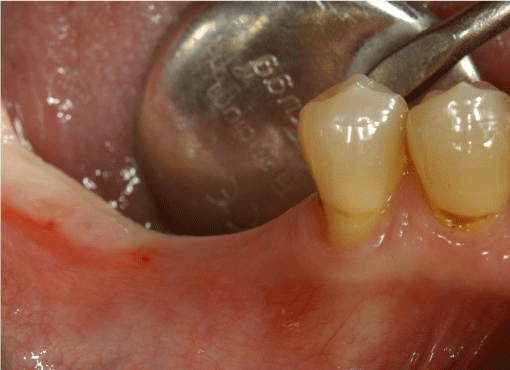
Pre-surgical view on the left side.
Preoperative evaluation orthopantomograph. In both sides, the upper cortical of the mandibular canal has been highlighted with a yellow line, while the mental foramen has been indicated with a yellow arrow.
Dental implants placement on the left side. Horizontal regeneration was required to obtain an adequate bone volume.
Dental implants placement on the right side. Vertical regeneration was needed around the exposed threads.
GBR with a bioabsorbable collagen membrane combined with a mixture of autologous bone and DBBM graft in a 1:1 ratio.
GBR with Titanium-reinforced e-PTFE Gore-Tex membrane combined with a mixture of autologous bone and DBBM graft in a 1:1 ratio.
Placement of the bioabsorbable collagen membrane over the grafted area.
Titanium-reinforced e-PTFE Gore-Tex membrane fixation with cortical pins on the lingual and buccal aspect.
Definitive zirconia crowns on the left side.
Definitive zirconia crowns on the right side.
Radiological 8-years follow up orthopantomograph.
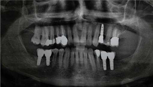
Clinical 8-years follow-up evaluation of the definitive restoration on the left side.
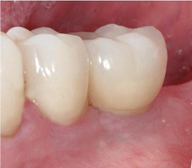
Clinical 8-years follow-up evaluation of the definitive restoration on the right side.
