Case Report
J Dent App. 2015;2(4): 207-209.
Multiple Bilateral 11 Supernumerary Teeth with Forth Molars in a Non-Syndromic Patient: An Unusual Case Report
Fatih Asutay1*, Ahmet Hüseyin Acar2, Mustafa Kirtay3, Hilal Alan4 and Yusuf Atalay5
1Department of Oral and Maxillofacial Surgery, Faculty of Dentistry, Afyon Kocatepe University, Afyonkarahisar, Turkey
2Department of Oral and Maxillofacial Surgery, Faculty of Dentistry, Bezm-i Alem Vakif University, Istanbul, Turkey
3Department of Oral and Maxillofacial Surgery, Faculty of Dentistry,Inönü University, Malatya, Turkey
4Department of Oral and Maxillofacial Surgery, Faculty of Dentistry, Inöönü University, Malatya, Turkey
5Department of Oral and Maxillofacial Surgery, Faculty of Dentistry, Afyon Kocatepe University, Afyonkarahisar, Turkey
*Corresponding author: Fatih Asutay, Afyon Kocatepe Üniversitesi, Di§ Hekimliği Fakültesi, Ağiz Di&s ve Çene Cerrahisi A.D, Güvenevler Mah. Inönü Bulvari No
Received: January 08, 2015; Accepted: March 11, 2015; Published: March 13, 2015
Abstract
Supernumerary teeth are known as the teeth in excess of the normal dentition. Multiple supernumerary teeth are usually observed as having syndromes. Conversely, the occurrence of multiple supernumerary teeth associated with any single syndrome is rare. This article presents an unusual case report, where a male patient who has 11 supernumerary teeth was without any syndromes. Patient also had 6 embedded permanent teeth (one premolar, one canine, four wisdom).
Introduction
Normally there should be 20 deciduous and 32 permanent teeth in the alveolar mechanism. The etiology of the term supernumerary teeth, also known as the hyperdontia, is completely unknown besides 2 available theories. The first theory suggests that tooth may occur by dividing the tooth bud. However the second theory implies that it may occur due to a local hyperactivity of the independent dental lamina. It also emphasized that there might be hereditary factors [1- 3].
Supernumerary teeth can be seen as single, multiple, bilateral or unilateral. Also it is classified based on shape; conical, tuberculate, supplemental or odontome and according to the location; mesiodens, paramolar, distomolar and parapremolar. Supernumeraries are usually observed in; maxillary midline (mesiodens), maxillary fourth molars, maxillary paramolars, mandibular premolars, maxillary lateral incisors, the mandibular fourth molars and maxillary premolars, respectively. It has been observed 10 times more in the upper jaw than the lower jaw [4].
Supernumerary teeth are often seen together with syndromes such as Gardner’s, Fabry Anderson, Ellis Van Creveld, Ehlers Danlos, Incontinentia pigmenti and Tricho-rhino-phalangeal Syndromes and developmental disorders such as Cleft lip and palate and Chondroectodermal dysostosis [5,6]. However, only a few cases have been reported without the correlation of a syndrome.
Supernumerary and impacted teeth can lead to malocclusion, root resorption, displacement or rotation, preclusion to eruption of the adjacent tooth, cyst formation, loss of vitality in the neighboring teeth, crowding, and aesthetic problems such as diastema [7,8].
The aim of this case report is to share clinical and radiological assessments of supernumerary and a large number of embedded permanent fourth molar teeth, which has no syndromic correlation with each other.
Case Report
Thirteen year old male patient was admitted to Inönü University, Faculty of Dentistry, Oral and Maxillofacial Surgery Department for a routine dental check and orthodontic examination. During the examination, a large number of embedded supernumerary teeth were observed in panoramic radiograph and then patient was referred tothe surgery department. Patient had no pain or any other complaints. There was no serious disease on his medical history.
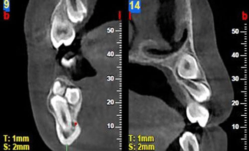
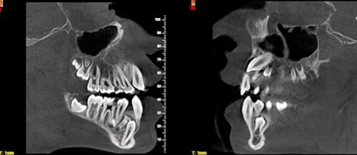
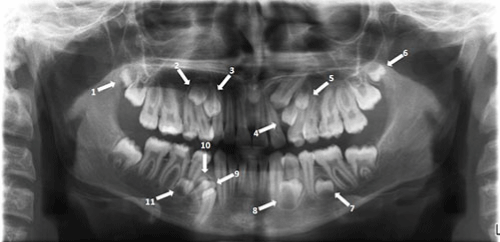
Computed-tomography (CT) and panoramic radiographs imaging were performed on the patient (Figures 1-3). 11 Supernumerary teeth were found on both maxilla (6 teeth) and mandibula (5 teeth). In addition, it was observed that left canine in the upper jaw, right first premolar in the lower jaw and all the wisdom teeth were embedded. Left lower first molar was extracted previously due to deep caries.

Figure 1: Coronal slices of CT.

Figure 2: Sagittal slices of CT.

Figure 3: Panoramic radiograph demonstrating multiple supernumerary teeth.
The patient signed the written consent of information before the surgery.
Discussion
The incidence of supernumerary teeth in the population was reported as 3.6% - 0,1% based on literature review [9-11]. However, the percentage of multiple supernumerary was found to be around 1% [12]. Supernumerary teeth can be seen in any part of the jaw. Rajab and Hamdan [13] have reported that 89.6% were seen in the premaxilla. Many studies have mentioned that the incidence of supernumerary teeth was observed in men more than women [13].
Although there are many theories on the formation of this situation, several authors have reported that genetic transmission might be responsible for that development. Some authors offered an autosomal dominant transition for Supernumerary teeth cases. There were a remarkable number of Supernumerary cases supporting this idea [4,14,15].
There are various estimates and theories related to the etiology. However, none of these has been proven. An autosomal dominant or recessive genetic inheritance, dichotomi and hyperactivity of the dental lamina could be counted among these [12,14].
An embedded supernumerary is often diagnosed incidentally during a routine examination. Periapical, occlusal or panoramic films could be used for diagnosis. However, more detailed information such as the proximity to the adjacent teeth and location of the subject in the bone could be reached through Computed Tomography (CT). Besides, CT plays an important role in treatment planning for this occasion [16].
Supernumerary teeth might cause premature space closure, diastema, pain, swelling, cystic formations, crowding, retention of deciduous teeth and pathological root resorption in the adjacent teeth [8,17]. Shetty and Sandler [18] reported that dentigerous cyst developed around the impacted supernumerary canine tooth. Enlargement around the follicles in the left upper canine tooth was observed in the case mentioned. Therefore, it could be stated that early detection and intervention, orthodontic and functional aspects of the situation are important.
There are two treatment options: surgical extraction or maintenance of the teeth and periodic monitoring for at least once a year. Early removal of supernumerary teeth is recommended when they cause problems such as delayed eruption or non-eruption of permanent teeth, displacement of the adjacent teeth, enhancement of various types of malocclusion, subacute pericoronitis, gingival inflammation, periodontal abscesses, cystic lesions, absorption of the root [19]. In the present case, it was decided to extract all supernumerary impacted upper and lower teeth, because the patient complained of malocclusion and orthodontic problems.
The timing of the surgical intervention is controversial. There are two opinions; first of these suggests the immediate removal after determination of the case. The second suggests to follow-up the completion of the root development of the adjacent teeth. Current literature suggests the extraction of the unerupted supernumerary teeth. Eruption rate of 54-75% have been observed after surgery. However, during or after surgery, procedure has the potential to damage the adjacent structures. Surgical approach could lead to root resorption and loss of vitality particularly in the mandibular premolar region [1,3]. We decided to remove all supernumerary teeth immediately because of the decision to commence the orthodontic treatment with the approval of the parents of the patient.
In the literature, a few multiple supernumerary reports are available that are not associated with any other syndrome or disease [1,5,14]. In this case, a large number of supernumerary teeth were accompanied by 2 pcs fourth molar and 6 pcs impacted permanent teeth (one canine, one premolar and four wisdom teeth).
Briefly, in cases of multiple supernumeraries, diagnosis and planning of the treatment should be done carefully by a multidisciplinary team. Imaging opportunities should be considered carefully to predict complications and to plan for the surgical approach.
Acknowledgement
All procedures followed were in accordance with the ethical standards of the responsible committee on human experimentation (institutional and national) and with the Helsinki Declaration of 1975, as revised in 2008.
References
- Nayak UA, Mathian VM, Veerakumar. Non-syndrome associated multiple supernumerary teeth: a report of two cases. J Indian Soc Pedod Prev Dent 2006; 24: S11-S14.
- Akgun OM. Non-syndrome patient with bilateral supernumerary teeth: Case report and 9-year follow-up. Eur J Dent 2013 7:123-126.
- Gunduz K, MuglaliM. Non-syndrome multiple supernumerary teeth: A case report. J Contemp Dent Pract 2007; 8: 81-87.
- Parihar A, Ram Kishore R, Vilas N, Madhu R. Multiple Impacted Permanent and Supernumerary Teeth: A Case Report. Journal of Clinical and Diagnostic Research 2010; 3287 - 3288.
- Turkkahraman H, Yilmaz HH,Cetin E. A non-syndrome case with bilateral supernumerary canines: report of a rare case. Dentomaxillofac Radiol 2005; 34:319-321.
- Moore SR, Wilson DF, Kibble J. Sequential development of multiple supernumerary teeth in the mandibular premolar region -- a radiographic case report. Int J Paediatr Dent 2002; 12: 143-145.
- Reddy GS. Nonsyndromic bilateral multiple impacted supernumerary mandibular third molars: a rare and unusual case report. Case Rep Dent 2013; 2013: 857147.
- Ün EC, Ezirganli S, Kirtay M, Özer K, Yağci S. Sendromsuz Çok Sayida Arti Küçük Azilar: 2 Olgu Sunumu. Süleyman Demirel Üniv Dis Hek Fak Derg 2011; 3: 72-75.
- Yusof WZ. Non-syndrome multiple supernumerary teeth: literature review. J Can Dent Assoc 1990; 56: 147-149.
- Schulze C. Incidence of supernumerary teeth. Dent Abstr 1961; 6.
- Niswander JD, Sujaku C. Congenital Anomalies of Teeth in Japanese Children. Am J Phys Anthropol 1963; 21: 569-574.
- Rajab LD, Hamdan MA, Supernumerary teeth: review of the literature and a survey of 152 cases. Int J Paediatr Dent 2002; 12: 244-254.
- Batra P, Duggal R, Parkash H. Non-syndromic multiple supernumerary teeth transmitted as an autosomal dominant trait. J Oral Pathol Med 2005; 34: 621-625.
- Sedano HO, Gorlin RJ. Familial occurrence of mesiodens. Oral Surg Oral Med Oral Pathol 1969; 27: 360-361.
- Anthonappa RP, Omer RS, King NM. Characteristics of 283 supernumerary teeth in southern Chinese children. Oral Surg Oral Med Oral Pathol Oral Radiol Endod 2008; 105: e48-54.
- Esenlik E, Sayin MO, Atilla AO, Ozen T, Altun C, et al. Supernumerary teeth in a Turkish population. Am J Orthod Dentofacial Orthop 2009; 136: 848-52.
- Shetty R, Sandler PJ. Keeping your eye on the ball. Dent Update 2004; 31: 398-402.
- Moore JR. Surgery of the mouth and jaws. Blackwell scientific publications, Oxford 1985; 373-75.
- Hogstrom A, Andersson L. Complications related to surgical removal of anterior supernumerary teeth in children. ASDC J Dent Child 1987; 54: 341-343.