
Research Article
J Dent App. 2015;2(5): 214-218.
Bacterial Leakage of Mineral Trioxide Aggregatebased and Resin-based Sealers in Teeth Ready for Root Retainers
Flávia Sens Fagundes Tomazinho*
Professor, Endodontia - Universidade Positivo, Brazil
*Corresponding author: Flávia Sens Fagundes Tomazinho, Professor, Endodontia - Universidade Positivo, Brazil
Received: January 29, 2015; Accepted: March 11, 2015; Published: March 13, 2015
Abstract
Aim: To perform an in vitro experiment to compare the sealing capability of a mineral trioxide aggregate (MTA) Fillapex sealer and an AH Plus sealer after root canal preparation for root canal pins.Methods: Forty-four lower premolar roots were biomechanically prepared, and the diameter of the cervical, middle, and apical thirds were standardized. The roots were sterilized, and the subsequent steps were conducted in a laminar flow booth. The specimens were filled by the lateral condensation technique with MTA Fillapex or AH Plus sealers, and the filling material was removed immediately after filling by leaving two different lengths (4 mm or 8 mm) of the remaining root filling. Four roots made up the positive and negative controls. The specimens were then inserted into a dual chamber device and placed in contact with sterile brain-heart infusion broth. The roots were inoculated with 1 × 10⁷ colony-forming units/mL of Enterococcus Faecalis, and the bacterial suspension was maintained in contact with the filling and renewed every 48 h, with daily observation for 60 days. The data were analyzed by the Kaplan-Meier survival test. The filling sealers and the remaining filling were analyzed with a Chi-square test at a significance level of 5%. Results: Comparison of the MTA Fillapex and AH Plus sealers and filler lengths relative to time of contamination was not significantly different (p = 0.519), despite the smaller frequency of infiltration that was observed in the roots filled with MTA Fillapex with the 8-mm remaining filling. Conclusion: Despite the infiltration of E. faecalisin all of the conditions, the length of the remaining root and the time until infiltration were positively correlated as the time that was required for bacterial infiltration was greater in the group with the 8-mm-long remaining material compared to the group with the 4-mm-long remaining material.
Keywords: Enterococcus faecalis; Dental infiltration; Root canal treatment
Abbreviations
MTA: Mineral Trioxide Aggregate
Introduction
One of the desirable physicochemical properties of endodontic sealers is that they adhere to the dentin walls of root canals [1], and then hermetic filling can be obtained through root canal sealing to avoid fluid percolation into the periapical tissue [2]. In addition, antibacterial properties are desirable in order to prevent endodontic reinfection.
The use of endodontic sealers that are associated with guttapercha is currently considered a standard procedure in endodontic filling [3], mainly due to the lack of adherence of the gutta-percha to the dentin walls. The draining properties of the endodontic sealer must also be taken into consideration so that the spaces between the filling material and the root wall can be filled, thus providing higher sealing quality [2].
The sealer capacity for maintaining the apical seal can become more evident when there is a need to perform a preparation for root retainers, as the material may move during the mechanical preparation [4].
During the interval between the preparation of the canal for the retainer and its installation, the coronal portion of the dental element remains, for the most part, sealed only by a temporary material. Such material typically provides low resistance and may move due to the movements and forces employed during chewing, thus leaving the canal passage exposed to the oral environment and enabling root canal contamination by microorganisms. The need to partially unfill the root canal and prepare it to receive a pin can accelerate bacterial microleakage once the sealed portion is around 3–5 mm. Resistant microorganisms such as Enteroccocus faecalis may remain in these spaces or even reach apical tissues through the dentin interface and filling material, thereby resulting in the acute deterioration of periapical lesions and the consequent failure of the endodontic treatment.
Several studies have considered coronal infiltration an important cause of endodontic treatment failure. It has been shown that filled roots that are exposed to the oral cavity are invariably contaminated by fluids, bacteria, and bacterial by-products. Long-term contamination may lead to failure of the endodontic treatment and compromise the prosthetic/restorative treatment as a whole [5].
The lack of endodontic sealers that have all of the ideal physicochemical properties has encouraged the development of a wide variety of materials, including materials that have been developed for greater adhesion, such as the AH Plus sealer, and those that have been developed for better performance of the biological properties and sealing [6], such as the MTA Fillapex sealer, which was developed from a mineral trioxide aggregate (MTA).
Therefore, the aim of this study was to perform an in vitro experiment to compare the sealing capability against E. faecalis of AH Plus and MTA Fillapex sealers after root canal preparation for root canal pins for a trial period of 60 days.
Material and Methods
This study was approved by the Human Research Ethics Committee of Positivo University (Protocol #088/11).
Selection and preparation of specimens
For this study, 44 single-rooted human permanent premolars with straight roots were selected. The sampled teeth had a minimum root length of 11 mm, a whole root apex, and no endodontic treatment, bone resorption, or calcifications. The teeth were kept in 0.1% Thymol solution at 4°C.
The specimens were washed in running water for 24 h to remove all traces of the Thymol solution, and the length of the root was standardized at 11 mm. The working length (WL) was standardized at 10 mm. The cervical portion of the root was prepared with a Gates Glidden drill #6. The roots were instrumented by manual instruments (K-file, DentsplyMaillefer, Ballaigues, Switzerland), with the crowndown technique, with the following file sequence: #80, # 70, and with an apical stop diameter of #60 for all elements. The irrigating solution used during the entire preparation was 2.5% sodium hypochlorite (AsferIndústriaQuímica Ltda., São Caetano do Sul, São Paulo, Brazil), and the final irrigation was performed with 10 mL of 17% ethylenediaminetetraacetic acid solution (Pharmacy-School Positivo University, Curitiba, Paraná, Brazil), which was followed by irrigation with 10 mL of distilled water and the drying of the canals with absorbent paper (Dentsply-Maillefer, Petrópolis, Rio de Janeiro, Brazil).
After the preparation, the samples were autoclaved at 120°C for 20 min (Cristófoli Autoclave Vitale 12 model, Campo Mourão, Paraná, Brazil). After this step, all of the procedures were conducted in a laminar flow chamber to maintain sterility.
The specimens were randomly, double blinded, divided into 2 experimental groups (n = 20) according to the endodontic sealer: Group I, AH Plus (Dentsply De Trey GmbH, Konstanz, Germany) or Group II, MTA Fillapex (Angelus, Londrina, Paraná, Brazil). Composition of the sealers is shown in Table 1.
![]()
Composition
Preparation mode
AH Plus� (Dentsply, Konstanz, Germany)
Paste A - bisphenol-A, bisphenol-F calcium tungstate, zirconium oxide, silica iron oxide pigments
Paste B - dibenzyldiamineaminoadamantane tricyclodecane-diaminecalcium tungstate, zirconium oxide, silica,
silicone oil
The components were
mixed in equal
portions of pastes A and B.
MTA Fillapex� (Angelus, Londrina, PR, Brazil)
Salicylate resin, diluting resin, natural resin, bismuth trioxide, nanoparticulated silica, MTA, pigments
The components were combined by using a self-mixing tip attached to a syringe.
Table 1: Tested sealers and their composition.
After the filling, the groups were subdivided into two subgroups (n = 10) according to the length of the remaining root filling: Subgroup A, 8 mm or Subgroup B, 4 mm. Four specimens were used for the positive and negative controls, with two teeth in each group.
The root canals were filled gutta-percha and sealer with the lateral condensation technique. The sealers were handled according to the manufacturer’ instructions.
To perform partial removal of the filling, Paivapluggers (Golgran, São Paulo,São Paulo, Brazil) were heated to redness at the predetermined lengths. The remaining cement was removed from cavity with cotton ball soaked in alcohol. Visibly could not be seen any amount outstanding of sealer in walls. Confirmation of the remaining filling length was conducted with a Hedströem file (Dentsply-Herpo, Petrópolis, Rio de Janeiro, Brazil) and rubber stop. The specimens were placed in an oven at 37°C for a period corresponding to three times the hardening time of each sealer (6 h for the MTA Fillapex sealer and 24 h for the AH Plus sealer).
Bacterial infiltration
The outer surfaces of the roots in the experimental and positive control groups were covered with two layers of cosmetic nail polish (Colorama, Maybelline, New York, NY, USA), except for 1 mm short of the root apex. Negative control teeth were completely sealed, including the apical foramen.
The bacterial infiltration underwent testing with the dual chamber method (Figure 1). The upper chamber was inoculated with 10 μL of brain-heart infusion broth with 1 × 10⁷ colony-forming units/mL of E. faecali s(ATCC 19433) while maintaining the bacterial suspension in contact with the filling. The BHI broth was renewed every 48 h. The specimens were kept at 37°C for 60 days, and the turbidity of the BHI broth was checked daily. When turbidity was verified, the day and specimen were recorded.
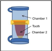
    Figure 1:  Dual-chamber system used to evaluate bacterial infiltration.
Chamber1 containing the bacterial agent, chamber 2 containing the sterile
substrate and the tooth interposed between them.
Statistical analysis
The data were statistically analyzed with the Kaplan–Meier survival test with SPSS software, version 15.0 (IBM Corporation, Armonk, NY, USA). The endodontic sealers and the remaining filling were analyzed with a Chi-square test at a significance level of 5%.
Results
Contamination frequency was higher in samples with a length of 4 mm than those of 8 mm, but there was no statistically significant difference (Table 2).
![]()
    Length of remaining root canal filling
    Groups
    Total
      N
    Contaminated samples
      N
    Success (without contamination)


    N
    Percent


    4
    AH Plus
    10
    8
    2
    20.0%


    MTA Fillapex
    10
    7
    3
    30.0%


    Overall
    20
    15
    5
    25.0%


    8
    AH Plus
    10
    6
    4
    40.0%


    MTA Fillapex
    10
    5
    5
    50.0%


    Overall
    20
    11
    9
    45.0%
Table 2: Frequency of contaminated and uncontaminated samples according to the material and length of filling.
Between the endodontic sealers tested, the MTA Fillapex group showed a lower contamination frequency compared to the AH Plus group.
The comparison of variables (endodontic sealer and remaining filler) in relation to the time of contamination was conducted with a Kaplan–Meier survival analysis, which was complemented by the Log-rank test (Mantel-Cox). No statistically significant difference was observed between the variables (p = 0.519). Table 3 shows the cumulative survival according to the time interval in days. For both materials used, there was a decrease in survival as time increased. It ranged from 1.000 and 0.900 in the first 15 days and from 0.500 to 0.200 in the last 60 days. The cumulative survival was lower in the samples with the 4-mm-long remaining filler, but there was no significant difference (Figures 2 and 3).
![]()
    Groups
    Length of remaining root canal filling (mm)
    Time (days)
    Uncontaminated at the beginning of the interval (lx)
    Contamination during the interval (dx)
    Ratio of cumulative survival at the end of the interval S(tx)
    Standard Error


    AH plus
    4
    1 to 15
    9
    1
    0.900
    0.095


    16 to 30
    6
    4
    0.600
    0.155


    31 to 45
    5
    5
    0.500
    0.158


    46 to 60
    2
    8
    0.200
    0.126


    8
    1 to 15
    10
    0
    1.000
    0.075


    16 to 30
    9
    1
    0.900
    0.095


    31 to 45
    6
    4
    0.600
    0.155


    46 to 60
    4
    6
    0.400
    0.155


    MTA
    4
    1 to 15
    10
    0
    1.000
    0.075


    16 to 30
    8
    2
    0.800
    0.126


    31 to 45
    6
    4
    0.400
    0.155


    46 to 60
    3
    7
    0.300
    0.145


    8
    1 to 15
    10
    0
    1.000
    0.075


    16 to 30
    9
    1
    0.900
    0.095


    31 to 45
    7
    3
    0.700
    0.145


    46 to 60
    5
    5
    0.500
    0.158
Table 3: Survival analysis by Kaplan–Meier test.
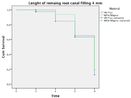
    Figure 2:  Survival analysis by the Kaplan–Meier test in the group with 4-mmlong
remaining filling material.
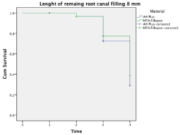
    Figure 3:  Survival analysis by the Kaplan–Meier test in the group with 8-mmlong
remaining filling material.
Discussion
The filling of the root canal with a biocompatible and inert material is considered an important part of the endodontic treatment. The use of endodontic sealers with gutta-percha has become standard in the filling of root canals in order to prevent marginal microleakage [7], which has been singled out as one of the main causes of endodontic failure as it compromises the sealing of the root canal system.
The infiltration of bacteria and its by-products can occur in a short period of time through the filling material, mainly in teeth with extensive coronal destruction and that will be subjected to the production of root canal retainers due to factors associated with the preparation of retainers, such as aseptic chain breakage during procedures that are performed without complete insulation, the possibility of filling displacement during removal of the coronal portion of the filling material, placement of temporary restorations between prosthetic work sessions, and the length of the remaining root canal filler [8].
Several methods have been used to assess the sealing ability of filling materials. Clinically, the bacterial infiltration model can provide a more accurate indicator of the sealing ability, and it has the benefit of using an etiologic agent of apical periodontitis [2].
In this study, E. faecali swas selected for the examination, as it is a pathogen that is frequently recovered from infected root canals and that is capable of invading dentinal tubules [9,10].
In this study, the root length, apical diameter, and preparation diameter of the coronal third of the root were standardized to allow for the same amount of bacterial inoculum to be placed in contact with the filling material inside the prosthetic space. The roots were autoclaved after the instrumentation, and all of the procedures were then conducted in a laminar flow chamber. The sterility of the testing device mounts was confirmed by the absence of medium turbidity in the negative control group.
Three items should be discussed regarding the results of this study: the bacterial infiltration that occurred in the remaining filling material, the time until infiltration, and the endodontic sealer used.
E. faecalis infiltration through the filling material occurred in all of the experimental groups, and this was probably due to the presence of gaps in the interface between the filling material and the wall of the root canal. These defects can be assigned to a number of variables, such as the internal anatomy of the root canal system, the biomechanical preparation, the properties of the irrigants, the physicochemical properties of the materials, and the filling technique [11]. The results of this study, in which bacterial infiltration occurred regardless of the length of the remaining root canal filling, are in line with those of previous studies [3,8].
For the time until the infiltration of E. faecalis, BHI turbidity was only observed on the 15th day of the experiment in the group sealed with the AH Plus sealer, and the MTA Fillapex group showed no difference according to the sealer used on the 17th day. Kuga et al. [12] evaluated the antimicrobial activity of these two sealers and observed similarities between them. However, the results of this study showed that the antimicrobial capabilities of these sealers were unable to completely prevent the infiltration of E. faecalis. Therefore, an important role of the filler is the formation of a physical barrier to prevent bacteria from reaching the apical region and periapical tissues [11]. Despite the absence of a statistical difference, the contamination frequency was higher in the filled samples with a length of 4 mm than in the samples with a length of 8 mm; this result corroborates the findings by Mozini et al. [8], in which there was a positive correlation between the length of the root remaining and the sealing efficacy, considering that the smallest remaining filler infiltrated considerably more than at the other lengths.
In this study, the hypothesis that the sealer could influence the endodontic apical sealing was tested. There are constantly new studies on AH Plus and MTA Fillapex with analyses of their properties. Previous studies have explained that the high bond strength that is obtained with epoxy resin-based sealers is due to their ability to form a covalent bond with an open epoxide ring in any amino group that is exposed to low-voltage curing light and collagen, thus providing long-term dimensional stability [13-15].
Studies have assessed the bond strength of the sealers used in this study to the root canal wall and observed that AH Plus was significantly superior to MTA Fillapex[16,17]. Another study on MTA found that the low-bond resistance of the MTA Fillapex was due to the low capacity of adhesion to dental extensions due to apatite formation by the MTA [18]. The resin components in MTA can affect its bonding to dentin[19], showing once again that the sealer composition can influence its ability to adhere to the root canal wall along with gutta-percha.
Although the bonding of the MTA Fillapex sealer to the dentin walls can be affected by its composition, there were no statistically significant differences found between the two sealers tested in this study. Contrary to the results reported by Razavian et al. [20], where the MTA Fillapex showed significantly higher amounts of microleakage with resin-based sealers.
Considering the limitations of the in vitro study, the results of this study showed that there was no difference in coronal infiltration by E. faecalis between the sealers tested. Apparently, there was a positive correlation between the length of the remaining root and the time until infiltration as the time that was required for bacterial infiltration was greater in the group with the 8-mm-long remaining material compared to the group with the 4-mm-long remaining material.
Acknowledgment
The authors would like to thank Dr. Juliana Feltrin de Souza Caparroz for her assistance with the statistical analysis.
References
- Haragushiku GA, Sousa-Neto MD, Silva-Sousa YT, Alfredo E, Silva SC, et al. Adhesion of endodontic sealers to human root dentine submitted to different surface treatments. Photomed Laser Surg 2010;28:405-410.
- Schilder H. Filling root canals in three dimensions. 1967. J Endod 2006;32:281-290.
- Brosco VH, Bernardineli N, Torres SA, Consolaro A, Bramante CM, et al. Bacterial leakage in obturated root canals-Part 2: a comparative histologic and microbiologic analyses. Oral Surg Oral Med Oral Pathol Oral RadiolEndod 2010;109:788-794.
- Cheung W. A review of the management of endodontically treated teeth. Post, core and the final restoration. J Am Dent Assoc 2005;136:611-619.
- Yucel AC, Güler E, Güler AU, Ertas E. Bacterial penetration after obturation with four different root canal sealers. J Endod 2006;32:890-893.
- Scarparo RK, Haddad D, Acasigua GA, Fossati AC, Fachin EV, et al. Mineral trioxide aggregate-based sealer: analysis of tissue reactions to a new endodontic material. J Endod 2010;36:1174-1178.
- Huffman BP, Mai S, Pinna L, Weller RN, Primus CM, et al. Dislocation resistance of ProRoot Endo Sealer, a calcium silicate-based root canal sealer, from radicular dentine. IntEndod J 2009;42:34–46.
- Mozini AC, Vansan LP, Sousa Neto MD, Pietro R. Influence of the length of remaining root canal filling and post space preparation on the coronal leakage of Enterococcus faecalis. Braz J Microbiol 2009;40: 174-179.
- Love RM. Enterococcus faecalis--a mechanism for its role in endodontic failure. IntEndod J 2001;34:399-405.
- Zoletti GO, Pereira EM, Schuenck RP, Teixeira LM, Siqueira JF Jr, et al. Characterization of virulence factors and clonal diversity of Enterococcus faecalis isolates from treated dental root canals. Res Microbiol 2011;162:151-158.
- Saleh IM, Ruyter IE, Haapasalo M, Ørstavik D. Survival of Enterococcus faecalis in infected dentinal tubules after root canal filling with different root canal sealers in vitro. IntEndod J 2004;37:193-198.
- Kuga MC, Faria G, Weckwerth PH, Duarte MA, De Campos EA, et al. Evaluation of the pH, calcium release and antibacterial activity of MTA Fillapex. Rev Odontol UNESP 2013;42:330-335.
- Lee KW, Williams MC, Camps JJ, Pashley DH. Adhesion of endodontic sealers to dentin and gutta-percha. J Endod 2002;28:684-688.
- Fisher MA, Berzins DW, Bahcall JK. An in vitro comparison of bond strength of various obturation materials to root canal dentine using a push-out test design. J Endod 2007;33:856-858.
- Vilanova WV, Carvalho-Junior JR, Alfredo E, Sousa-Neto MD, Silva-Sousa YT. Effect of intracanalirrigants on the bond strength of epoxy resin-based and methacrylate resin-based sealers to root canal walls. IntEndod J 2012;45:42-48.
- Baechtold MS, Mazaro AF, Crozeta BM, Leonardi DP, Tomazinho FS, et al. Adhesion and formation of tags from MTA Fillapex compared with AH Plus® cement. RSBO. 2014;11:71-76.
- Tedesco M, Felippe MC, Felippe WT, Alves AM, Bortoluzzi EA, et al. Adhesive interface and bond strength of endodontic sealers to root canal dentine after immersion in phosphate-buffered saline. Microsc Res Tech 2014;77:1015-1022.
- Sagsen B, Ustün Y, Demirbuga S, Pala K. Push-out bond strength of two new calcium silicate-based endodontic sealers to root canal dentine. IntEndod J 2011;44:1088-1091.
- Assmann E, Scarparo RK, Böttcher DE, Grecca FS. Dentin bond strength of two mineral trioxide aggregate-based and one epoxy resin-based sealers. J Endod 2012;38:219-221.
- Razavian H, Barekatain B, Shadmehr E, Khatami M, Bagheri F, et al. Bacterial leakage in root canals filled with resin-based and mineral trioxide aggregate-based sealers. Dent Res J 2014;11:599-603.