Research Article
J Dent App. 2015;2(5): 223-228.
Challenging the Clostridium botulinum Toxin type A (BoNT/A) with a Selection of Microorganisms by Culture Methods and Extended Storage of used Vials to assess the Loss of Sterility
Martin Turner, Sim K Singhrao*, Sarah R Dennison, L H Glyn Morton and St John Crean
Oral & Dental Sciences Research Group, School of Medicine and Dentistry, University of Central Lancashire, Preston, PR1 2HE, UK
*Corresponding author: S. K. Singhrao, Oral & Dental Sciences Research Group, School of Medicine and Dentistry, University of Central Lancashire, Preston, PR1 2HE, UK
Received: January 09, 2015; Accepted: March 27, 2015; Published: March 30, 2015
Abstract
In 2002, botulinum toxin type A (BoNT/A) was approved by the US Food and Drug Administration (FDA) for cosmetic use. However, there may be procedural differences between the ways in which a clinician handles, applies and stores the product compared to the suggested guidelines of the manufacturer for handling and storage. To this end vials (N = 12) of BoNT/A were tested for the incidence of microbial contamination followed by challenging the product with a selection of microorganisms by culture methods and by using a calcein release assay to contaminate multi-dose vials at the single concentration used for facial aesthetics. A culture, droplet method was used to count microorganisms challenged with the therapeutic product and to compare viability levels in appropriate controls as well as measuring their lytic properties via an existing cell-free system involving calcein release. Counts of test organisms within the droplets, with the product and the controls without the product were undertaken using Image J software. The result from the incidence of in-vial contamination was inconclusive. Bacterial levels between controls and product challenged groups demonstrated no differences in the growth of viable microorganisms following immediate contact (p =0.05). The cell-free calcein release assay demonstrated differences at all time points for low levels of lysis in each case with bacterial lipid extract and were statistically significant (p = 0.011). Although these data appear to correlate with the minimum inhibitory concentration, the additives and vial integrity are also likely to contribute to the maintenance of BoNT/A sterility.
Keywords: BoNT/A; Lysis; Bacteria; Lipid extract
Introduction
Botulinum toxins are a group of neurotoxins derived from Gram positive, anaerobic, spore forming, rod shaped bacteria known as Clostridium botulinum, Clostridium butyricum and Clostridium baratii. Seven serologically distinct subtypes of the toxin from C. botulinum strains are reported [1]. They are designated the code A, B, C1, D, E, F and G and there is a high degree of amino acid sequence homology among the various serotypes [2]. Of these, serotype A is the most potent neurotoxin of all and has the longest duration of action in a biological system. The main site of action of the toxin serotypes in vivo are peripheral cholinergic nerve endings, including the neuromuscular junction and both sympathetic and parasympathetic nerve terminals. Despite the potency of the protein, it can be used safely and effectively to manage a number of clinical conditions. Botulinum toxin type A (BoNT/A) was first licensed in 1992 as Botox Cosmetic® (Allergan, Inc., Irvine, CA., USA) for use in the reduction of glabellar lines. Carruthers & Carruthers [3] described the effect of BoNT/A (called onabotulinum toxin by Allergan) upon rhytids. The biological activity of BoNT/A is measured by the Speywood units which are determined by the median LD50 dose given as an intraperitoneal injection to Swiss-Webster mice.
Botulinum toxin can enter and act on virtually any cell by a variety of mechanisms including false receptor implantation, liposome transport, and increasing membrane permeability [4]. The mechanism for activity of the toxin is mediated by dissociation of the 150 kDa core protein of BoNT/A from its toxin-complex to enter the neuron by receptor mediated endocytosis [2, 5, 6]. Once inside the nerve, the disulphide bond is cleaved and the light and heavy chains of the toxic protein separate and disperse into the cytoplasm. The light chain has protease enzyme activity and, depending on the serotype, may act to cleave any of its three substrate SNARE (Soluble N-ethyl-maleimide-sensitive-factor Attachment Receptor) proteins: synaptosomal associated protein 25 (SNAP-25), synaptobrevin and syntaxin. The binding of BoNT/A, for example with the light chain with SNAP-25 protein, prevents fusion of acetylcholine containing vesicles at the presynaptic membrane site. This results in the blockage of acetylcholine being released at the neuromuscular junction and loss of neurotransmitter prevents further signaling at this site and impairs normal functioning of the muscle.
Currently, BoNT/A is used therapeutically for the treatment of cervical dystonia, spasticity, blepharospasm, strabismus, hemifacial spasm, relief of musculoskeletal and neuropathic pain, mesenteric hypertrophy, hyperhidrosis and in facial aesthetics [7-9].
Although the action of BoNT/A at the neuromuscular junction has been described, the additional actions upon other cholinergic nerve endings including noci-receptors has not been fully elicited [10]. BoNT/A also has an effect upon modulators of pain including substance P, glutamate and calcitonin gene-related peptide but the full mechanism is poorly understood [11]. The high toxicity of BoNT/A limits in vivo human studies due to practical and ethical issues.
Calcein release assay is a sensitive method that avoids culture methodologies and is widely used to compliment the minimum inhibitory (MIC) and/or minimum lethal (MLC) concentrations experimentation [12]. The advantage of using this method is that the experiment can be repeated for the individual phospholipid components of the test microorganisms. We selected the following organisms Escherichia coli, Enterococcus faecalis, Staphylococcus aureus, Staphylococcus epidermidis, and Candida albicans in which literature indicated the major lipid constituents for these microbes were dioleoylphosphatidylethanolamine (DOPE), dioleoylphosphatidylglycerol (DOPG), cardiolipin and lysylphosphatidylglycerol (LyslPG) [13]. These commercially synthesized lipids were also tested alongside of the freshly extracted lipids to evaluate antimicrobial activity of the selected test organisms.
The purpose of this study was to investigate whether BoNT/A with the excipients included in Azzalure® (Ipsen Biopharm Ltd, Wrexham, UK.), may/may not affect growth of common human body commensal microorganisms. It is also possible that the integrity of the Azzalure® packaging, itself is instrumental in maintaining sterility of the vial (due to toxicity of the product stored in it), and/or if it is the nature of the purified endotoxin that has antimicrobial activity. The rationale being that if the product becomes contaminated while in clinical use, the likely contaminants will come from the practitioners, their clothing or the immediate environment.
Materials and Methods
Ethical approval in the form of a written letter, was obtained for this study from the Research Ethics Committee, before commencing laboratory investigations in which all experimental procedures met the ethical guidelines of our academic institute.
Vial contents, storage, and integrity
Twelve previously reconstituted vials as per manufacturers protocol of BoNT/A, specifically Botox® (Allergan, USA) and Azzalure® (Ipsen Biopharm Ltd., Wrexham, UK) in which the product had been used up for their therapeutic application were collected from the north of England and South Wales, UK. These vials were destined for disposal but, in view of planning ahead for this study, the empty vials were collected from various locations of UK, in a cool box and thereafter kept in a locked refrigerator for a period ranging from 24h to 12 months by the lead author (MT). At the start of the laboratory investigation, all the tubes appeared moist with traces of the reconstituted product left inside the vials. All vials were rinsed with 400μl sterile saline under class II safety cabinet conditions and 100μl aliquots were inoculated separately onto plates of nutrient agar, yeast extract agar, Sabouraud agar, and tryptone soya agar plates. Nutrient agar plates were incubated for 48 h at 37°C, and malt extract agar pl ates were incubated at 30°C and Sabouraud agar plates and tryptone soya agar were incubated for 5 days at 22°C.
Culture maintenance and dilution profiles/regimes
Pure cultures of common human body flora such as E. coli NCTC 10418, E. faecalis NCIMB 775, S. aureus NCIMB 6571, S. epidermidis NCIMB 8558, and C. albicans NCYC 147, were cultured aerobically in the laboratory on nutrient agar plates and incubated for 48h at 37°C, and malt extract agar pl ates for yeast were incubated at 30°C for 5 days and Sabouraud agar plates and tryptone soya agar were incubated at 22°C for 5 days. These commensals are implicated as contaminants where there are poor cross infection control measures. The cultures were originally purchased from the National Collection of Type Cultures (NCTC) operated by Public Health England, National Collection of Yeast Cultures (NCYC), and National Collection of Industrial, Food and Marine Bacteria (NCIMB) and maintained in a teaching microbiology laboratory at our academic institute.
E. coli, E. faecalis, S. aureus, S. epidermidis, were maintained on nutrient agar (Oxoid Ltd, Basingstoke, UK) plates and C. albicans was maintained on malt extract agar (Lab M Ltd, Bury, UK) as described above.
Liquid cultures bacteria and yeast: E. coli, E. faecalis, S. aureus, and S.epidermidis were inoculated into freshly prepared, sterile nutrient broth (Oxoid) as per manufacturer’s instructions. All bacterial liquid cultures were incubated overnight at 37°C in a Gallenkamp orbital shaker incubator set at 180 rpm (Gemini BV, Apeldoorn, The Netherlands). A discrete colony of C. albicans was inoculated into 10 ml of sterile malt yeast glucose peptone (MYGP) broth (malt extract 1.5g, yeast extract 1.5g, glucose 5g, peptone 2.5g/500ml distilled water). The liquid culture was incubated overnight at 30°C in the Gallenkamp orbital shaker incubator set at 180rpm as before.
Dilution profiles/regimes: All cultures were pelleted and washed three times in ¼strength sterile Ringer’s solution tablets purchased from Lab M Ltd (Bury, UK) with re-suspension of cells in between each centrifugation step, using a Sigma 3-16PK bench top centrifuge (Sigma-Aldrich, Dorset, UK) at 8,200 rpm for 10 min at 4°C. The resultant cell pellet was re-suspended in Ringers solution prepared using tablets as described before (Lab M Ltd., UK) and held on ice for testing the antimicrobial activity of Azzalure ® (BoNT/A trade name, Ipsen Biopharm Ltd, Wrexham, UK) and for extracting lipids (see section on total lipid bacterial extract below).
In order to obtain an initially acceptable countable number of colonies, the suspensions were serially diluted down to 1x10-12 in sterile saline (0.9% sodium chloride solution) and drops of a fixed volume (25 and 50μl) E. coli, E. faecalis, S. aureus, S. epidermidis, and C. albicans, diluted cell suspension were pipetted onto the surface (in triplicate), of freshly poured, dried and pre-labeled agar plates for the cultivation of the test microorganisms for colony count of ~30-300. Drops of the yeast were incubated at 30°C for 24h and bacteria at 37°C for 24h. Following incubation the plates were checked for discrete colonies using a colony counter (Stuart Scientific, Fisher Scientific UK Ltd, Loughborough, UK). The plates were photographed with a Canon 450D digital SLR camera (Jessops, Leeds, UK), fitted with a standard 18-55mm lens. Colony forming units per milliliter (cfu/mL) were calculated according to the appropriate dilution factor using Image J software to count colonies/drop for each bacteria/yeast.
Assessment of Azzalure®antimicrobial activity using culturable methods: Following determination of the range of dilution in which discrete colonies could be estimated for subsequent experiments, bacteria and yeast were diluted with 0.9% sterile saline as for the dilution profiles. The Azzalure® (Ipsen Biopharm Ltd, Wrexham, UK) was reconstituted, according to the manufacturer’s instructions just before use, with 0.9% sterile saline to give a final concentration of 2 Speywood units/millilitre (2U/mL) of Azzalure® per 25μl of saline.
For discrete colonies for each organism in the range of 30-300 colonies/drop predetermined dilution for E. faecalis was 1x10⁻⁹, and for E. coli, S. epidermidis, S aureus, it was 1x10⁻¹⁰ and C. albicans was 1x10⁻⁴. A fixed volume of the cell suspension (25μl) and an equal volume (25μl) of 2U/mL Azzalure® (= 50μl final drop volume) in triplicate was applied onto their respective solid agar medium. Control plates consisted of a fixed volume (25μl) of the same diluted cell suspension to which 25μl sterile saline was added and placed as a drop (50μl) in triplicate, onto fresh media plates as for the Azzalure® test plates . Further controls were performed by spreading 100μl of the mixture prepared by adding 50μl of the reconstituted 2U/mL Azzalure® and 50μl sterile saline. The plates were exposed to the air in the microbiology laboratory for up to 4h. The plates were incubated at 30°C and at 37°C for 24h.
All experiments were performed in triplicate and on three different occasions. Nutrient agar plates were incubated at 37°C for 24h and the yeast extract plates were incubated at 30°C for 24h as before. Following incubation the plates were checked for differences in the number of colonies using a colony counter and photographed.
Calcein release cell-free assay
A previously reported lipid extraction method [14,15] was used to extract total lipids from all test microorganisms (E. coli, E. faecalis, S. aureus, S. epidermidis and C. albicans). Essentially, a small volume (1 ml) of each of the cell suspensions in Ringer’s solution, (see section on dilution profiles/regimes above) was transferred into sterile 1.5 mL Eppendorf® micro centrifuge tubes and centrifuged for 10 min in the Sigma 1-14, microcentrifuge (Sigma-Aldrich Ltd, Dorset, UK) at 14,000 rpm to form a bacterial cell pellet. Each cell pellet was re-suspended and mixed thoroughly by vortex mixing in 0.4 ml of Ringer’s solution to which was added, 1.5 ml of a 1:2 (v/v) chloroform - methanol mixture. 0.5 ml of Milli-Q-water (specific ≈ 18.2 MΩ cm) was then added to the tubes and mixed again by vortexing for 5 min before separating two phases of the solvents by centrifugation using a Sigma 1-14 centrifuge (Sigma-Aldrich, Dorset, UK) at 2850rpm, for 5 min and 4°C. The top aqueous phase was removed and subsequently discarded and the lower organic layer was dried under N2 gas to ensure that all the solvent had evaporated prior to its use with creating a lipid film.
The lipid films prepared above were suspended in 70 mM calcein. The suspension was thoroughly mixed by vortexing for 5 min prior to sonication for 30 mins. The calcein loaded liposomes were purified from free calcein and the vesicles that failed to entrap calcein by using a Sephadex G75 gel filtration column (Sigma-Aldrich, Dorset, UK). Prior to separation, the gel filtration column was equilibrated for 13 h in a buffer containing 20mM HEPES, 150 mM NaCl and 1.0 mM EDTA (pH 7.4) . Following specimen loading, the column was eluted with 5 mM HEPES at pH 7.5.
Calcein release assay was performed at 20°C in the dark, by adding 2ml of buffer containing 20 mM HEPES, 150 mM NaCl and 1.0 mM EDTA (pH 7.4) with 20μl of calcein containing liposomes and a 20μl aliquot of Azzalure® at the fixed concentration used for facial aesthetics (2U/mL of Azzalure®). To measure total calcein release, 20μl of Triton X100 was used to dissolve the liposomes. The fluorescence intensities of released calcein was measured using a FP- 6500 spectrofluorometer (JASCO, Tokyo, Japan), with an excitation wavelength of 490nm and an emission wavelength of 520nm. The percentage of dye leakage was then calculated. The percentage lysis was calculated using the following equation where A = absorbance.
The whole experiment was then repeated for the individual phospholipid components of the test microorganisms by preparing 91.0 μm unilamellar liposomes. In order to investigate the main individual phospholipids of bacterial membranes DOPE, DOPG, cardiolipin and LyslPG were purchased from Avanti Polar Lipids and were used without further purification. Phospholipids (7.5 mg) were dissolved in chloroform, evaporated under a stream of nitrogen, placed under vacuum for overnight and then rehydrated using 70mM calcein. These calcein containing liposomes were purified using gel filtration chromatography as for extracted lipids for functional testing by Azzalure®.
Statistical Analysis
The data from each species of bacteria and yeast tested for growth following contact with BoNT/A (Azzalure®) did not conform to normality and hence, the non-parametric Kruskal-Wallis test was performed and statistical analysis based on the null hypothesis that there was no difference between the mean calcein release across the different model membranes tested was undertaken using an ANOVA test (Minitab 16 statistical analysis software, Minitab Inc., State College, PA, USA).
Results
Vial contents, storage, and integrity
Following exposure of the reconstituted Azzalure® coated plates to the air (for 4h), all plates remained free of any air borne contaminants when checked by incubation of the plates at 30°C and at 37°C for up to 3 days.
One white/cream coloured colony, was observed on the nutrient agar from vial 10. The bacterial colony from vial 10 resembled the colonies growing on nutrient agar plates with known cultures of S,aureus. Its molecular identity was not examined. No growth was observed from any of the other vials (numbers 1-9 and 11-12).
The effect of BoNT/A on the growth of bacteria and yeast
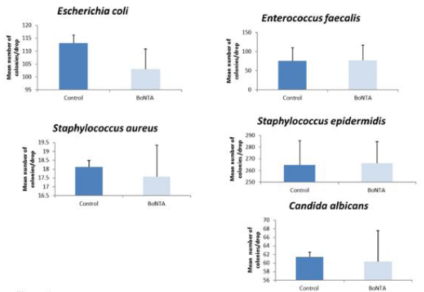
There was no statistical difference in any of the microorganism tested by immediate contact with the product p = 0.05 compared to those that contacted saline in controls (Figure 1 ). The colony counts varied between the three different sets of experiments as indicated by large error bars.

Figure 1: Each organism tested following immediate contact with the product compared with the saline control. There was no statistical difference in any of the microorganism tested C. albicans p = < 0.05.
The lytic ability of azzalure® calcein release assay
An antimicrobial compound can kill bacteria through many different mechanisms. The calcein release assay is an accepted methodology and in this study we used it to further determine to what extent Azzalure® may cause membrane disruption. The use of large unilamellar bacterial lipid extract liposomes loaded with calcein enables the membrane disruption to be assessed. In general, the probe is used to assess the increase in fluorescence when the calcein (florophore) is released due to membrane leakage.
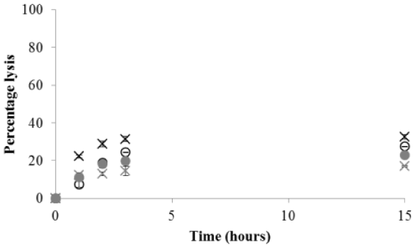
Figure 2 shows low levels of calcein leakage in the presence of Azzalure®. These data correlate with the MIC data, suggesting that Azzalure® and/or its additives have low levels of antimicrobial activity. Whilst these differences in calcein release after 2hours for bacterial lipid extract remain statistically significant (p = 0.011) the levels of release remained low for each organism. However, after 15hours there was a significant difference in calcein leakage for Azzalure® in the presence of liposomes prepared from the lipid extract [F = 51.420; p = 0.0001]. For liposomes formed from E. coli lipid membrane extract, Azzalure® caused greater levels of leakage (34%) and was statistically significant compared to E. faecalis, S. aureus, and C. albicans (p = 0.05). However, there was no significant difference in calcein leakage for liposomes formed from E. coli and S. epidermidis lipid extract (p = 0.29).

    Figure 2:  Percentage calcein leakage of botox for E. coli (grey crosses), E.
faecalis (light grey crosses), S. aureus (solid grey circles), S. epidermidis
(black crosses) and C. albicans (grey circles) lipid extract vesicles. The
values shown in the figure are the average and standard deviations of four
experiments.
The bacterial lipid membrane composition varies from bacterium to bacterium. PG is known as a major component in Grampositive bacteria whereas PE is identified as the major membrane component in Gram-negative bacteria [12]. Hence, the main lipid components of the organisms employed were investigated within these systems. Figure 3 shows that Azzalure® has a preference for anionic lipid LyslpePG, cardiolipin and do PG. Of these, PG is the main phospholipid component of all the test microorganisms. There was a significant difference in the mean calcein release after 15hours incubation in all of the model commercial lipid vesicles [F = 71.482; p = 0.0001] as shown in Figure 3. However, further statistical analysis demonstrated that there was no difference in the percentage of calcein released in the presence of Azzalure® following 15h incubation in all of the freshly extracted bacterial lipids and of the model commercial lipid liposomes. However in the extracted yeast lipid, there was a significant difference in lysis (p = 0.05).

    Figure 3:  Percentage calcein leakage of botox for LysylPG (black crosses),
Cardiolipin (grey crosses), DOPE (grey circles) and DOPG (black circles)
vesicles. The values shown in the figure are the average and standard
deviations of four experiments.
Discussion
As C. botulinum toxin type A or BoNT/A is used in facial aesthetics, in which the manufacturer’s very stringent and strict guidelines for product use must be followed, it was postulated that the manufacturer’s guidelines may be seeking to reduce the risk of contamination and/or loss in the efficacy of the re-constituted therapeutic product if used outside the recommended window of use of 4h. Since, literature supports the efficacy of BoNT/A protein to remain intact after reconstitution and storage up to one month and longer at 4°C and up to six months at -20°C [8,14,15] this investigation determined whether multi-use vials are susceptible to becoming contaminated during longer storage and/or following deliberate contamination of Azzalure® (Ipsen, UK), with known commensal human body flora. In addition, excipients are included in Azzalure® (Ipsen, UK), and their role as additional additives to the therapeutic product is unclear to the end user. This investigation employed, the cost effective, droplet method of Miles and Misra [16] designed to provide a rapid, reproducible method of counting microorganisms challenged with Azzalure® (Ipsen, UK). The procedure was performed under controlled laboratory conditions, to compare levels of microbial viability with appropriate controls.
Next we verified our results by washing out of reconstituted therapeutic product Botox® and Azzalure® from vials after the product had long been used up for periods of a few hours to 12 months. Overall, our results are in agreement with the findings of the previous two investigations in that 11 out of 12 vials maintained sterility [17,18] and that cross contamination is possible. The reasoning for the one vial (vial number 10) which demonstrated growth of white/ cream coloured single bacterial colony was likely the result of a splash/cross contamination during pipetting of S. aureus. However as no attempt was made to find the exact molecular identity of the bacterium, the contaminating organism and contamination of the vial remains unresolved.
Literature suggests that BoNT/A can adversely affect virtually any cell-type [4] and the saline (other than bacteriostatic saline) used for reconstituting the product may add to the stress of fewer bacteria and yeast cells if they were to contaminate the vials during handling. Additional “additives” that have the ability to reduce vial wetting [19] and the prevention of resistance to liquid ingress, can occur [20], and these help to create an environment within the vial which may limit the survival of contaminating microorgamisms. How long this environment could resist contamination is unknown but the results of this study suggest that after 12 months of appropriate storage, except for the one vial in which bacterial growth was recorded, no significant contamination had occurred in the remaining used vials. This leads to the conclusion that majority of the vials tested here remained sterile . This is likely to be due to the integrity of the Azzalure® packaging, itself rather than the purified endotoxin that demonstrated little antimicrobial activity.
Next we challenged Azzalure® (Ipsen, UK) with selected laboratory strains of test microorganisms to investigate any effect on their growth and viability. The selected test organisms included common commensals of the human body which have been implicated as contaminants where poor infection control has been observed. The effect of Azzalure® (Ipsen, UK) at the therapeutic dose, recommended for non-surgical facial aesthetics, and as measured by Speywood units/millilitre (U/mL) was tested on the selected organisms and was compared to controls without the product but under the same experimental conditions.
The experimental procedure used to evaluate the antimicrobial activity of Azzalure® (Ipsen, UK) demonstrated that statistically, there was no significant difference between the cfu/drop between the control and test samples for all five microorganisms.
In order to investigate the lytic ability of BoNT/A, a calcein release assay was undertaken using bacterial lipid extract and commercial lipids such as, DOPE, DOPG, cardiolipin and LyslPG [13]. The phospholipid composition of a cell membrane is essential in the selectivity of an antimicrobial compound [12]. The variation in the lipid composition and compound interaction is also dependent on phospholipid packing characteristics and stability [12]. The calcein release assay showed that BoNTA had a preference for anionic phospholipid headgroups PG, which is the main component of S. aureus, S.epidermidis and E. faecalis membranes. In a membrane with reduced negative charge the Azzalure®–NH2 may be attracted to the positive charges of the phospholipids in a bacterial membrane. In contrast, in the presence of phosphatidylethanolamine (PE), a reduced level of calcein lysis was observed indicating that since the lipid composition of E. coli membranes includes 91% PE, 3% PG, and 6% CL [13], this high level of PE content offers a protection against leakage. It is possible that the local interaction of the BoNT/A to the membrane requires binding of the BoNT/A to the target cell membrane. Furthermore, this initial interaction appears important for its binding to the neurotransmitter vesicles [4]. One of the excipients added to Azzalure® is albumin and is believed to encourage microbial growth. This inference is made from the reported observations that an infectious episode in the human host for example, decreases the normally high concentration of albumin during acute-phase [21]. A possible reason for this down regulation is for the innate immune responses to act to inhibit growth of bacteria invading the host.
In conclusion, except for the one out of 12 vials tested, majority of the vials remained sterile. The vial and stopper seem to be effective against microbial contamination even when the contents were accessed multiple times in our hands. It is therefore the integrity of the packaging of the multi-use vial over the antimicrobial activity of the product that protects the vial from being easily infected. This leads to the conclusion that BoNT/A is safe and effective if manufacturer’s instructions are followed.
References
- Hill KK, Smith TJ. Genetic diversity within Clostridium botulinum serotypes, botulinum neurotoxin gene clusters and toxin subtypes. Curr. Top Microbiol. Immunol 2013; 364: 1-20.
- Poulain B, Popoff MR, Molgó J. ‘How do the Botulinum Neurotoxins block neurotransmitter release: from botulism to the molecular mechanism of action’. The Botulinum J 2008; 1: 14–87.
- Carruthers JD, Carruthers JA. Treatment of glabellar frown lines with C. botulinum - A exotoxin. J Dermatol Surg Oncol 1992; 18: 17-21.
- Simpson LL. Identification of the characteristics that underlie botulinum toxin potency: Implications for designing novel drugs. Biochimie 2000; 82: 943–953.
- Brin MF, Botulinum toxin: chemistry, pharmacology, toxicity, and immunology. Muscle Nerve Suppl 1997; 6: S146-168.
- Binz T, Rummel A. Cell entry strategy of clostridial neurotoxins. J. Neurochem 2009; 109: 1584-1595.
- Clark RP, Berris CE. Botulinum toxin: a treatment for facial asymmetry caused by facial nerve paralysis. Plast Reconstr Surg 1989; 84: 353-355.
- Winter L, Spiegel J. Botulinum toxin type-A in the treatment of glabellar lines. Clin Cosmet Investig Dermatol 2009; 22: 1-4.
- Kim J. Contralateral botulinum toxin injection to improve facial asymmetry after acute facial paralysis. Otol Neurotol 2013; 34: 319-324.
- Mustafa G, Anderson EM, Bokrand-Donatelli Y, Neubert JK, Caudle RM. Anti-nociceptive effect of a conjugate of substance P and light chain of botulinum neurotoxin type A. Pain 2013; 154: 2547-2553.
- Gazerani P, Au S, Dong X, Kumar U, Arendt-Nielsen L, et al. Botulinum neurotoxin type A (BoNTA) decreases the mechanical sensitivity of nociceptors and inhibits neurogenic vasodilation in a craniofacial muscle targeted for migraine prophylaxis. Pain 2010; 151: 606-616.
- Dennison SR, Phoenix DA, Snape TJ. Synthetic oligoureas of metaphenylenediamine mimic host defence peptides in their antimicrobial behavior. Bioog Med Chem Lett 2013; 23: 2518-2521.
- Ratledge C, Wilkinson SG. (eds) Microbial lipids 1988; l 1. Academic Press, London.
- Bligh EG, Dyer WJ. A rapid method of total lipid extraction and purification. Can J Med Sci 1959; 37: 911-917.
- Rose HG, Oklander M. Improved Procedure for the Extraction of Lipids from Human Erythrocytes. J Lipid Res 1965; 6: 428-431.
- Hexsel DM, De Almeida AT, Rutowitsch M, De Castro IA, Silveira VL, et al. Multicenter, double-blind study of the efficacy of injections with botulinum toxin type A reconstituted up to six consecutive weeks before application. Dermatol Surg 2003; 29: 523-529.
- Parsa AA, Lye KD, Parsa FD. Reconstituted botulinum type A neurotoxin: clinical efficacy after long-term freezing before use. Aesthetic Plast. Surg 2007; 31: 188-191.
- Miles AA, Misra SS. The estimation of the bactericidal power of the blood. J. Hyg 1938; 38: 732-749.
- Menon J, Murray A. Microbial growth in vials of Botulinum toxin following use in clinic. Eye (Lond) 2007; 21: 995-997.
- Alam M, Dover JS, Arndt KA. Pain associated with injection of botulinum A exotoxin reconstituted using isotonic sodium chloride with and without preservative: a double-blind, randomized controlled trial. Arch Dermatol 2002: 138: 510-514.
- Pickett A, Perrow K. Formulation composition of botulinum toxins in clinical use. J Drugs Dermatol 2010; 9: 1085-1091.
- Kirsch LE. Pharmaceutical container/closure integrity. VI: A report on the utility of liquid tracer methods for evaluating the microbial barrier properties of pharmaceutical packaging. PDA J Pharm Sci Technol 2000; 54: 305-314.
- Gabay C, Kushner I. Acute-phase proteins and other systemic responses to inflammation. N Engl J Med 199: 340: 448-454.
Citation: Turner M, Singhrao SK, Dennison SR, Glyn Morton LH and St John Crean. Challenging the Clostridium botulinum Toxin type A (BoNT/A) with a Selection of Microorganisms by Culture Methods and Extended Storage of used Vials to assess the Loss of Sterility . J Dent App. 2015;2(5): 223-228. ISSN:2381-9049