Case Report
J Dent App. 2015;2(6): 243-245.
Management of Massive Peripheral Ossifying Fibroma in the Right Lingual Vestibule of Mandible
Jitender Batra*, Pradeep Kumar, Monika Chahar, Gyanander Attresh and Vikas Berwal
Department of Oral & Maxillofacial Surgery, Post Graduate Institute of Dental Sciences, Pt. B.D Sharma U.H.S, Rohtak, Haryana, India
*Corresponding author: Dr. Jitender Batra, Department of Oral & Maxillofacial Surgery, Post Graduate Institute of Dental Sciences, Pt. B.D Sharma U.H.S, Rohtak, Haryana, India
Received: February 02, 2015; Accepted: April 27, 2015;Published: April 29, 2015
Abstract
Brief Background: Peripheral ossifying fibroma (POF) is a common solitary gingival overgrowth thought to arise from the gingival corium, periosteum or periodontal ligament. POF is an occasional growth of the anterior region of mandible and accounts for 3.1% of all oral tumors and 9.6% of the gingival lesions. About 60% of these tumors occur in maxilla and more than 50% of all cases of maxillary POFs are found in the incisors and canine areas.
Material and Method: A 25 year-old female patient reported with complaint of non-tender growth over right lingual vestibule of lower jaw and was diagnosed histopathologically as a case of peripheral ossifying fibroma. The case was managed with surgical resection.
Discussion: Dental calculus, plaque, microorganisms, dental appliances, and restorations are considered to be the irritants triggering the lesion. The lesion though usually smaller than 1.5 cm in diameter can reach a much larger size and can cause separation of the adjacent teeth, resorption of the alveolar crest, destruction of the bony structure and cosmetic deformity. The treatment of choice for this lesion is complete surgical excision with the removal of the irritating factors.
Conclusion: Due to higher recurrence rate of the lesion, early detection and complete surgical resection of these lesions followed by long term follow-up bear importance in clinical management.
Keywords: Peripheral Ossifying fibroma; Fibro-osseous lesions; Mandible
Introduction
Peripheral ossifying fibroma is non-neoplastic enlargement of gingiva that is thought to be reactive in nature. POF is an occasional growth of the anterior region of mandible and accounts for 3.1% of all oral tumors and 9.6% of the gingival lesions [1]. About 60% of these tumors occur in maxilla and more than 50% of all cases of maxillary POFs are found in the incisors and canine areas [2]. Due to their histopathological as well as clinical resemblance, peripheral ossifying fibroma are thought to arise as pyogenic granuloma which undergoes fibrous maturation and subsequent calcification. Trauma and sometimes local irritating factors i.e. ill-fitting denture, calculus and faulty restoration may initiate the development of peripheral ossifying fibroma [3]. It affects both the gender but female predilection is more than the male. Racial predominance is 71% in white in contrast to 36% in black. Peak incidence occurs in second and third decade of life [4]. In this case report; we highlight a case of young female with unusual growth on the right lower lingual vestibule in relation to root stumps of first molar.
Case Report
A 25 year-old systemically healthy female patient referred to department of Oral and Maxillofacial Surgery, Postgraduate Institute of Dental Sciences, Rohtak complaining of a solitary symptomatic growth over lingual right side of lower jaw since 8 months which started as a small peanut sized growth and increased in size slowly.
Extraorally, there was no swelling on right site of face. Face was apparently symmetrical. An intraoral clinical examination revealed sessile growth approximately 4*3.7* cm, extending from lower right canine area anteriorly to retromolar area posteriorly (Figure 1). Growth was painless, hard in consistency and non tender. No paresthesia associated with lower lip. Electric pulp testing of dental vitality proved negative for lower right 46. Concerned tooth was associated with grade-2 mobility. Oral hygiene of patient was fair and mouth opening was normal.

    Figure 1:  Showing the growth on lingual aspect of right side of mandibular
molar region.
Orthopantogram findings depicted carious 46. There was a radiolucent line along distal root of right first lower molar showing widening of periodontal ligament (Figure 2). Incisional biopsy was taken, and report was suggestive of a fibro-osseous lesion mostly peripheral ossifying fibroma.
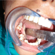
    Figure 2:  Orthopantogram showing carious 46 and radiolucency along its
distal root.
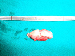
In present case, sessile base of growth was assessed. Growth was incised with the help of no. 15 B.P blade to expose the bony part. Bony growth was excised with help of chisel from its sessile base (Figure 3). Smoothening of rough bony surface was done by trimmer. Bleeding was observed from lingual cortical plate after ostectomy. Bleeding was controlled by using bone wax. Involved tooth was extracted following hemostasis. Excised specimen was sent for histopathological examination.
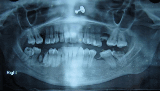
Figure 3: Excised growth.
Histopathological report showed stratified squamous keratinized epithelium with stroma laminar calcification with mild inflammatory component and diagnosis in consistence with clinical diagnosis of peripheral ossifying fibroma (Figure 4). Patient is under regular follow up. Figure 5 reveals the healing at 3 months post-operatively.
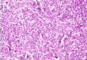
    Figure 4:  Histopathological picture showing squamous keratinized epithelium
with stroma laminar calcification with mild inflammatory component.
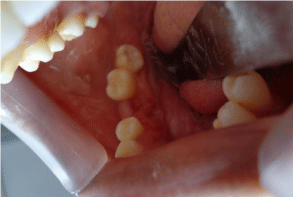
Figure 5: Healing at 3 months follow up.
Discussion
Ossifying fibroma is classified as, and behaves like, a benign bone neoplasm. It is often considered to be a type of benign fibro-osseous lesion (FOLs) which affect the jaws and craniofacial bones. FOL refers to a group of pathologic processes in which normal bone is replaced by fibroblasts and collagen fibers containing variable amounts of mineralized material (bone and cementum). This group encompasses fibrous dysplasia, benign fibro osseous neoplasms ossifying fibroma), and a heterogeneous group of reactive lesions (osseous dysplasia, POF) [5].
There are two types of ossifying fibromas: the central type and the peripheral type. The central type arises from the endosteum or the periodontal ligament adjacent to the root apex and causes the expansion of the medullary cavity. The peripheral type occurs solely on the soft tissues covering the tooth-bearing areas of the jaws [6]. Pathogenesis of peripheral ossifying fibroma is uncertain, they are thought to arise from periosteal and periodontal membrane [7].
The POF and ossifying fibroma (OF) are the lesions that exhibit similar histomorphologic features and both originate from periodontal ligament cells. But a POF is a reactive lesion where as an OF is a benign neoplastic lesion included in the group of benign fibro-osseous lesions of the jaws and both POF and OF show different proliferative activities [8].
Gingival lesions that imitate POF are peripheral giant cell granuloma, pyogenic granuloma, fibroma, calcifying epithelial odontogenic cyst, calcifying odontogenic cyst, etc. POF has also been described by various synonyms such as peripheral cemento ossifying fibroma, peripheral odontogenic fibroma (PODF) with cementogenesis, peripheral fibroma with osteogenesis, peripheral fibroma with calcification, fibrous epulis, calcifying fibroblastic granuloma, etc. [9]. Almost 60% of the lesions occur in the maxilla and mostly occur anterior to molars. The lesion is most common in the second decade of life affecting mainly females which was also seen in our case [10].
Dental calculus, plaque, microorganisms, dental appliances, and restorations are considered to be the irritants triggering the lesion [11]. The lesion though usually smaller than 1.5 cm in diameter can reach a much larger size and can cause separation of the adjacent teeth, resorption of the alveolar crest, destruction of the bony structure and cosmetic deformity [12]. In reported case, a sessile growth was of size 4*3.7 cm approximately, present in mandibular premolar region, with mobility of associated tooth. Panoramic radiograph depicted widening of periodontal ligament which shows its origin from periodontal ligament.
POF is definitively diagnosed through a histopathological examination. The histopathological examination usually shows the following features: benign fibrous connective tissue with varying fibroblast, myofibroblast and collagen content, sparse to profuse endothelial proliferation, and mineralized material that may represent mature, lamellar or woven osteoid, cementum-like material, or dystrophic calcifications. Acute or chronic inflammatory cell infiltration can also be observed in these lesions [13]. In our case, histopathological report showed stratified squamous keratinized epithelium stroma laminar calcification with mild inflammatory component which confirm the diagnosis of lesion as peripheral ossifying fibroma. The treatment of choice for this lesion is complete surgical excision with the removal of the irritating factors. In present case lesion was excised with extraction of involved tooth.
Due to the high rate of recurrence (8% to 20%), close postoperative monitoring is required in all cases of POF [14]. POF recurs due to the incomplete removal of the lesion, the failure to eliminate local irritants and difficulty in accessing the lesion during surgical manipulation as a result of the intricate location of the lesion (usually an interdental area) [15].
Conclusion
It is difficult to differentiate between most of the reactive gingival lesions clinically, particularly in the initial stages. Thus every excised tissue should be sent for the histological examination for confirmation of the clinical diagnosis. Since the recurrent rate of peripheral ossifying fibroma is high, regardless of the surgical technique employed, it is important to eliminate the etiological factors and long postoperative follow up is mandatory in all the cases.
References
- Keluskar V, Byakodi R, R.shah N. Peripheral ossifying fibroma. J Indian Acad oral Med Radiol 2008; 20: 54-56.
- Neiville BW, Damn DD, Allen CM. Text book of oral maxillofacial pathology 2nd ed. Philadelphia WB Saunders 2004; 451-452.
- Delbem AC, Cusha RF, Silea JZ, Soubhia AMP. Peripheral cement-ossifying fibroma in child. A follow up of 4 years. Report of a case. Eur J dent 2008; 2: 134-137.
- Buchner A, Hansen LS. Histomorphologic spectrum of peripheral ossifying fibroma; Oral Surg Oral med Oral Pathol 1987; 83: 452-461.
- Reichart PA, Philipsen H, Sciubba JJ. The new classification of Head and Neck Tumours (WHO) – any changes? Oral Oncol 2006; 42: 757–758.
- Speight PM, Carlos R. Maxillofacial fibro-osseous lesions. Curr Diagn Pathol 2006; 12: 1–10.
- Sousa SC, Mesquita RA, Araujo NS. Proliferative activity in peripheral ossifying fibroma and ossifying fibroma. J Oral Pathol Med 1998; 27: 64-67.
- Kendrick, Woggoner WF. Managing peripheral ossifying fibroma. J dent child 1996; 63: 135-138.
- Walters JD, Will JK, Hatfield RD, Cacchillo DA, Raabe DA. Excision and Repair of the Peripheral Ossifying Fibroma: A Report of 3 Cases. J Periodontal 2001; 72: 939-944.
- Buchner A, Hansen LS. The Histomorphologic Spectrum of Peripheral Ossifying Fibroma. Oral Surg Oral Med Oral Pathol 1987; 63: 452-461.
- Mesquita RA, Orsini SC, Sousa M, de Araújo NS. Proliferative activity in peripheral ossifying fibroma and ossifying fibroma. J Oral Pathol Med 1998; 27: 64-67.
- Poon CK, Kwan PC, Chao SY. Giant peripheral ossifying fibroma of the maxilla: report of a case. J Oral Maxillofac Surg 1995; 53: 695-698.
- Kumar SK, Ram S, Jorgensen MG, Shuler CF, Sedghizadeh PP. multicentric peripheral ossifying fibroma . J oral sci 2006; 48: 239-43.
- Farquhar T, Maclellan J, Dyment H, Anderson RD. Peripheral ossifying fibroma: a case report. J Can Dent Assoc 2008; 7: 809-812.
- Shetty DC, Urs AB, Ahuja P, Sahu A, Manchanda A, et al. Mineralized components and their interpretation in the histogenesis of peripheral ossifying fibroma. Indian J Dent Res 2011; 22: 56-61.
