Case Report
J Dent App. 2015;2(6): 251-253.
Radiographic Revelation of Foreign Body in Primary Tooth: A Rare Case Report
Palit MC¹*, Agrawal J² and Saha S¹
¹Department of Pedodontics & Preventive Dentistry, Sardar Patel Post Graduate Institute of Dental and Medical Sciences, Lucknow, U.P. India
²Department of Oral Medicine and Radiology, Gitam Dental College and Hospital, Visakhapatnam, A.P. India
*Corresponding author: Palit MC, Department of Pedodontics & Preventive Dentistry, Sardar Patel Post Graduate Institute Of Dental And Medical Sciences, Lucknow, U.P. India
Received: February 23, 2015; Accepted: April 27, 2015; Published: April 29, 2015
Abstract
Children have a habit of ingestion or aspiration of foreign object, which pose severe danger to the child as well as parents. However the insertion of objects into the tooth is not very common and most of the time parents are unaware as children fear to reveal. Such objects act as a site of infection and may lead to discoloration of the tooth as well as abscess.
In the present case child presented with a discoloured maxillary right lateral incisor. Radiograph revealed presence of a metallic object inside the root canal of the same tooth, which neither the patient was aware nor the child disclosed during history taking. Hence early diagnosis and treatment is necessary to avoid future dreadful situation to child as well as patient. Parents should also be instructed to maintain a nonjudgmental and non-authoritarian approach to handle children, so that kids won’t be fearful to discuss their problems.
Keywords: Deciduous tooth; Foreign object; Stapler pin; Radiographs
Introduction
Accidental ingestion of foreign body by children is a pediatric emergency. It causes panic for children as well as parents. Children have a tendency to ingest marbles, toys, coins, keys and pins [1]. However insertion of foreign body in tooth by children is highly uncommon phenomenon. Open carious tooth in oral cavity acts as a site for insertion of foreign body leading to pain, swelling and abscess [2]. Proper history, detailed clinical examination and radiographs are necessary to detect the cause and give an appropriate diagnosis. A radiograph is also necessary to detect the correct size, type and position of the foreign object. This case report describes occurrence of stapler pin in deciduous maxillary right central incisor and its subsequent management.
Case Report
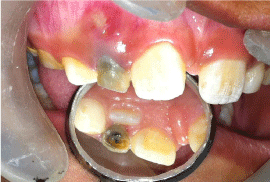
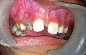
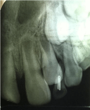
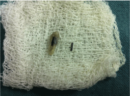
8-year-old girl with her parents reported to our dental department with the chief complaint of pain in upper right front teeth past one week. Parents also gave the history of pus discharge from the same tooth past three days. On clinical examination there was blackish discoloration on right deciduous lateral incisor i.e. 52. The pulp chamber of the tooth 52 was open with food lodgment (Figure 1). Labial vestibule in relation to the same tooth had a draining sinus (Figure 2). Intraoral periapical radiograph in relation to 52 showed a radiopaque twisted wire like unusual object in the pulp chamber as well as root canal (Figure 3). On enquiring the possible history of a metallic object inside the root canal parents mentioned that the child has a habit of putting stapler pins inside the mouth. As the permanent lateral incisor was erupting during the time of presentation of the case, we decided to extract 52 after informing the parents and taking their consent. Extraction was done under local anesthesia. After extraction the foreign object was removed from the tooth and it was found to be a stapler pin (Figure 4).

Figure 1: Right deciduous maxillary incisor with open carious lesion.

Figure 2: Discoloration and draining sinus i.r.t. 52.

    Figure 3:  Intraoral periapical radiograph revealed a radiopaque twisted wire
like unusual object in the pulp chamber as well as root canal.

Figure 4: Stapler pin retrieved from tooth.
Discussion
Children have a predisposition to put variety of objects into the oral cavity. These can cause ingestion or aspiration posing difficulty for parents [3]. If the objects are sharp or pointed, they pose a danger even during their removal by endoscopic means. Rupture of the common carotid artery, aortic pseudo-aneurysms, esophageal tears and fistula, pericarditis and cardiac tamponade have also been reported which are very serious complications [4,5].
Insertion of objects into the tooth is not very common and most of the time children happen to hide it from parents due to fear. In the present case parents were aware that the child has a habit of putting stapler pins into the mouth, but had never imagined the insertion of pin inside the tooth. When the pin was revealed in radiograph they were horrified. Child did not mention during the history taking that she had a habit of lodging stapler pin inside the tooth. Foreign bodies inside the tooth can act as a focus for infection and result in pain, bleeding and swelling. Case has been reported where placement of jewelry chain piece into a maxillary central incisor has resulted in actinomycosis [6]. Grossman [7], Gelfman [8], and Harris [9] reported retrieval of indelible ink pencil tips, brads, a tooth pick, adsorbent points, tomato seed, pins, wooden toothpick, a pencil tip, plastic objects, toothbrush bristles and crayons from the root canals of anterior teeth left open for drainage. Hence, it is important that an attempt is made to remove the foreign object, but by a dentist. Attempts at removal by the child or parent may result in accidental aspiration or ingestion. In the present case, the child did not reveal the incident till the stapler pin showed up on the X-ray.
Mcauliffe et al. have reported five radiographic methods to localize metallic foreign body in oral cavity [2]
- Parallax views (either horizontal or vertical);
- Vertex-occlusal views;
- Triangulation techniques;
- Stereo radiography; and
- Tomography.
In the present case we have used intraoal periapical radiograph, which clearly revealed metallic object inside the root canal of 52. For the management of such emergency removal of the foreign object from the root canal or extraction of the tooth by a specialist should be considered. Barbed broach, H file or the Masserman kit consisting of a number of differently sized trepans, which cut a narrow space around the object allowing it to be released, can also be used to remove the pin from the root canal. The Steglitz forceps have been described for use of removal of silver points from the root canal [10].
In the present case as the child was eight years old, the tooth was a deciduous maxillary lateral incisor, radiograph revealed a erupting permanent lateral incisor, discoloration of the tooth, mobility and draining sinus in relation to the labial vestibule in relation to 52. Hence a decision was made to extract the tooth after taking the parents consent. Similar treatment plan of extraction of primary tooth, after radiographic diagnosis of foreign object has also been reported by Holla G et al. (2010) and Pereira T (2013) [11,12].
Timely diagnosis and management of foreign object embedded in the tooth should be done to as early as possible to avoid complication as we did for our case. Advising parents to keep small objects out of reach of children can do prevention of such emergency. Pediatricians should also warn the parents that child could even try to put small tiny objects inside the open carious tooth. Management of open carious lesions hence should be done as early as possible to avoid further complications. Parents should also be instructed to maintain a nonjudgmental and non-authoritarian approach to handle children, so that kids will not be fearful to discuss their problems with parents. Written warnings by manufacturing companies should also spell out this message.
References
- Van As AB, du Toit N, van As AB, du ToitN, Wallis L, et al. The South African experience with ingestion injury in children. Int J Pediatr Otorhinolaryngol. 2003; 67: S175-S178.
- Macauliffe N, Drage NA, Hunter B. Staple diet: a foreign body in a tooth. Int J Paediatr Dent. 2005; 15: 468–471.
- Sai Prasad TR, Low Y, Tan CE, Jacobsen AS. Swallowed foreign bodies in children: Report of four unusual cases. Ann Acad Med Singapore. 2006; 35: 49-53.
- Spitz L, Kimber C, Nguyen K, Yates R, de Leval M. Perforation of the heart by a swallowed open safety pin in an infant. J R Coll Surg Edinb. 1998; 43:114-116.
- Gun F, Salman T, Abbasoglu L, Celik R, Celik A. Safety pin ingestion in children: A cultural fact. PediatrSurg Int. 2003; 19: 482-484.
- Goldstein BH, Sciubba JJ, Laskin DM. Actinomycosis of the maxilla: Review of literature and report of case. J Oral Surg. 1972; 30: 362–366.
- Grossman JL, Heaton JF. Endodontic case reports. Dent Clin North Am. 1974; 18: 509-527.
- Zillich RM, Pickens TN. Patient-included blockage of the root canal. Report of a case. Oral Surg Oral Med Oral Pathol. 1982; 54: 689-690.
- Harris WE. Foreign bodies in root canals: Report of two cases. J Am Dent Assoc. 1972; 85: 906-911.
- Lumley PJ, Walmsley AD. The removal of foreign objects from root canals. Dent Update. 1990; 17: 420-423.
- Holla G, Baliga S, Yeluri R, Munshi AK. Unusual objects in the root canal of deciduous teeth: A report of two cases. Contemp Clin Dent. 2010; 1: 246-248.
- Pereira T, Pereira S. An unusual object in the root canal of a primary tooth - a case report. Int J Paediatr Dent. 2013; 23: 470-472.