Case Presentation
J Dent App. 2015; 2(7): 261-263.
The Concurrence of Radix Paramolaris and C Shaped Root Canal Anatomy in Adjacent Teeth: A Rare Finding
Vasudeva A, Sinha DJ, Tyagi N, Tyagi SP and Vasudeva A*
Department of Conservative Dentistry and Endodontics, Kothiwal Dental College and Research Centre, India
*Corresponding author: Agrima Vasudeva, Department of Conservative Dentistry and Endodontics, Kothiwal Dental College and Research Centre, India
Received: May 29, 2015; Accepted: September 09, 2015; Published: September 11, 2015
Abstract
For a successful endodontic treatment thorough knowledge of root canal anatomy and its variations is of utmost importance .One of such variations is the presence of a third root mesiobucally, called Radix Paramolaris. Another anatomic variation is C shaped morphology in which instead of the typical shape of the pulp chamber with three root canals, there are C-shaped orifice of the root canal. This case report describes a rare case with the concurrence of two root canal anatomical variations in adjacent teeth i.e 36 (Radix Paramolaris) and 37 (C shaped canal configuration).
Keywords: C shaped; Radix paramolaris
Introduction
For a successful endodontic treatment, knowledge of the root canal anatomy is of utmost importance. Complex root canal anatomy is often the culprit that results in endodontic failures. Thus, a clinician must be aware of the variations from the normal root canal anatomy.
Usually two different roots namely mesial and distal are found in the mandibular molars. Sometimes a third disto-lingual root may be present [1]. This third root may arise from a part of the apical third of the mesial root or, occasionally, from distal root [1] Carabelli first reported this macrostructure in 1844 namely radix entomolaris (RE) [2]. Sometimes it can be present mesio-bucally and is known as radix paramolaris (RP) [3,4].
The C-shaped root canal configuration was first documented in endodontic literature by Cooke and Cox in 1979 and is so named for the cross-sectional morphology of the root and root canal of the molar teeth [5]. In these cases instead of the typical shape of the pulp chamber with three root canals, there are the C-shaped orifices of the root canal on the mandibular molars. Their main anatomical feature is the presence of a fin connecting the individual root canals whereas the orifice may appear as a single ribbon-shaped opening with a 180° arc [6].
Endodontic textbooks state that the C-shaped canal is not uncommon, and this is confirmed by studies in which frequencies ranging from 2.7% to 8% have been reported [5]. Most often, the C-shape of the pulp chamber is used to describe the mandibular second molars, the maxillary first molars (0.92%) and the maxillary second molars (4.9%) [7-9].
Case Presentation
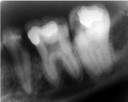
A female patient aged 35 years reported to the Department of Conservative Dentistry and Endodontics, Kothiwal Dental College and Research Centre with the chief complaint of pain in the lower left back teeth. Pain was severe and continuous in nature. On clinical examination, decay was seen with respect to 35, 36 and 37. On radiographic examination, radiolucency was seen involving the pulp in all the teeth indicating irreversible pulpitis (Figure 1).

Figure 1: Preoperative radiograph.
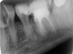
36 was tender on percussion and patient was more symptomatic in respect to that tooth. So, root canal procedure was initiated in 36 prior to 37 and 35. Access was gained to the pulp chamber after administration of local anesthesia (2% Lignocaine with 1:100000 epinephrine), under rubber dam isolation. Access preparation was done with endo-access bur and canal orifices were located with DG 16 endodontic explorer. Three canals were located but the dentinal map seems to be slightly extending in a mesiobuccal direction and thus an extra canal orifice was located. Patency was confirmed with K-file 10. Working length was established with the use of apex locator (Root ZX, J. Morita Inc.) To check the presence of extra canal, an angulated working length radiograph was taken which confirmed the presence of an extra root. The canals were cleaned and shaped with hand K-files and nickel titanium rotary ProTaper files (Dentsply Maillefer, Switzerland). The canals were sequentially irrigated using 5.25% Sodium hypochlorite and 17% EDTA during the cleaning and shaping procedure. The canals were thoroughly dried and obturation was done using F2 Pro Taper Gutta-percha and AH Plus sealer (Dentsply, Maillefer, Switzerland) (Figure 2).

Figure 2: Post obturation radiograph.
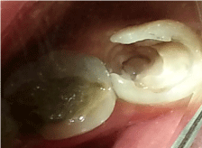
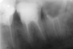
In the next visit root canal procedure was performed in relation to 37. On opening the root canal and removal of tissue from the pulp chamber the canal system appeared ‘C’ shaped (Figure 3). Deep orifice preparation was done and careful probing with small files characterized the C- shape more accurately. The working length intraoral periapical radiographs revealed the presence of a C shaped canal (Fan’s classification type III). The canals were sequentially irrigated using 5.25% Sodium hypochlorite and 17% EDTA during the cleaning and shaping procedure. The canals were thoroughly dried and obturation was done using thermoplasticized obturation technique (Thermafil, Dentsply) and AH Plus sealer (Dentsply, Maillefer, Switzerland). 35 was also root canal treated (Figure 4). Post obturation restorations were done wrt 35, 36 and 37.The patient was reviewed after a month and was found to be asymptomatic and thereby was advised to get the teeth crowned.

Figure 3: Clinical picture of C shaped morphology wrt 37.

Figure 4: Post obturation radiograph.
Discussion
This is a rare case as till date no report have been published indicating the presence of two root canal anatomical variations in two adjacent teeth i.e 36 ( Radix Paramolaris) and 37 ( C shaped canal configuration).
The exact etiology of behind the formation of radix paramolaris is still unknown. The prevalence of RP as reported in the literature is 0%, 0.5% and 2% for the first mandibular molar, second molars and third molars, respectively [10].However, some studies have reported RP in first mandibular molars [11,12].
To achieve a correct diagnosis minimum of two diagnostic radiographs are necessary using buccal object rule. Access cavity preparation should be modified usually from a triangular to a trapezoidal shape. The modification should be done following the dentinal map [13].
The location of RP is (mesio) buccal. Dimensions of the RP may vary from a normal root with a root canal, to a conical short extension. Types A and B are described by Carlsen and Alexandersen [11]. RP whose cervical part was located on the mesial root complex was referred to as Type A and when it was centrally located, between the mesial and distal root complexes it was referred to as Type B. In the case reported RP was Type A of Carlsen and Alexandersen classification. An additional root is usually associated with an increased number of cusps and root canals [12].
The presence of an RE or an RP has clinical implications in endodontic treatment. An accurate diagnosis of these supernumerary roots can avoid complications or a ‘missed canal’ during root canal treatment. Because the (separate) RP is mostly situated in the same buccolingual plane as the mesiolingual root, a superimposition of both roots can appear on the preoperative radiograph, resulting in an inaccurate diagnosis [14].
C-shaped canal is most frequently found in the mandibular second molar, although it could be found in the mandibular first premolar, the mandibular first molar, the maxillary first molar, and the maxillary second molar [15]. When considering the C-shaped mandibular second molar, it has been documented that this morphologic configuration has significant ethnic variation. In the white population the incidence ranges from 2.7%–7.6%; the Lebanese population has a reported 19.1% incidence. Incidence in the Chinese population reached a reported 31.5%, and incidence was 44.5% in Koreans. These types of configurations have a bilateral incidence that may well reach 70%. Also the mandibular premolars with C-shaped configuration appear to have a higher incidence rate in the Chinese population [16].
The shape and the number of roots are determined by Hertwig’s epithelial sheath, which bends in a horizontal plane below the amelocemental junction and fuses in the centre leaving openings for roots. Fused roots may form either by coalescence owing to cementum deposition with time, or as a result of failure of Hertwig’s epithelial sheath to develop or fuse in the furcation area. A C-shaped canal appears when fusion of either the buccal or lingual aspect of the mesial and distal roots occurs. This fusion remains irregular, and the two roots stay connected by an interradicular ribbon [17].
The first classification of the C-shaped root canals was done by Melton and co-authors in 1991[5]. Later based on it, Fan [8] made an anatomic classification: 1. Category I (C1) - continuous C-shaped root canal from the orifice to the apex of the root; 2. Category II (C2) -one main root canal and a smaller one; 3. Category III (C3) – two or three root canals; 4. Category IV (C4) - an oval or a round canal; 5. Category V (C5) - no canal lumen or there is one close to the apex.
Once recognized, the C-shaped canal provides a challenge with respect to debridement and obturation, especially because it is unclear whether the C-shaped orifice found on the floor of the pulp chamber actually continues to the apical third of the root. The difficulties arise from the fact that with the C-shaped root canals it is the possible to have a thin net of anastomoses in root canal system [8,9,18] .
The mesiobuccal and distal canal spaces were prepared normally. However, the isthmus was not prepared with larger than no. 25 files; otherwise, strip perforation was likely to occur. Extravagant use of small files and 5.25% sodium hypochlorite is a key to thorough debridement of narrow canal isthmuses. A copious volume of irrigant allowed for more cleansibility and effectively removes tissues from narrow C-shaped canal ramifications [5]. Obturation of ‘C’ shaped canal requires technique modification, sealing the isthmus area is difficult if lateral condensation is used, hence thermoplasticized gutta percha obturation was done.
Conclusion
Successful and predictable endodontic treatment requires knowledge of biology, physiology and root canal anatomy. Teeth with variable root canal anatomy pose a challenge. Thus, for a successful endodontic treatment thorough knowledge of root canal anatomy and its variations, careful interpretation of the radiograph, close clinical inspection of the floor of the chamber, and proper modification of access opening are necessary.
References
- Parthasarathy B, Manje Gowda PG, Sridhara KS, Subbaraya R. Four canalled and three rooted mandibular first molar (Radix Entomolaris)-Report of 2 cases. J Dent Sc Res. 2011; 2: 1-5.
- Bolk L. Bemerküngen über Wurzelvariationen am menschlichen unteren Molaren. Zeiting fur Morphologie und Anthropologie. 1915; 17: 605–610.
- Carlsen O, Alexandersen V. Radix entomolaris: Identification and morphology. Scan J Dent Res. 1990; 98: 363-73.
- Sinha D J, Sinha AA. Additional Roots: Challenge to the Endodontist. IJCD. 2014; 2: 7-10.
- Jafarzadeh H, Wu YN. The C-shaped Root Canal Configuration: A Review. J Endod. 2007; 33: 517–523.
- Fan B, Min Y, Guanfan Lu, Jun Yang, Cheung GSP, Gutmann JL. Negotiation of C-Shaped Canal Systems in Mandibular Second Molars. J Endod. 2009; 35: 1003-1008.
- Cook HG, Cox FL. C-shaped canal configurations in mandibular second molar. J Am Dent Assoc. 1979; 99: 836-839.
- Fan B, Cheung GS, Fan M, Gutman JL, Bian Z. C-shaped canal system in mandibular second molars: Part I - Anatomical features. J Endod. 2004; 30: 899-903.
- Fan B, Cheung GS, Fan M, Gutman JL, Fan W. C-shaped canal system in mandibular second molars: Part II - Radiographic features. J Endod. 2004; 30: 904-908.
- Visser JB. Beitrag zur Kenntnis der menschlichen Zahnwurzelformen. Hilversum: Rotting. 1948: 49 –72.
- Carlsen O, Alexandersen V. Radix paramolaris in permanent mandibular molars. Identification and morphology. Scan J Dent Res. 1991; 99: 189–195.
- Sperber GH, Moreau JL. Study of the number of roots and canals in Senegalese first permanent mandibular molars. Int Endod J. 1998; 31: 117–122.
- Nagesh Bolla, Balaram D, Naik, Sarath Raj Kavuri, Sanjay Krishna Sriram. Radix Entomolaris: A Case Report. Endodontology. 121-124.
- Shailendra Gupta, Deepak Raisingani, Rishidev Yadav. The Radix Entomolaris and Paramolaris: A Case Report. J Int Oral Health. 2011; 3: 43-50.
- Jin GC, Lee SJ, Roh BD. Anatomical study of C-shaped canals in mandibular second molars by analysis of computed tomography. J Endod. 2006; 32: 10–13.
- Jorge NR, Martins, Sergio Quaresma, Maria Carlos Quaresma, Jared Frisbie-Teel. C-shaped Maxillary Permanent First Molar: A Case Report and Literature Review. J Endod. 2013; 39: 1649-1653.
- Fouzan KS. C-shaped root canals in mandibular second molars in a Saudi Arabian population. Int Endod J. 2002; 35: 499 –504.
- Loh HS. Incidence and features of the mandibular second molars in Singaporen Chinese. Aust Dent J. 1991; 36: 442-444.