
Case Presentation
J Dent App. 2015; 2(7): 264-268.
Anterior Aesthetic Rehabilitation – How to get Success again after the Unexpected Failure: Case Report
Toniollo MB¹*, Aguiar SC² and Corrêa MH³
¹Department of Dental Materials and Prosthodontics, Professor of the Specialization Course in Dental Implants of the National Association of Dental Studies, Brazil
²Professor Coordinator of the Specialization Course in Dental Implants of the National Association of Dental Studies, Brazil
³Implant Specialist by the National Association of Dental Studies, Brazil
*Corresponding author: Marcelo Bighetti Toniollo, Department of Dental Materials and Prosthodontics, Professor of the Specialization Course in Dental Implants of the National Association of Dental Studies, Brazil
Received: May 14, 2015; Accepted: September 10, 2015; Published: September 12, 2015
Abstract
Modern society, with the contemporary dentistry, aim by a proper function accompanied by extreme aesthetics. In order to get increasingly beautiful, harmonic and natural results, the search for development of rehabilitation indirect materials is constant, such as pure ceramic crowns (metal free) and high aesthetic content systems such as IPS E.Max. However, fateful events, such as root fracture, still remain unpredictable in the everyday context, and must be thought on how to have a proper resolution. In this paper it was described a case in which success first achieved, through metal free crowns on the upper incisors, gives way to a fateful situation of longitudinal root fracture of one of the elements rehabilitated. Thus, it is argued in this article one of the viable alternatives to be traced through modern procedures, that brings predictability, to circumvent the failure and go back to get the expected result and patient expectation. The conciliation of surgical and prosthetic procedures, with the aid of reverse planning and modern techniques, for the sake of good results and with respect to the involved tissues, allow obtaining harmonious and satisfactory work.
Keywords: Dental aesthetics; Dental implants; Ceramics
Introduction
The ceramics have reports of very old usage, but dentistry has focused on this stuff for two centuries. Negative points such as hardness and brittleness or friability became manageable against the high aesthetic and biological quality for the prosthetic work [1]. Technological advances have branched ceramics in numerous types and ways of obtaining, from the conventional mixture of powder and liquid, injected system, infiltrated by Glass and Machined (CAD/ CAM). Until the mid 1980s the only aesthetic option was the metal ceramic crowns. Advances in dental materials and implants led to the use of pure ceramic crowns (metal free) generating high aesthetic gain and biological affinity [2]. One of the systems that have gained prominence in studies and clinic routine is the ceramics reinforced by lithium disilicate, which provides its adequate strength [3,4]. Within this context, the IPS E.Max was consolidated as an option of choice among professionals in front of their great aesthetic and functional qualities. The versatility of these systems for metal free ceramics allowed wide dissemination and extremely viable applicability. For their full and proper use was also need the development of adhesive systems in order to enable effective union between substrate and rehabilitation [5,6]. Along the evolution of materials for indirect restorations, implant dentistry has made great advances in functional and aesthetic concepts, thus allying the use of metal free restorations and adding value among all the characteristics mentioned above. Another highlight along the dental implants refers to the possibility of their conciliation to the immediate restoration, installed together the implants [7]. This technique, which differs from the immediate load [8,9], provides numerous aesthetic and functional advantages, such as improved healing and formation of soft tissue emergence profile and better osseointegration of the implant [10,11]. Furthermore, new approaches with respect to the peri implant tissues (tissue care) and use of prosthetic connections that allow the Switch Platform philosophy and better mechanical stability to the system brought real gains to the final outcome. Even with all the evolutions of contemporary dentistry unexpected factors may occur and cause catastrophic failure. One of the most damaging setbacks in the oral cavity involving teeth is the longitudinal root fracture, there is no solution beyond the extraction. So with the conciliation of the various specialties and the best possible techniques that involve them, always with respect for the correct principles and concepts, the present case report shows that is possible to obtain clinical, functional and aesthetic success, harmonizing rehabilitation with nature, even after a failure episode involving teeth fracture.
Case Presentation
The present case report performed in male patient, 45 years, consisting of the rehabilitation of 4 upper incisors, began by making metal free crowns (pure ceramic IPS E.Max). Patient had unsatisfactory direct resins in elements 11, 12 and 21, and temporary crown in element 22, and showed desire to reinvigorate the look and functionality of that area. The involved elements already had satisfactory endodontic treatment, and element 22 had satisfactorily metallic pin installed. So it was opted for maintaining the metallic pin and make aesthetic fiberglass pins in the other teeth, (Figure 1A). Conventional procedures such as preparation, provisional phase, molding and aesthetic proves were made, and the total of 4 metal free E.Max crowns were finalized (Figure 1B). The teeth were treated with phosphoric acid (Figure 1C) and adhesive system (Figure 1D), and the crowns were treated with hydrofluoric acid (Figure 2A) and silane (Figure 2B) for subsequent adhesive cementation (RelyX ARC, 3M ESPE - Sumaré, SP) (Figure 2C). The entire procedure had no problems, and the resolution was aesthetic and functional appropriated (Figures 2D and 2E). After about 8 months of completion of treatment, the patient returned to dental office complaining of sensitivity in the vestibular region of the element 22, being found early fistula originating from root fracture (Figures 3A and 3B). The choice of treatment in longitudinal root fracture case is the element extraction and immediate implant with immediate restoration. It is also necessary evaluate the need or not in use biomaterial and membrane. Initially the crown was removed leaving only the fractured root (Figure 3C). This time it was possible to clearly identify the area of root fracture and closed the diagnosis and cause of the fistula. To better prognosis after surgical procedure and better tissue behavior with the most possible respect it opted by the root extraction without performing relaxing incisions, using only tooth extractor (Neodent - Curitiba, Paraná) and delicate instruments. To this, there was intraradicular preparation with specific bit (Figure 4A), an adaptation of the traction component to the root (Figure 4B) and thus atraumatic root extraction with the lowest possible injury of surrounding tissues (Figures 4C, 4D and Figure 5A). The irrigation and curettage were performed, in addition to the proof of the surgical guide, which was made according to the standards of previously rehabilitation (Figure 5B). Entire sequence of drilling and bone preparation was performed following the manufacturer’s recommendation to be installed implant with Morse taper prosthetic connection, measuring 3.5x13mm - Drive (Neodent, Curitiba - PR). Completed implant installation in the planned position (Figure 5C) and obtained the minimum torque of 40 N/cm for immediate restoration [12] it was verified the necessary height for a transmucosal component for cemented prosthesis, universal pillar measures 3.3x6x6.5mm, torqued with 20N/cm (Figure 5D). In order to maintain the gingival outline and the “red aesthetic” preserved was inserted biomaterial inorganic mineral bone (Bio-oss, Geistlich Pharma - Switzerland) and collagen membrane of slow absorption (Lumina Coat, Criteria - São Carlos, SP) in gap area between the implant and vestibular bone (Figure 6A and 6B). Thus, the area showed up ready to arrest the provisional tooth with the use of a plastic cylinder on the installed pillar (Figures 6C and 6D). The technique of making the provisional teeth in the region of 22 was following the Technique of wall and adapted facet - Palhares and Toniollo [7] (Figure 7A). Thus, it was obtained good immediately aesthetic results, and the patient can leave the surgical procedure very similar to that when he came in.
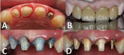
    Figure 1:  A-metallic pin and aesthetic fiberglass pins, occlusal view, B-4 metal
free E.Max crowns finalized, C-treatment with phosphoric acid, D-treatment
with adhesive system.
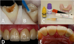
    Figure 2:  A-treatment with hydrofluoric acid, B-treatment with silane,
C-products used for cementation, D and E-finalized case.

Figure 3: A and B-fistula originating from root fracture, C-fractured root.
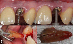
    Figure 4:  A-intraradicular preparation, B-traction component adaptation, C
and D-atraumatic root extraction.
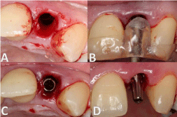
    Figure 5:  A-atraumatic root extraction, B-surgical guide positioned,
C-completed implant installation, D-component for cemented prosthesis
installed.
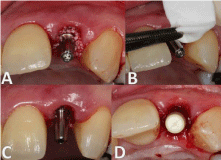
    Figure 6:  A-inorganic mineral bone inserted, B-collagen membrane inserted,
C and D-plastic cylinder for provisional tooth installed on the pillar.
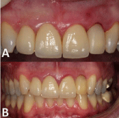
    Figure 7:  A-provisional tooth in the region of 22 immediately after the surgical
procedure, B-provisional tooth in the region of 22 after six months of surgical
procedure.
After six months recommended for implant osseointegration, the patient returned for further treatment, being possible to observe good condition for tissue behavior, as well as maintaining levels of soft tissue surrounding the manipulated area (Figure 7B), thanks to the immediate installation of provisional restoration after surgical procedure. The prosthetic component was removed and it was also found good aspect of the cervical gingival tissue with adequate copy of the emergence profile modeled by provisional restoration (Figure 8A). Performing an analysis of existing prosthetic space, it was opted in exchange for another universal pillar with other measures (3.3x6x4.5mm) in order to lead the cervical margin more intra-sulcular, and use a component with index positioning (Figure 8B). To use the same temporary tooth it was made internal wear and posterior relining. Thereafter were made conventional molding procedure of the prosthetic component installed and adjustments of shape, contour, texture and color of the element on the implant, with reference to the previously rehabilitation on tooth (Figure 8C). The completion of the case proved to be very interesting, since the expectation to maintenance the optimum result obtained before the fracture was big (Figure 8D, Figures 9A, 9B and 9C and Figure 10A and 10B). Note that comparing the level of soft tissue condition and gingival margin of the restoration on the tooth element 22 (Figure 11A and 11A1) and over implant (Figure 11B and 11B1) could be maintained satisfactorily and healthy, with good maintenance of the gingival outline without gingival retraction or modification of any transition between restoration on the tooth for restoration over implant.

    Figure 8:  A-cervical gingival tissue of 22 areas, B-exchange of prosthetic
component, C-adjustments of shape, contour, texture and color of the element
on the implant based on previously rehabilitation on tooth, D-finalized case.
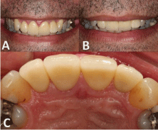
Figure 9: A, B and C-finalized case.

Figure 10: A and B-finalized case.
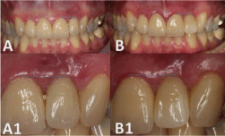
    Figure 11:  A and A1-level of soft tissue condition and gingival margin of the
restoration on tooth 22, B and B1-level of soft tissue condition and gingival
margin of the restoration on implant 22.
Discussion
In fact dentistry has made great advances, specifically in the use of indirect prosthetic procedures, combined or not with implantology, bringing invaluable gains in dentistry routine. The new ceramic systems allow excellent esthetic gain without resorting to an adequate mechanical strength, since the strengthening of crystals of leucite or lithium disilicate brought adequate strength and compatibility with teeth. Moreover, its evolution, with more opaque pellets and makeup possibilities allowed rehabilitation not only on aesthetic substrates, but also on darkened substrates or even metal pins [1,13,14]. Occurrences as dental fractures are still present, and always will be, bringing a surprise component that makes us reflect on how to promote proper solvability of these events. In this sense implantology brings the ability to return the patient to their functional and aesthetic needs. But for that to happen properly, and yet this patient coming from a previous treatment of high aesthetic quality, it is necessary that several factors are properly assessed and decide on which treatment plan should be followed [15]. Of paramount importance and indispensable condition to obtain favorable results, working within the predictability, is the use of the surgical guide, since improper positioning of the implant leads to poor prosthetic resolution and failure case [16]. In this case the clinical results obtained by all procedures, such as immediate implant and immediate restoration, surgical guide, inorganic mineral bone graft and collagen membrane, beyond the minimally invasive surgery and maintaining the stability of peri implant tissues, provided optimal recovery of the area. We note that this was only possible through the use of modern techniques, concepts of tissue care and discharge planning meticulously, always focusing on a favorable prognosis and predictability of a high standard. Furthermore, results like this only became possible by the advent of concepts of Switch Platform and internal Morse taper connection, which refer to the greater respect for peri implant tissues and a new mechanical and aesthetic approach [12, 17]. The main objective of the case, which was the maintenance of red and white high aesthetic previously obtained in rehabilitation on teeth, could only be achieved due to the conciliation of concepts arising from the surgical and prosthetic areas harmonically [18]. That’s how the reverse planning is effective within the clinical professionals practice, making the predictability big allied in routine of modern dental procedures.
Conclusion
Through the literature review and the results obtained in the resolution of the presented case, it can be concluded that:
- The advent of implant dentistry together with all the developments about its study brought extremely viable resolubility, both from a functional and aesthetic standpoint.
- Respect for peri implantar biology, as well as putting into practice the concepts of tissue care and health maintenance of tissue with minimally invasive procedures interfere directly in the final results.
- Knowledge of prosthetic concepts such as reverse planning combined with knowledge of surgical tissue manipulation and surgical guide optimize predictability and good prognosis of the treatment.
Acknowledgment
The authors acknowledge the brilliant work and partnership of Dent Lab Laboratory - Laboratory of Prosthodontics, Ribeirão Preto - SP, responsible for all laboratory technical part, which was crucial for the correct and proper completion of the case, both aesthetically as functional standpoint.
Also records here thanks to Dr. Márcio Henrique Corrêa and the patient in question, by giving in availability in the case record.
References
- Hart DA, Powers WH. Flexural strength and modulus of opalescent porcelain. J Dent Res. 1994; 73: 235.
- Rossato DM, Saade EG, Saad JRC, Porto-Neto ST. Aesthetic all-ceramic dental crowns for anterior teeth: a case report. Rev Sul Bras Odontol. 2010; 7: 494-498.
- Nishioka RS. Adhesive prostheses without metal using IPS Empress 2. Rev Assoc Paul Cir Dent. 2002; 56:277-279.
- Denry IL. Cerâmicas. In: Craig RG, Powers JM. Materiais dentários restauradores. São Paulo: Santos. 2004; 551-574.
- Holmes JR, Sulik WD, Holland GA, Bayne SC. Marginal fit of castable ceramic crowns. J Prosthet Dent. 1992; 67: 594-599.
- Denry I, Kelly JR. State of the art of zirconia for dental applications. Dent Mater. 2008; 24: 299-307.
- Palhares D, Corsini CB, Toniollo MB. Technique development for fidelization and time optimization in capturing of immediate provisional on optimal positioning. Full Dentistry in Science. 2014; 6:73-80.
- Degidi M, Piattelli A. Immediate functional and non-functional loading of dental implants: a 2- to 60-month follow-up study of 646 titanium implants. J Periodontol. 2003; 74: 225-241.
- Cochran D, Morton D, Weber PH. Relatórios do consenso e procedimentos clínicos recomendados sobre protocolos de carga para implantes dentários endoˇsseos. Int J Oral maxillofac implants. 2006; 19:109-115.
- Tselios N, Parel SM, Jones JD. Immediate placement and immediate provisional abutment modeling in anterior single-tooth implant restorations using a CAD/CAM application: a clinical report. J Prosthet Dent. 2006; 95: 181-185.
- Acchili A, Tura F, Euwe E. Immediate/early function with tapered implants supporting maxillary and mandibular posterior fixed partial dentures: preliminary results of a prospective multicenter study. J Prosth Dent. 2007; 97:52-58.
- Garber DA, Belser UC. Restoration-driven implant placement with restoration-generated site development. Compend Contin Educ Dent. 1995; 16: 796, 798-802, 804.
- Anusavice KJ. Materiais dentários. 10. ed. Rio de Janeiro: Guanabara Koogan. 1998: 311.
- Conceição EM, Sphor AM. Fundamentos dos sistemas cerâmicos. Porto Alegre: Artmed, 2005.
- Mecall RA, Rosenfeld AL. Influence of residual ridge resorption patterns on implant fixture placement and tooth position. Int J Periodont Rest Dent. 1991; 11: 8-23.
- Botinno MA. Anterior aesthetic implants. Implantnews. 2006; 6: 560-568.
- Salama H, Salama M. The role of orthodontic extrusive remodeling in the enhancement of soft and hard tissue profiles prior to implant placement: a systematic approach to the management of extraction site defects. Int J Periodontics Restorative Dent. 1993; 13: 312-333.
- Touati B. Custom-guided tissue healing for improved aesthetics in implant-supported restorations. Int J Dent Symp. 1995; 3: 36-39.