
Case Presentation
J Dent App. 2015; 2(7): 274-277.
Mineral Trioxide Aggregate Obturation in Retreatment with Regenerative Adjuncts of Bioceramic Allograft in Large Periapical Defect: A Case Report with 5 Year Follow Up
Zoya A, Ali S*, Tewari RK, Mishra SK, Kumar A and Iftekhar H
Department of Conservative Dentistry & Endodontics, Dr. Z. A. Dental College, Aligarh Muslim University, India
*Corresponding author: Ali S, Department of Conservative Dentistry and Endodontics, Dr. Z. A. Dental College and Hospital, Aligarh Muslim University, India
Received: February 09, 2015; Accepted: September 10, 2015; Published: September 12, 2015
Abstract
Endodontic overfills and incomplete obturation is usually associated with an array of local complications and results in persistent periapical infection and inflammation. Endodontic challenges further aggravates in such cases if the tooth actually is young with incomplete root formation, i.e. presence of blunderbuss canal. Herein, a case of wide open apices in maxillary central incisors with over extrusion of gutta percha and associated large periapical lesion is presented. Mineral Trioxide Aggregate (MTA) was used to obturate the blunderbuss canals and an alloplastic graft, Biphasic Calcium Phosphate (BCP) were filled in the bony defect after surgical curettage. At the 6th month postoperatively, periapical osseous healing was satisfactory. The advantages and indications of MTA as obturation material and bone graft in periodontal regeneration are discussed. Clinically and radiographically the case was followed for next 5 years to assess the outcome of MTA obturation.
Keywords: Biphasic calcium phosphate; Blunderbuss canal; Retreatment; MTA obturation
Abbreviations
BCP: Biphasic Calcium Phosphate; GP: Gutta Percha; MTA: Mineral Trioxide Aggregate.
Introduction
Traumatic injury or caries in young permanent teeth results in pulpal inflammation or necrosis and subsequent incomplete development of dentinal wall and root apices [1]. Such type of canals often called “blunderbuss” as they have divergent and flaring foramen. The problems associated with these canals are: weaker roots prone to fracture, poor crown to root ratio, and more susceptible to periodontal involvement [2].
Complete debridement, disinfection and three dimensional obturation of these canals is a daunting task as they lack apical constriction [3]. Thus, treatment in these cases are aimed at developing the apical stop for confinement and condensation of obturating material.
Apexification and apical barrier technique are the two commonly employed methods for management of immature permanent teeth with open wide apices. Although apexification using calcium hydroxide has very high success rate [4] but decrease in fracture resistance of the tooth, longer duration of treatment, multiple appointments, and high patient compliance are the disadvantages. Apical barrier technique using MTA is becoming the standard for treatment of immature roots with necrotic pulps. In this technique an artificial stop is created that would enable the root canal to be filled immediately [5]. With the advantages of shorter treatment time, development of a good apical seal, and MTA induced hard-tissue deposition in periradicular area [6], apical barrier technique have shown long term success [7].
Prevention of development of endodontic disease and resolution of periapical pathosis is only possible when the filling materials obturate the canal three dimensionally and prevents further ingress of bacteria, entombs remaining microorganisms, and block their nutrient supply [8,9]. MTA provides remarkable physiochemical and bioactive properties, offering an outstanding edge when used as canal obturation material [9].
Apical periodontitis is an immunoinflammatory reaction resulting in destruction of periradicular tissues in response to etiologic agents of endodontic origin [10]. Although, nonsurgical root canal therapy, apical microsurgery, and extraction are the different treatment modalities for the management of periapical inflammatory lesion. Nonetheless, surgical intervention allows thorough debridement which leads to a faster healing rate and also permits placement of different biomaterials (Biphasic calcium phosphate, Platelet rich fibrin, Calcium enriched mixture etc.) to improve the new bone formation especially for large defects [11].
This case reports presents the successful management of large periapical lesion associated with overextended filling materials followed by MTA obturation in blunderbuss canals of maxillary central incisors.
Case Presentation
A 17 year old female presented with chief complaint of pain and swelling in relation to upper front teeth along with blackening of crown margins (Figure 1). Her medical history was not significant. The patient gave history of treatment by a general practitioner for the same teeth. Clinically, the teeth 11 and 21 were covered with a defective prosthesis revealing plaque and calculus at the margins. Teeth 12 and 22 were tender on percussion. Tooth 12 was having grade II mobility along with root exposure. Radiographic examination revealed that both central incisors were having wide open apices with large periapical radiolucencies. Single cones of gutta percha points were overextended periapically in both 11 and 21 (Figure 2).
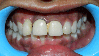
Figure 1: Presenting clinical photograph.
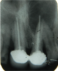
Figure 2: Preoperative radiograph.
After removal of prosthesis (Figure 3), oral prophylaxis was completed and intracanal filling material was removed using Haedstromfiles (DentsplyMaillefer, Ballaigues, Switzerland). Access cavities were refined using Endo-Z (Dentsply/Tulsa Dental, Tulsa, OK). The root canals were instrumented with K-file (DentsplyMaillefer, Ballaigues, Switzerland) upto working length, irrigated with 1% sodium hypochlorite and 0.9% normal saline (Fresenius, Kabi, India) as final rinse, and then dried with paper points. Calcium hydroxide (Ultracal XS; Ultradent, South Jordan, UT) was placed in the root canals for 3 weeks. All the treatment options were described to the patient and surgical approach was planned in the next appointment after taking patient consent.
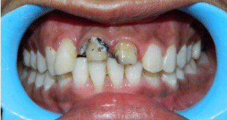
Figure 3: Condition after removal of crowns.
At second appointment i.e. after 3 weeks, calcium hydroxide was flushed out using 1% sodium hypochlorite and normal saline. The canals were dried with paper points. Localanesthetic (1.8 ml 2% lidocaine with 1:100,000 epinephrine) was used to give infraorbital nerve block. A full-thickness rectangular mucoperiosteal flap was reflected by a sulcular incision starting from distal of tooth 12 to distal of tooth 22 to with the aim to expose periapical the periapical region. Cortical plate was absent in relation to teeth 11 and 12. Pathological mass (Figure 4) of size approximately 2x1 inches in dimension was curetted out and osseous defect and root canals of tooth 11 and 21 was copiously irrigated with betadine and sterile saline solution (Figure 5).
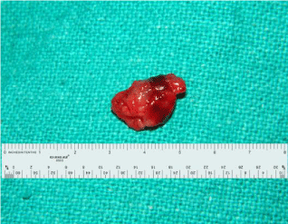
Figure 4: Pathologic mass curetted out of bony defect.
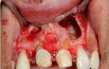
Figure 5: Clean bony cavities after curettage of pathologic tissue.
Gauze pieces were placed in bony defect and root canals were dried. White MTA (Pro root MTA; Tulsa Dental, Johnson City, TN) was mixed according manufacture’s instruction and packed into the canals with open apices i.e. tooth 11 and 21 using amalgam carrier and hand pluggers. These canals were completely obturated with MTA which was condensed both coronally and apically using straight and root end condensers (Figure 6). The tooth 22 with closed apex was obturated by gutta percha and eugenol based sealer.
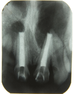
    Figure 6:  Radiograph after surgical curettage and MTA obturation of root
canals.
The walls of the cavity were curetted again to induce bleeding and a nanocrystalline graft material Sybo Graf™Plus (Eucare Pharmaceuticals, Chennai, India) was placedin the defects and adapted along all the surfaces of the defect (Figure 7). The graft material was packed with light pressure upto the level of surrounding cortical plate. The flap was gently repositioned and a slight pressure was applied to adapt it to the bony margins. Once it was confirmed that the flap was being supported by the graft material, the flap was sutured using 4-0 silk. Post-surgical instructions were given and medication was prescribed. The sutures were removed after 5 days, and satisfactory healing was observed.
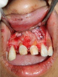
Figure 7: Bioceramic graft material packed in the bony defect.
As the mobility of tooth 12 did not diminish, owing to the deep pocket and much bone loss therefore it was extracted subsequently. After healing of extraction socket porcelain fused to metal bridge extending from tooth 13 to tooth 22 was fixed with dual cure resin cement (Paracore, Coltene Whaledent, Altstätten, Switzerland).
Radiograph were taken during follow up visits at 1 month (Figure 8a), 9 month (Figure 8b), 24 month (Figure 8c), and 5 year (Figure 8d). During these visits patient does not complaint of pain, swelling, or discomfort.

    Figure 8:  Follow up radiographs after (a) 1 month, (b) 9 months, (c) 24
months and (d) 5 years.
Discussion
Nonvital teeth with immature roots make treatment more challenging with a reduced prognosis because the wide open apices does not likely permit the standard root canal protocol like biomechanical preparation, disinfection, three dimensional obturation. 12 Furthermore, the thin and fragile roots and associated chronic periapical lesion further aggravates the condition. Thus, these difficulties can be managed by minimal instrumentation of canal, formation of hard tissue barrier at apex, placement of an artificial apical barrier, and by reinforcing the weakened root against fracture [12].
This case report describes the MTA obturation of blunderbuss canals and surgical management of associated large periapical lesion.
Several in vitro cell culture studies demonstrated that periapically extruded Gutta percha (GP) can be toxic to human dental pulp cells [13], mouse fibroblasts [14], and associated with delayed healing at periapex [15]. Extruded GP points also form a favorable substrate for microbial colonization in the form of biofilm and houses a wide range of bacterial species on their surfaces thereby sustaining periapical inflammatory processes and present as long-standing periapical infection [16]. Thus, in the present case overfilling, inadequate obturation, and lack of fluid impervious seal might be the contributing factors in the development of such large periapical pathology.
Apexification and nonsurgical treatment of periapical lesion using calcium hydroxide was not chosen in this case due to several reasons: long duration procedure with multiple appointment, requirement of patient compliance, presence of resistant microbial flora in previously treated cases, unpredictable prognosis, temporary coronal seal may results in reinfection between the appointments, and calcium hydroxide weaken the resistance of dentin to fracture [17].
Trope and others have recommended low concentration of sodium hypochlorite for root canal irrigation and debris removal in teeth with open apices because there may be chances of periapical extrusion resulting in cytotoxicity [12,18,19]. Therefore, 1% NaOCl was employed in this case. However, the reduced concentration can be compensated by the higher volumes of irrigant used.
Several leakage studies [20,21] have shown that gutta-percha is highly susceptible to microleakage when a deficient coronal seal is present irrespective of the obturation technique used. MTA provides superior marginal adaptation compared to other conventional materials as it forms a mineralized dentin – MTA interstitial layer and its particles occludes and penetrate the dentinal tubules [22,23] entombing the bacteria within. These kinds of characteristics could possibly produce challenging environment for bacterial survival. Both bacteriostatic and bactericidal potential of MTA makes it potent against Enterococcus faecalis and Candida albicans and thus refractory endodontic diseases [24]. Therefore, MTA obturation was preferred over other methods in this case as has been advocated by Bogen et al [9].
Numerous alloplastic materials are proposed for filling bony defects. These grafts prevent hematoma formation, act as scaffold, and facilitate bone regeneration. Therefore, with the aim of regeneration of new bone in large periapical defect, biphasic calcium phosphate (BCP) ceramic was used in this case. BCP is biphasic consist of soluble Hydroxyapatite (70%) and soluble β-Tricalcium phosphate (30%). The faster resorption of β-Tricalcium phosphate helps in the process of bone mineralization however, slower resorbability of Hydroxyapatite helps for bone in-growth providing a scaffold for calcification. Advantages of using BCP are: biocompatibility, osteoconductive effect, similar structure of apatite like in bone, and cost effectiveness [25].
After taking radiographs during the follow up visits it was observed that MTA in root canal and BCP in the periapical defect facilitate significant, progressive, and predictable periapical healing along with osseous regeneration.
Conclusion
The presented report describes the successful management with long term follow-up of a retreatment case of blunderbuss canals associated with large periapical lesion by MTA obturation and BCP ceramic.
On the basis of the findings of studies addressed in the literature and the 5-year clinical and radiographic outcomes of this case, it might be concluded that (1) MTA, a well-known osteoinductive and cementogenic material demonstrate superior healing rates when used for obturation of root canal system in retreatment cases compromised by microleakage, inadequate disinfection and obturation, and large periapical lesions. (2) Large bony defects might and should be filled with biocompatible, bioactive, and osteoconductive materials that allows regenerative tissue process rather repair and ensures faster healing.
This case report adds some knowledge to the limited literature available on long term clinical outcome of MTA obturation. However, further experimental studies are necessary to investigate the effects of MTA as obturation material and co-administered biomaterials on periapical healing and endodontic success.
References
- Trope M. Treatment of the immature tooth with a non-vital pulp and apical periodontitis. Dent Clin North Am. 2010; 54: 313-324.
- Fuks AB, Camp J. Crown fractures: A practical approach for the clinician. Berman LH, Blanco L, Cohen S, editors. In: A clinical guide to dental traumatology. St. Louis, MO: Mosby. 2011: 27-126.
- Hargreaves K, Law A. Cohen's pathways of the pulp. 10th ed. St. Louis, MO: Mosby Elsevier. 2011.
- Cvek M. Prognosis of luxated non-vital maxillary incisors treated with calcium hydroxide and filled with gutta percha. A retrospective clinical study. Endod Dent Traumatol 1992; 8: 45–55.
- Morse DR, Larnic JO, Yesilsoy C. Apexification: review of the literature. Quintessence Int. 1990; 21: 589–598.
- Shabahang S, Torabinejad M. Treatment of teeth with open apices using mineral trioxide aggregate. Pract Periodont Aesthet Dent. 2000; 12: 315–320.
- Sachdeva GS, Sachdeva LT, Goel M, Bala S. Regenerative endodontic treatment of an immature tooth with a necrotic pulp and apical periodontitis using platelet-rich plasma (PRP) and mineral trioxide aggregate (MTA). A case report. Int Endod J. 2014: 902-914.
- Sundqvist G, Figdor D. Essential endodontology: Prevention and treatment of apical periodontitis. 2nd ed. Oxford, UK: Blackwell Science. 1998.
- Bogen G, Kuttler S. Mineral trioxide aggregate obturation: Review and case series. J Endod. 2009; 35: 777-790.
- Marton IJ, Kiss C. Protective and destructive immune reactions in apical periodontitis. Oral MicrobiolImmunol. 2000; 15: 139-150.
- Yoshikawa G, Murashima Y, Wadachi R. Guided bone regeneration (GBR) using membranes and calcium sulphate after apicectomy. A comparative histomorphometrical study. Int Endod J. 2002; 35: 255–263.
- Trope M. Treatment of immature teeth with non-vital pulps and apical periodontitis. Endod Topics. 2006; 14: 51-59.
- Das S. Effect of certain dental materials on human pulp in tissue culture. Oral Surg Oral Med Oral Pathol. 1981; 52: 76-84.
- Pascon EA, Spangberg LS. In vitro cytotoxicity of root canal filling materials: 1. Gutta-percha. J Endod. 1990; 16: 429-433.
- Nair PN, Sjogren U, Krey G, Sundqvist G. Therapy-resistant foreign body giant cell granuloma at the periapex of a root-filled human tooth. J Endod. 1990; 16: 589–595.
- Noiri Y, Ehara A, Kawahara T. Participation of bacterial biofilms in refractory and chronic periapical periodontitis. J Endod. 2002; 28: 679–683.
- Andreasen JO, Farik B, Munksgaard EC. Long-term calcium hydroxide as a root canal dressing may increase risk of root fracture. Dent Traumatol. 2002; 18: 134–137.
- Cvek M, Nord CE, Hollender L. Antimicrobial effect of root canal debridement in teeth with immature roots. A clinical and microbiologic study. Odontol Revy. 1976; 27: 1–10.
- Spangberg L, Rutberg M, Rydinge E. Biologic effects of endodontic antimicrobial agents. J Endod. 1979; 5: 166–175.
- Madison S, Wilcox LR. An evaluation of coronal microleakage in endodontically treated teeth. Part III—in vivo study. J Endod. 1988; 14: 455–458.
- Jacobson HL, Xia T, Baumgartner JC, Marshall JG, Beeler WJ. Microbial leakage evaluation of the continuous wave of condensation. J Endod. 2002; 28: 269–271.
- Torabinejad M, Watson TF, Pitt Ford TR. Sealing ability of a mineral trioxide aggregate as a retrograde root filling material. J Endod. 1993; 19: 591–595.
- Komabayashi T, Spangberg LS. Particle size and shape analysis of MTA finer fractions using Portland cement. J Endod. 2008; 34: 709–711.
- Santos AD, Moraes JCS, Arau jo EB, Yukimitu K, Vale rio Filho WV. Physico-chemical properties of MTA and a novel experimental cement. Int Endod J. 2005; 38: 443–447.
- Bolukbasi N, Yeniyol S, Tekkesin MS, Altunatmaz K. The use of platelet-rich fibrin in combination with biphasic calcium phosphate in the treatment of bone defects. A histologic and histomorphometric study. Curr Ther Res Clin Exp. 2013; 75: 15-21.