Case Presentation
J Dent App. 2015; 2(8): 278-281.
Cad-Cam Fabricated One-Piece Ceramic Post and Core for Teeth Supporting Fixed Partial Dentures: Report of Two Cases
Bankoğlu Güngör M*, Karakoca Nemli S, Doğan A, Tamam E and Turhan Bal B
Department of Prosthodontics, Gazi University, Turkey
*Corresponding author: Bankoğlu Güngör M, Department of Prosthodontics, Gazi University, Ankara, Turkey
Received: March 09, 2015; Accepted: September 10, 2015; Published: September 12, 2015
Abstract
Endodontically treated teeth with inadequate tooth support can be rehabilitated either prefabricated or custom fabricated post and core materials. Custom post-core restorations are fabricated by casting or computer aided design and computer aided manufacturing (CAD-CAM) systems. It is recommended that teeth treated with post and core restorations should be one-piece and adequate fit of the post into the root canal should be achieved for supporting fixed partial dentures. The purpose of this clinical report is to represent chairside treatment procedures of two patients who received all ceramic fixed partial dentures with one-piece CAD-CAM generated ceramic post core restored abutment teeth. In the first case, it is presented that maxillary central teeth restored with one-piece ceramic post and core for supporting fixed partial denture on the anterior region. In the second case, it is presented that mandibular premolar tooth restored with one-piece zirconia post and core for supporting fixed partial denture on the posterior region. Both cases were treated with CAD-CAM fabricated fixed partial dentures after the cementation of posts and cores. During one-year follow-up period, the patients were satisfied with their prostheses esthetically and functionally. No complication was observed during the follow up period.
Keywords: CAD-CAM; Ceramic; Zirconia FPD; Post-core
Introduction
Different types of posts such as titanium, gold plated, chromenickel or gold-cast, ceramic prefabricated posts are inserted into root canals to support and strengthen the tooth structure [1]. Although wide variety of post-core restoration technologies and materials have been introduced into the dental market, there is no consensus on the most appropriate treatment choice for post core systems [1]. Prefabricated posts have good biomechanical and biocompatibility properties; however they cannot be customized for the compact adaptation to the prepared post cavity. On the other hand; using composite resins for core material may have a higher failure rate because of the weak bonding between the prefabricated post and composite core [2]. One of the production method of ceramic restorations is CAD-CAM. There are different methods for preparing ceramic CAD-CAM post core systems: the core can be constructed separately and adhesively luted to post and tooth, one piece post and core complex can be constructed, and the core can be constructed using a heat pressed technique [1,3]. Using these methods, disadvantages of direct composite core might be avoided. Functional requirement is an important aspect to be considered when selecting materials and techniques to restore endodontically treated teeth. Because anterior and posterior teeth are subjected to different forces [4]. On the other hand, endodontically treated teeth used as abutments for fixed and removable partial dentures are subjected to large horizontal and torque forces during function [5]. However, to the best of authors’ knowledge, current literature includes no evidence on the use of all ceramic post-core restorations for FPD abutment teeth.
The purpose of this clinical report is to represent treatment procedures of two patients who received all ceramic FPDs with onepiece CAD-CAM generated ceramic post core restored abutment teeth.
Case Presentation
This article presents two patients who referred to Gazi University, Faculty of Dentistry, and Department of Prosthodontics to be treated with post core restorations. Descriptions of Case 1 and Case 2 are presented in Table 1. Radiographic examinations were indicated successful endodontic treatment characterized by healthy apical sections, and adequate root canal fillings. Clinical examinations were revealed missing coronal portion for both patients. The treatment details were discussed and patients signed an informed consent. The clinical study was approved by the Ethics Committee of Ankara University, Faculty of Dentistry by grant no. 36290600/21.
    Patient
    Age
    Tooth Number
    Region
    Number of the supporting teeth
    Restorative Materials


    Post-core
    Crown


    Case 1
    23
    11, 21
    Maxillary Anterior
    11,13
    Lithium isilicate
      (IPS e.max CAD)
    Zirconia (InCoris ZI; framework) Veneer porcelain (VITA)


    Case 2
    42
    35
    Mandibular Posterior
    35,37
    Monolithic rconia
      (InCoris TZI)
    Monolithic zirconia
      (InCoris TZI)
Table 1: Description of the cases.
Case 1
In the Case 1, one-piece post-core restorations were used to support anterior fixed partial dentures (Figure 1a and 1b). The root canal lengths were measured from the radiograph and the root filling materials were determined to be removed. The post spaces were enlarged with post drills and undercuts at the post space were eliminated (Figure 2a). The edges of the teeth were rounded to facilitate the scanning and milling process. Micro brushes were adapted into the root canals. Impressions of the post spaces and adjacent teeth were taken with condensation silicone elastomer (Zeta Plus; Zhermack, Badia Polesine, Italy) by using double mix technique. Cerec Optispray (Sirona, Germany) was applied to the impressions and homogeneous and thin anti reflective coatings were obtained. Then, digital impressions were made with an extra oral scanner (inEos X5; Sirona, Germany) to generate virtual models. Special software (in Lab SW 4.2.1.61068, Sirona Dental Systems, Bensheim, Germany) was used to design the one-piece post and core restorations. “Crown” was selected as restoration type. Margins were drawn on the virtual models. After a proposal for the restorations was calculated by the software, some modifications were made on this design (Figure 2b). Spacer thickness was not determined. Post-core restorations were processed in a milling unit (inLab MC XL, Sirona Dental Systems, Bensheim, Germany). When the milling process was finished, the lithium disilicate posts were fully crystalized in a furnace (Programat P300, Ivoclar Vivadent, Schaan, Liechtenstein) according to the crystallization firing program recommended by the manufacturer. For the cementation procedure, the posts surfaces were conditioned with 9% hydrofluoric acid ( Ultradent Porcelain Etch, South Jordan, Utah, USA) for 90 s and subsequent silanization ( Ultradent Silane, South Jordan, Utah, USA) was performed for 60 s. Then, one-piece post-core restorations were cemented with a dual-cure resin cement (Panavia F 2.0, Kuraray, Japan), furthermore crown heights were modified (Figure 2c) with diode laser (SIROlaser Advanced; Sirona Dental Systems, Bensheim, Germany). After this step, the prepared teeth and cemented post-cores were scanned with an intraoral scanner (Omnicam; Sirona Dental Systems, Bensheim, Germany) to design and mill the restorations. “Framework” was selected as the restoration type. The maxillary and mandibular arches, and occlusion were scanned and virtual models were generated. Model axes were determined and margins were drawn on the virtual models (Figure 3a). After the frameworks were designed (Figure 3b), the occlusal and proximal contacts were checked. Spacer thickness of 50 μm was determined. The framework were milled in the milling unit (inLab MC XL, Sirona Dental Systems, Bensheim, Germany) and sintered in dry conditions with a holding time of 120 min in sintering furnace. Then, the frameworks were veneered with veneering porcelain with conventional methods according to manufacturers’ recommendations. When the restorations were fabricated, fit of the restorations were checked and premature contacts in centric occlusion and lateral excursions were avoided. Then, the restorations were cemented. Firstly the bonding agent (TRI-S Bond Plus; Clearfil, Kuraray, Japan) applied onto the teeth surfaces. Cementations were performed with transparent dual-cure resin cement (Bifix, Voco, Cuxhaven, Germany) according to manufacturer’s instructions. Excess cement was removed. Restorations were cured for 20 s from each side and all margins were finished and polished with abrasive disks (Figure 3c). During one-year follow-up period, the patient was satisfied with the prosthesis esthetically and functionally. No complication was observed during the follow up period.

Figure 1: a) Initial view of Case 1. b) Initial panoramic radiograph of Case 1.

    Figure 2:  a) Preparing root canals in Case 1. b) Design of the post-core
restorations of Case 1. c) Laser modification of gingiva of Case 1 and
cementation of post-core restorations.

    Figure 3:  a) Drawing margins of frameworks of Case 1 on the virtual model.
b) Design of the frameworks of Case 1. c) Final restoration of Case 1.
Case 2
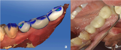
In the Case 2, one-piece post was used to supported posterior fixed partial denture (Figure 4a). One-piece post was designed and fabricated as described in Case 1 (Figure 4b, 4c, and 4d) from monolithic zirconia. Milled zirconia post-core were cemented in same conditions with Case 1, however any surface treatment was not applied to surfaces of zirconia for cementation. After the cementation of post-core (Figure 5a and 5b), the teeth were scanned with an intraoral scanner (Omnicam; Sirona Dental Systems, Bensheim, Germany) to design and mill the restorations. “Bridge Restoration” was selected as the restoration type. The prepared teeth, opposite arch, and occlusion (buccal bite) were scanned and virtual models were generated. After drawing margins, three unit fixed partial denture was designed from monolithic zirconia (Figure 6a). After then, the restoration was milled and sintered. Some modifications were intraorally made on the milled restoration, fit of the restoration was checked and premature contacts were avoided. The restoration was cemented after glazing the external surfaces of the restoration. The cementation procedure of fixed partial denture was same as in the first case (Figure 6b). During one-year follow-up period, the patient did not state any complications regarding esthetical or functional properties.

    Figure 4:  a) Initial periapical radiograph of Case 2. b) Drawing margin of postcore
restoration of Case 2 on the virtual model. c) Design of the post-core
restoration of Case 2. d) Milled post-core restoration of Case 2.

    Figure 5:  a) Digital model showing cemented post-core restoration of Case 2.
b) Cementation of post-core restoration of Case 2.

    Figure 6:  a) Design of the restoration in Case 2. b) Final restoration of Case
2.
Discussion
The restoration of endodontically treated teeth is extensively studied but still controversial issue in dentistry [4]. Treatment planning for a post-core restoration should include several considerations such as remaining tooth structure, functional aspects, the use or nonuse of posts, design and material of the post and the core, and coronal restoration [4,5]. Endodontically treated teeth those are used as abutments for fixed partial dentures show different properties from single standing teeth with a crown restoration [4,5]. The higher fracture risk of these teeth has been reported [6]. Thus, custom posts are recommended for abutment teeth of FPDs [7]. In addition, proper alignment among the support teeth can be achieved using these posts [8]. In the second case of present report, an endodontically treated premolar with extensive coronal hard tissue loss was used as abutment of a posterior FPD. One piece custom designed zirconia post and core which provided a restoration with greater toughness, maximal adaptability to the canal, and adequate esthetics was fabricated [8]. The FPD was also fabricated from monolithic zirconia using CAD-CAM system at one appointment. Thus the patient was treated with a highly esthetic, preciously adapted, and rapidly produced restoration. In the first case, lithium disilicate one-piece post-cores were fabricated for endodontically treated roots of the two central incisors. Loss of extensive coronal tooth structure required to establish a strong adhesive bond between post-core and root canal, therefore lithium disilicate which is an etchable ceramic was chosen as the post-core material. After luting post-cores, a zirconia reinforced all ceramic FPD was fabricated.
Fabricating one-piece post-core restoration using CAD-CAM system was described by Liu et al [2]. Which include preparing a wax pattern of post-core, digitizing the pattern using a scanning, and processing data using CAD-CAM software. However, at the impression phase of the root canal, it is generally difficult to remove the resin or wax pattern from the root canal for taking the direct impression of the prepared root cavity furthermore this process is time consuming and needs technical effort. The technique described in this report offers the advantage of fabricating post and core structure by directly scanning the impression of post space and full arch. Thus dimensional changes of the gypsum model and the pattern material such as resin, wax can be eliminated and post-core is digitally designed on a 3-D model instead manual preparation of the pattern. Consequently, more precious and time saving fabrication process can be achieved compared with conventional methods [9]. In the present cases one-piece custom post-core restorations for abutment teeth were treatment of choice. Although there is no consensus on the superiority of custom post and cores when compared with prefabricated posts, it was stated that one-piece posts are more reliable than prefabricated posts with direct cores as the number of interfaces decreases [10].
Conclusion
One piece post and cores made of zirconia and glass ceramic were fabricated to support all-ceramic fixed partial dentures in the present cases. This article represents fabricating one-piece milled ceramic post and core for endodontically treated teeth supporting fixed partial dentures. Post-core structures fabricated from high strength ceramic materials, zirconia and lithium disilicate, using CAD-CAM technology supported all ceramic fixed partial dentures without compromising esthetic. In addition, the post and core preciously fitted into a prepared post space and anatomically correct core could be fabricated. The survival rate of these restorations should be further investigated.
References
- Ozcan N, Sahin E. In vitro evaluation of the fracture strength of all-ceramic core materials on zirconium posts. Eur J Dent. 2013; 7: 455-460.
- Liu P, Deng XL, Wang XZ. Use of a CAD/CAM-fabricated glass fiber post and core to restore fractured anterior teeth: A clinical report. J Prosthet Dent. 2010; 103: 330-333.
- Butz F, Lennon AM, Heydecke G, Strub JR. Survival rate and fracture strength of endodontically treated maxillary incisors with moderate defects restored with different post-and-core systems: an In vitro study. Int J Prosthodont. 2001; 14: 58-64.
- Faria AC, Rodrigues RC, de Almeida Antunes RP, de Mattos Mda G, Ribeiro RF. Endodontically treated teeth: characteristics and considerations to restore them. J Prosthodont Res. 2011; 55: 69-74.
- Tang W, Wu Y, Smales RJ. Identifying and reducing risks for potential fractures in endodontically treated teeth. J Endod. 2010; 36: 609-617.
- Nyman S, Lindhe J. A longitudinal study of combined periodontal and prosthetic treatment of patients with advanced periodontal disease. J Periodontol. 1979; 50: 163-169.
- Willershausen B, Tekyatan H, Krummenauer F, Briseño Marroquin B. Survival rate of endodontically treated teeth in relation to conservative vs post insertion techniques -- a retrospective study. Eur J Med Res. 2005; 10: 204-208.
- Awad MA, Marghalani TY. Fabrication of a custom-made ceramic post and core using CAD-CAM technology. J Prosthet Dent. 2007; 98: 161-162.
- Bosch G, Ender A, Mehl A. A 3-dimensional accuracy analysis of chairside CAD/CAM milling processes. J Prosthet Dent. 2014; 112: 1425-1431.
- Fernandes AS, Shetty S, Coutinho I. Factors determining post selection: a literature review. J Prosthet Dent. 2003; 90: 556-562.