
Case Presentation
J Dent App.2015; 2(8): 291-294.
Spindle Cell Lipoma on the Floor of the Mouth
Yalçin M¹, Atilgan SS¹, Laçin N¹, Atalay Y²*
¹Department of Oral and Maxillofacial Surgery, Dicle University, Turkey
²Department of Oral and Maxillofacial Surgery, Afyon Kocatepe University, Turkey
*Corresponding author: Atalay Y, Department of Oral and Maxillofacial Surgery, Afyon Kocatepe University, Faculty of Dentistry, Turkey
Received: March 05, 2015; Accepted: September 10, 2015; Published: September 12, 2015
Abstract
Spindle Cell Lipoma (SCL) is a benign lipomatous histological variant of lipoma which is rarely observed in the oral cavity. It presents as a circumscribed mass in the buccal mucosa, tongue, floor of the mouth or hard palate. In English literature there are nearly 10 case reports of SCL on the mouth floor, all of which were treated with local excision. A case is here presented of intraoral spindle cell lipoma on the floor of the mouth of a 49-year old female. Although oral SCL is rare, it should be considered in the differential diagnosis of lipamatous neoplasms occurring on the mouth floor.
Keywords: Lipoma; Spindle cell lipoma; Oral cavity; Mouth floor
Introduction
Lipomas rarely occur in the oral and maxillofacial region. One kind is the Spindle Cell Lipoma (SCL), which is typically seen as a benign lipomatous neoplasm in the posterior neck and back of older males, and accounts for approximately 1.5% of all lipoma cases [1]. In the oral cavity, the buccal mucosa, tongue, and floor of the mouth are common locations of lipomas [2]. They present as a slow-growing, painless, asymptomatic yellowish, nodular, sub mucosal mass and are covered by normal mucosa. Oral lipomas may cause mastication and speech difficulties [3]. SCL is a distinct histological variant of lipoma derived from prelipoblastic mesenchymal cells [4]. SCL is a rare benign neoplasm, and oral SCL is extremely rare. In this paper, a case is reported of SCL localized to the floor of the mouth in a 49-year old female, and the clinical, histopathological and immune histochemical findings of this case are presented.
Case Presentation
A 49-year old female patient was referred to the Department of Oral and Maxillofacial Surgery with the complaint of swelling on the floor of the mouth, which had been ongoing for 2 years (Figure 1). The patient complained of occasional discomfort while speaking. The growth was gradual in onset, and had slowly increased in size over a period of 2 years to reach its present size. The well circumscribed swelling was not tender on palpation, soft in consistency and mobile. In the intraoral examination, a smooth well-defined sessile nodule of similar color to the surrounding mucosa was detected on the anterior floor of the mouth (Figure 1). It was superficially located and non-infiltrating in front of the sublingual caruncle (opening of the submandibular and sublingual duct) on the floor of the mouth (Figure 1). Based on the history and clinical findings, a provisional diagnosis of lipoma was considered. Excisional biopsy was made under local anesthesia. The yellowish, well-circumscribed soft tissue mass with a lobulated surface 1.5 x 1 x0.5 cm (length x width x thickness) in size was totally excised and sent for microscopic examination in 10% buffered formalin (Figure 2,3,4). In the microscopic examination of H and E stained slides, using a light microscope, a well-circumscribed nodular mass was seen. The histopathology of the soft tissue section showed an encapsulated lesion composed of abundant mature adipocytes arranged in lobules, infrequent blood capillaries, collagen-forming CD34-positive spindle cells and wiry collagen in a variable myxoid background. The lobules were separated by fibrous connective tissue septa. The adipocytes appeared polygonal in shape with clear cytoplasm and the placement of a peripheral flat nucleus that was compressed against the cell membrane (Figure 5A, 5B). Based on clinical and histopathological findings, the swelling was finally diagnosed as SCL. No recurrence has been detected since surgery (Figure 6).
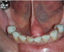
Figure 1: Preoperative clinical situation: Swelling on the floor of the mouth.
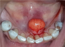
Figure 2: Yellowish, well-circumscribed soft tissue mass.
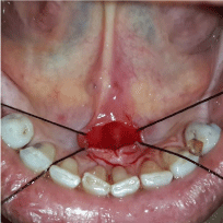
Figure 3: Surgery site after excision of mass.

Figure 4: Macroscopic examination revealing a nodular lesion.
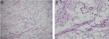
    Figure 5: A-5B: The histological features revealed fat cells, well vascular
network, spindle cells (H&E).
Discussion
Lipomas usually occur in the shoulders, neck and axilla, whereas this benign tumor is rarely seen in the oral cavity. Intraoral lipomas are rare and statistically constitute approximately 1-4% [4] and SCL constitute approximately 1.5% of all adipose tumors [5]. Intraoral lipomas are benign mesenchymal neoplasms that originate in mature adipose cells with a differential diagnosis of other soft tissue lesions such as mucoceles, dermoid cysts, ranulae, thyroglossal duct cysts, pleomorphic adenomas, angiolipomas, fibrolipomas and malignant lymphomas. In the oral cavity, the buccal mucosa, tongue, and floor of the mouth are the most common locations [5]. Histologically, they can be classified as simple lipoma, fibrolipoma, SCL, intramuscular or infiltrating lipoma, angiolipoma, pleomorphic lipoma, myxoid lipoma and atypical lipoma [3]. The most common site for SCL is the tongue (37%), followed by the cheek/buccal mucosa (31%), and floor of the mouth (15%) but other locations such as the lip, hard palate, alveolar ridge and maxilla have also been reported (Table 1) [6].
![]()
Author/ Year
Age(years)
Sex
Location
Size (mm)
Follow-up (Years)
Mc Daniel et al. 1984
33
52
F
M
Floor of mouth Tongue
10
2
8
Christopoulos et al. 1989
58
M
Hard palate
20
2
Levy & Goding 1989
74
F
Floor of mouth
45
--
Lombardy & Odell 1994
68
F
Tongue
15
--
Tosios et al. 1995
55
M
Cheek
40
2
Khoo & Lian 1995
23
M
Cheek
50
--
Piatteli et al. 1999
75
M
Cheek
20
--
Dutt et al. 1999
42
F
Tongue
30
--
Agoff et al. 2001
61
F
Buccal� vestibule
Said-Al- Naief et al. 2001
66
53
M
F
Tongue
Tongue
Darling et al. 2002
69
M
Alveolar ridge
5
2
Atik et al. 2002
45
M
Tongue
20
Kaku et al. 2003
75
M
Tongue
30
Matsuura et al. 2005
78
M
Tongue
10
Piatteli et al. 2005
50
M
Floor of mouth
10
3
Kawasaki et al. 2006
42
F
Cheek
50
--
Coimbra et al. 2006
29
55
84
88
F
F
F
M
Floor of mouth
Lower lip
Floor of mouth
Buccal mucosa
15
6
10
10
3
1
2
Billings et al. 2006
55
84
88
45
67
31
75
F
F
M
M
M
F
F
Lower lip
Floor of mouth
Cheek
Tongue
Tongue
Tongue
Tongue
6
10
10
9
10
3
5
4
2.25
1.5
Imai et al. 2008
72
M
Tongue
15
1
Veccio et al. 2009
72
M
Cheek
25
Stokes et al. 2011
35
M
Maxilla
15
0.7
Manor et al. 2011
23
M
Upper lip
24
5
Caldeira et al. 2011
38
60
M
M
Cheek
Hard palate
50
23
Chandrashekahar et al. 2012
58
M
Buccal mucosa
Manor et al. 2012
45
43
M
M
Tongue
Buccal mucosa
15
25
2
1
Junior et al. 2013
64
F
Tongue
22
Abdulaziz Al Sheddi et al. 2014
68
M
Mandibular mucogingival junction
20
1
Yalçin et al. 2014
49
F
Floor of mouth
15
1
Table 1: Features of the 41 spindle cell lipoma reported in the oral cavity.
SCL is a benign neoplasm and was originally described by Enzinger and Harvey [7]. SCL typically occurs as a painless, slow growing and solitary mass. SCL appears to be well-circumscribed and may demonstrate a thin capsule with a fatty, mucoid or shiny surface, with consistency varying from soft to firm according to the collagen content [8, 9].Microscopic examination showed mature typical fat cells, spindle cells, thick collagen bundles, myxoid interstitial material. The spindle cells are characterized by oval or elongated nuclei and cytoplasm is sparse , poorly defined and eosinophilic [10]. Spindle cells do not react with antibodies to S-100 and factor VIII or to a- smooth actin muscle or desmin. A nerve sheath , endothelial or muscular origin can therefore be eliminated [11, 12]. In contrast, spindle cells show an immunoreactivity to CD34 and vimentin [13]. CD34 is rarely seen in well-differentiated liposarcoma and can prevent the misdiagnosis of SCL as liposarcoma [6]. In contrast to liposarcoma, SCL has a superficial location, a well-defined mass, a uniformity and association of the spindle cells with mature and regular collagen fibers, and no lipoblasts or mitotic (Figure 6). The differential diagnosis of SCL is important because it can easily be misdiagnosed as a malignant lesion such as a liposarcoma. Liposarcoma in sites of easily accessible areas such as the tongue should be classified as atypical lipomas whilst similar tumors arising in inaccessible sites where complete excision of the lesion is difficult to perform should be designated as liposarcoma [14].

Figure 6: The final intra-oral view.
The clinical features of the case reported are suggestive of lipoma. Hematoxylin and eosin stained sections showed an expansive mesenchymal neoplasm composed of mature typical fat cells, connected by a fine vascular network, with spindle cells and thick fibrous bundles permeating the fat cells. Spindle cells have little cytoplasm and elongated nuclei in a regular arrangement. No lipoblastic activity is observed in SCL and there are seldom mitotic figures.
Conclusion
In summary, SCL is a benign soft-tissue neoplasm that is an uncommon variant of lipoma. The lesion is typically slow-growing in nature, encapsulated and usually symptom free. Surgical excision is the elective treatment of choice in cases where the lesion is encapsulated and can be easily removed from surrounding tissues. The relapse of this variant is uncommon, but long-term follow-up is essential.
Acknowledgment
The authors report no conflict of interests related to this case report.
References
- Fletcher C, MARTIN BATES E. Spindle cell lipoma: a clinicopathological study with some original observations. Histopathology. 1987;11:803-17.
- Júnior OC, de Aguiar ECG, Sartori JHF, de Oliveira Lima F. Spindle cell lipoma of the tongue: A case report of unusual occurrence. Journal of oral and maxillofacial pathology: JOMFP. 2013;17:148.
- Ayasaka N, Chino Jr T, Chino T, Antoh M, Kawakami T. Infiltrating lipoma of the mental region: report of a case. British Journal of Oral and Maxillofacial Surgery. 1993;31:388-90.
- Piattelli A, Perrotti V, Fioroni M, Rubini C. Spindle cell lipoma of the floor of the mouth: Report of a case. Auris Nasus Larynx. 2005;32:205-7.
- PATH F. Spindle-cell variant of intralingual lipoma-report of a case with literature review. The Journal of laryngology and otology. 1999;113:587-9.
- E, Sion-Vardy N, Brennan PA, Bodner L. Spindle Cell Lipoma of the Oral Cavity: A Clinico-Pathologic Analysis of 35 Reported Cases. Surgical Science. 2013;4:196.
- Enzinger FM, Harvey DA. Spindle cell lipoma. Cancer. 1975;36:1852-9.
- Barnes L. Tumors and tumor-like lesions of the soft tissues. Surgical pathology of the head and neck. 1985;1:725-880.
- Weiss SW, Goldblum JR, Folpe AL. Enzinger and Weiss’s soft tissue tumors; Elsevier Health Sciences: 2007.
- Allen PW. Tumors and proliferations of adipose tissue: a clinicopathologic approach; Masson Pub. USA: 1981.
- Tosios K, Papanicolaou SI, Kapranos N, Papadogeorgakis N. Spindle cell lipoma of the oral cavity. International journal of oral and maxillofacial surgery. 1995;24:363-4.
- Lombardi T, Odell E. Spindle cell lipoma of the oral cavity: report of a case. Journal of oral pathology & medicine. 1994;23:237-9.
- J. MULVANY ACS, JOHN P, COLLINS, NICHOLAS. Spindle cell lipoma of the breast. Pathology. 1999;31:288-91.
- Moore P, Goede A, Phillips D, Carr R. Atypical lipoma of the tongue. The Journal of Laryngology & Otology. 2001;115:859-61.