
Case Report
J Dent App. 2016; 3(1): 309-311.
Correction of Unilateral Posterior Crossbite with Functional Mandibular Shift: A Case Report
Agrawal P*, Kulkarni S and Swamy N
Department of Pediatric and Preventive Dentistry, Sri Aurobindo College of Dentistry, Devi Ahilya VishwaVidhyalaya, Indore, Madhya Pradesh, India
*Corresponding author: Agrawal P, Post Graduate Student, Department of Pediatric and Preventive Dentistry, Sri Aurobindo College of Dentistry, Indore, Madhya Pradesh, India
Received: June 13, 2016; Accepted: August 26, 2016; Published: August 29, 2016
Abstract
Unilateral Posterior Crossbite with functional shift of the mandible is a common condition. Early treatment of the condition may prevent adverse effects on temporomandibular joint and masticatory muscles. Many treatment options have been discussed in the literature so far. During the deciduous and early mixed dentition stages, smaller forces can be used to achieve sutural expansion. When expansion is carried out during the late deciduous dentition, the first permanent molars usually erupt into satisfactory transverse positions, without crossbite. This case paper aims to discuss the management of this condition in primary dentition in a 5 year old child using quad helix.
Keywords: Mandibular shift; Posterior crossbite; Functional shift; Unilateral crossbite
Introduction
Posterior Crossbite is defined as any abnormal buccal–lingual relation between opposing molars, premolars or both in centric occlusion [1]. The reported incidence ranges from 7% to 23% [2- 4]. Higher incidence rates may result when cases with edge-to-edge transverse discrepancy are included [4]. The most common form of posterior crossbite is a unilateral presentation with a functional shift of the mandible toward the Crossbite side (FXB); it occurs in 80% to 97% of cases [2,4].
The prevalence of FXB at the deciduous dentition stage is 8.4% and 7.2% at the mixed dentition stage [4]. Spontaneous self corrections are seen in 0% to 9% of cases. Similarly, frequency of spontaneous development of crossbite not present earlier is 7%.
Etiology of FXB is usually a combination of dental, skeletal and neuromuscular functional components [5,6]. Smaller maxillary to mandibular intermolar width, due to genetic or environmental factors, intubation in infancy, mouth breathing causing airway obstruction due to hypertrophied adenoid, finger sucking and non nutritive sucking for >4yrs are associated with increased mandibular intercanine width, decreased maxillary intercanine width and greater lower facial height.
Crossbites in the early mixed dentition are believed to be transferred from primary to permanent dentition and can have longterm effects on growth and development of teeth and jaws [7]. FXB may lead to abnormal mandibular movements and strain the orofacial structures, causing adverse effects on the temporomandibular joints and masticatory system [8,9]. Spontaneous correction of such malocclusion has been reported to be too low to justify non intervention [10,11], and the rate of self-correction was shown to range from 0% to 9% [10,12]. Therefore, interceptive treatment is often advised to normalize the occlusion and create conditions for normal occlusion.
Various treatments have been suggested for posterior crossbite correction such as rapid maxillary expansion [13,14] and slow expansion with a quad-helix or a removable expansion plate [15]. This case report aims at discussing the management of unilateral posterior crossbite with mandibular shift in primary dentition in a 5 year old child using quad helix.
Case Presentation
A 5-year-old boy was brought by his parents to the department of Pedodontics with complain of thumb sucking habit. Extraorally, he had a balanced face with a pleasant profile, maxillary dental midline coincident with the facial midline. The chin was deviated to the right by 7 mm from the facial midline, and the entire maxillary right posterior segment was tipped palatally. Unilateral (right) posterior crossbite was evident and expressed as a result of functional shifts in the transverse dimensions (to the right side). He presented in the primary dentition stage with mesial step molar relationships. The mandibular dental midline deviated from the maxillary dental midline (designated as the mesial of the maxillary right central incisor) by 3 mm to the right in centric occlusion (Figure 1,2).
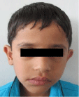
Figure 1:
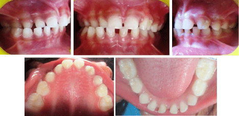
Figure 2:
Quad helix modified as a blue grass appliance to prevent tongue thrusting habit was delivered to the child (Figure 3). After one week quad helix was activated and the patient was recalled for regular check up after every 15 days. After 2 months of follow up slight over correction of the crossbite was achieved (to prevent relapse). The appliance was de-cemented and patient was kept under observation (Figure 4,5).
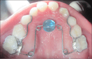
Figure 3:
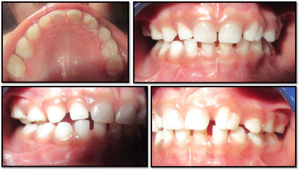
Figure 4:
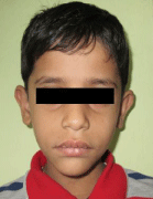
Figure 5:
Discussion
On extraoral evaluation, the patient displayed chin deviation to the right in centric occlusion. Facial asymmetry with chin deviation to the crossbite side is a known concurrent finding in cases affected by FXB [16]. Therefore, the treatment was provided to help avoid growth imbalance of both skeletal and dentoalveolar structures. In cases of unilateral crossbite, determining the correct treatment approach for each individual case is the key to treatment success and stability. The clinician must first distinguish crossbites of dental origin from those of skeletal origin.
There is a growing body of evidence that untreated crossbites will lead to permanent growth alteration, making early treatment crucial. Evidence from tomographic studies has shown that the condyles in child crossbite patients are related asymmetrically within the fossa, but that symmetry is restored after early treatment. Symmetry of the mandible and its rotational position relative to the cranial base is altered in adult patients with untreated posterior crossbites. It has been concluded that muscle, skeletal and joint adaptation in crossbites occurs early in development. Once these adaptations are firmly established in adulthood, treatment may require a combined orthodontic and surgical approach [16,17]. Sutural expansion is more stable than dental tipping; therefore, all efforts should be directed towards maximal sutural opening. During the deciduous and early mixed dentition stages (patients below 8 years of age) smaller forces can be used to achieve sutural expansion. When expansion is carried out during the late deciduous dentition, the first permanent molars usually erupt into satisfactory transverse positions (i.e., without crossbite).
Treatment during the late mixed dentition is difficult because of exfoliating deciduous teeth. Older patients in the early permanent dentition stage (12 years and above) require greater force for expansion and a faster rate of expansion because of growth-related changes in suture biology. Treatment of FXB by maxillary expansion is, therefore, best carried out during the late deciduous or early mixed dentition stages. Removal of functional interferences has been shown to be useful only in patients below age of 5, with success rates ranging from 27% to 64%.
In a study of 76 4-year-old children with posterior crossbite, Lindner reported 50% correction after functional grinding. The greatest chance of correction after selective grinding occurred when the maxillary intercanine width was at least 3.3 mm greater than the corresponding mandibular intercanine width.
Expansion with the quad-helix would not control the palatal tipping of the right posterior segment mesial to the first molar (especially the primary maxillary right canine). Therefore, a removable appliance would better control the palatal tipping of canine and maxillary right first molar. Removable appliances utilize palatal anchorage, and the ability to move a selected block of teeth [18]. But they have the disadvantages of poor oral hygiene, and cooperation as young patients, lose appliance and thus increase treatment cost, relapse of the previous expansion and lower success rates. Fixed appliances are typically favoured for expansion due to reduced cost and treatment time. Treatment and retention time using the quad helix was a fifth and cost was a third that of the removable expansion plate [4].
The mandibular incisors underwent continuous spontaneous alignment throughout the treatment period. This can be attributed to the anterior crossbite correction that allowed the maxillary incisors to overlap the mandibular ones and enabled the latter to tip back to their original places. Such spontaneous alignment is an example of how early treatment can produce additional favorable effects on the developing dentition. It should be noted that cases with symmetrical arches could benefit from symmetric expansion even in the presence of unilateral crossbite and mandibular shift. Minor expansion of the opposite side unavoidably occurred. Thus, the expansion of both sides must be carefully monitored in such cases. However, for the involved side - overcorrection is usually recommended in posterior crossbite cases; Treatment of FXB involves expansion of the maxillary arch, removal of occlusal interferences and elimination of the functional shift. Slow maxillary expansion can be used during the deciduous or early mixed dentition stages with a W arch, Quad helix, fixed expander (Haas, Hyrax or Super screw) or a removable expander. The rate of expansion is a quarter revolution of the screw every second or third day and the estimated time to correct the crossbite is 6–12 weeks [4].
Overexpansion is appropriate, to the point where the lingual cusps of the upper molars contact the buccal cusps of the lower molars. The appliance should be left in place to serve as a retainer for an additional 4–6 months (or for a period at least equal to that required to correct the crossbite). When a screw is used as the active mechanism, it can be stabilized with a ligature wire or with composite to prevent relapse. The rate of expansion is 1–2 quarter revolutions of the screw per day, and the estimated time to treat the crossbite is 2–6 weeks [19].
Potential side effects: In rapid maxillary expansion, some spontaneous increase occurs in the intercanine width of the mandibular permanent dentition. This also occurs to a minimal degree with slow maxillary expansion in the early mixed dentition stage [19]. The mere act of sutural expansion can cause forward movement of the maxilla. This can be useful in Class III cases, especially when maxillary protraction is used in conjunction with maxillary expansion. It has been shown that correction of crossbite with functional shift in the mixed dentition can be successful in 84% to 100% of cases [2,8,12,15,19].
The type of appliance, follow-up period, and criteria used for the definition of success also affect the reported success rate 4. If unilateral posterior crossbite is planned to be corrected in the mixed dentition, this report states that treatment with the quad-helix is an appropriate and successful method.
References
- Moyers R. Handbook of orthodontics. 2nd ed. Chicago: Year Book Medical Publishers, Inc; 1966: 332–341.
- Thilander B, Lennartsson B. A study of children with unilateral posterior crossbite, treated and untreated, in the deciduous dentition — occlusal and skeletal characteristics of significance in predicting the long-term outcome. J Orofacial Orthop. 2002; 63: 371–383.
- Infante PF. An epidemiologic study of finger habits in preschool children as related to malocclusion, socioeconomic status, race, sex and size of community. ASDC J Dent Child. 1976; 43: 33–38.
- Kennedy DB, Osepchook M. Unilateral Posterior Crossbite with Mandibular Shift: A Review. J Can Dent Assoc. 2005; 71: 569–573.
- Allen D, Rebellato J, Sheats R, Ceron AM. Skeletal and dental contributions to posterior crossbites. Angle Orthod. 2003; 73: 515–524.
- Oulis CJ, Vadiakas GP, Ekonomides J, Dratsa J. The effect of hypertrophic adenoids and tonsils on the development of posterior crossbite and oral habits. J Clin Pediatr Dent. 1994; 18: 197–201.
- McNamara JA Jr. Early intervention in the transverse dimension: is it worth the effort? Am. J. Orthod Dentofacial Orthop. 2002; 121: 572–574.
- Troelstrup B, Moller E. Electromyography of the temporalis and masseter muscles in children with unilateral cross-bite. Scand J Dent Res. 1970; 78: 425–430.
- Ingervall B, Thilander B. Activity of temporal and masseter muscles in children with a lateral forced bite. Angle Orthod. 1975; 45: 249–258.
- Kutin G, Hawes RR. Posterior cross-bites in the deciduous and mixed dentitions. Am J Orthod. 1969; 56: 491–504.
- Lindner A, Henrikson CO, Odenrick L, Modeer T. Maxillary expansion of unilateral cross-bite in preschool children. Scand J Dent Res. 1986; 94: 411–418.
- Thilander B, Wahlund S, Lennartsson B. The effect of early interceptive treatment in children with posterior crossbite. Eur J Orthod. 1984; 6: 25–34.
- Sandikcioglu M, Hazar S. Skeletal and dental changes after maxillary expansion in the mixed dentition. Am J Orthod Dentofacial Orthop. 1997; 111: 321–327.
- Erdinc AE, Ugur T, Erbay E. A comparison of different treatment techniques for posterior crossbite in the mixed dentition. Am J Orthod Dentofacial Orthop. 1999; 116: 287–300.
- Bjerklin K. Follow-up control of patients with unilateral posterior cross-bite treated with expansion plates or the quad-helix appliance. J. Orofac. Orthop. 2000; 61: 112–124.
- Pirttiniemi P, Kantomaa T, Lahtela P. Relationship between craniofacial and condyle path asymmetry in unilateral cross-bite patients. Eur. J. Orthod. 1990; 12: 408–413.
- Pinto AS, Buschang PH, Throckmorton GS, Chen P. Morphological and positional asymmetries of young children with functional unilateral posterior crossbite. Am J Orthod Dentofacial Orthop. 2001; 120: 513–520.
- Littlewood SJ, Tait AG, Mandall NA, Lewis DH. The role of removable appliances in contemporary orthodontics. Br Dent J. 2001; 191: 304–310.
- Sarver DM, Johnston MW. Skeletal change in vertical and anterior displacement of the maxilla with bonded rapid palatal expansion appliances. Amer J Orthod Dentofacial Orthop. 1989; 95: 462–466.