
Special Article - Dental Implants
J Dent App. 2016; 3(2): 315-318.
Two-Implant-Retained Overdentures using Locator Attachments in Completely Edentulous Patients with Severely Resorted Mandible: A Report of Two Cases
Kaneko T*, Nakamura S, Hino S, Horie N and Shimoyama T
Department of Oral and Maxillofacial Surgery, Saitama Medical Center, Saitama Medical University, Japan
*Corresponding author: Kaneko T, Department of Oral and Maxillofacial Surgery, Saitama Medical Center, Saitama Medical University, 1981 Kamoda, Kawagoe, Saitama 350-8550, Japan
Received: August 29, 2016; Accepted: September 20, 2016; Published: September 22, 2016
Abstract
Implant-retained overdentures (IODs) offer advantages in terms of lessinvasive surgery, ease of cleaning, and cost-effectiveness. Use of an IOD supported by two implants (two-IOD) has been advocated as the minimal standard of care for the treatment of an edentulous mandible. We describe two cases in which a two-IOD was applied using Locator attachments for completely edentulous patients with severely resorbed mandibles. Case 1 showed a severely atrophic mandible requiring major bone augmentation for the fixed implant prostheses. Case 2 had a severely atrophic mandible and complex medical history. Both cases were treated with a two-IOD using Locator attachments for reasons of treatment cost, minimizing surgical damage, and systemic condition of the patient. Outcomes of prostheses were evaluated using chair side scoring to measure masticatory function.
Keywords: Two-implant-retained overdenture; Locator attachment; Dental implants; Severely atrophic mandible
Abbreviations
CAD/CAM: Computer Aided Design/Computer Aided Manufacturing; CT: Computed Tomography; IOD: Implant- Retained Overdenture
Introduction
A completely edentulous jaw might result in systemic problems such as dyspepsia and poor nutritional status, as well as incomplete mastication and dysphagia in the oropharyngeal region. Under conditions of oral dysfunction due to an edentulous jaw, dental implants can restore both esthetic and functional deficits and have demonstrated significant improvements in quality of life for edentulous patients [1]. For treatment variation of implants, both fixed and removable prostheses can be successfully applied to edentulous jaws. Implant-retained overdenture (IOD) is favorable in terms of reducing the invasiveness of surgery [2], decreasing the costs [3], and facilitating hygiene rehabilitation [4]. Although these prostheses can be anchored to the implants with splinted attachments such as a bar or unsplinted attachments such as ball anchors, Locators, double crowns, and magnets [5], bar attachment needs more space within the denture than unsplinted attachments, and is often difficult to accommodate with anchorage attachments. In addition, fabrication and results of prostheses are sensitive to the skill of the dental technician [6]. In contrast, an IOD using unsplinted attachment is more suitable for a large number of patients because of the smaller space requirements within prostheses and the simple fabrication process [6-8]. In the McGill Consensus Statement on overdentures, the two-IOD has been advocated as the minimal standard of care in the treatment of an edentulous mandible [2].
This article describes two cases of two-IOD using Locator attachments in completely edentulous patients with severely resorbed mandible and systemic disease.
Case Presentation
Case 1
The patient was a 71-year-old man who was referred to our department for dental implant therapy due toa chief complaint of masticatory disturbance caused by unstable complete lower dentures. He had past histories of well-controlled diabetes mellitus and hypertension. Oral examinations revealed that both the maxilla and mandible were edentulous, and the alveolar ridges in the region of the mandibular molars were flattened (Figure 1). Orthopantomography showed severe resorption of mandibular bone, and the alveolar crest was extremely close to the mandibular canal (Figure 2). The patient was informed of the details of the treatment options, including implantfixed prostheses associated with bone augmentation and IOD. Because of the surgical damage and time required for bone augmentation and treatment cost, two-IOD was selected for treatment, and Locator attachments (Kerator Overdenture attachment system; KJ Meditech, Gwang-iu, Korea) were adopted as the retention device to improve denture stability. Prior to implant surgery, conventional upper and lower full dentures were newly fabricated. A radiographic template was then prepared from the duplicate dentures and data from computed tomography (CT) were utilized in image analysis software (S implant Pro TM; Materialise Dental NV, Leuven, Belgium) to gather diagnostic information for proper implant placement. CT showed that alveolar crest height was severely reduced in the molar regions, especially in the left molar region (Figure 3A and 3B). Bilateral mental for amina were observed in the alveolar crest sites. However, sufficient bone had been maintained in the canine-lateral incisor area to make implant insertion possible (Figure 4), and two implants (Osseo speed TX; Dentsply IH AB, Mölndal, Sweden) were placed with sufficient initial stability (Figure 5). Abutment connection was performed 2months after implant insertion (Figure 6), and a nylon male cap was incorporated into the denture base. After setting the two-IOD, denture stability was markedly enhanced, and occlusal force and masticatory function were significantly improved (Figure 7). As of more than 3years since setting the two-IOD, no problems have been observed.
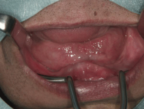
    Figure 1:  Preoperative oral findings. Alveolar ridges in the region of the
mandibular molars are flattened.
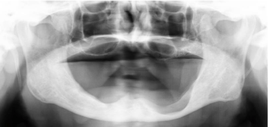
    Figure 2:  Preoperative panoramic radiography. Alveolar crest height is
severely reduced in the molar regions, especially in the left molar region.
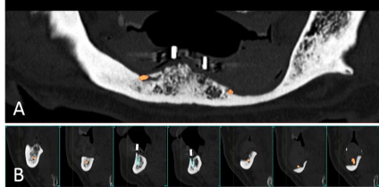
    Figure 3:  Preoperative panoramic and cross-sectional computed tomography
and simulation of implant placement. A: Panoramic image; B: Cross-sectional
image.

    Figure 4:  Preoperative three-dimensional computed tomography and
simulation of implant placement.
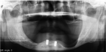
    Figure 5:  Postoperative panoramic radiography. Two implants were placedin
bilateral canine-lateral incisor area.
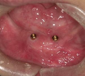
    Figure 6:  Connection of Locator attachments was performed two months
after implant insertion.
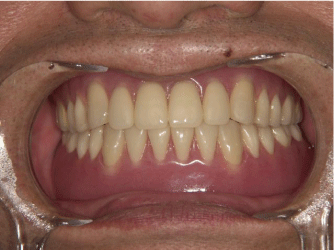
Figure 7: Clinical view after setting the two-IOD.
Case 2
The patient was a 73-year-old woman who presented to our department due to a masticatory disturbance caused by unstable lower dentures. The patient strongly hoped to undergo installation of a fixed implant prosthesis using dental implants. She had a past history of well-controlled diabetes mellitus and hypertension, chronic cardiac and renal failure, and hepatic cirrhosis. Oral examination revealed that the alveolar ridges in the regions of the mandibular molars were flattened. Radiography showed advanced bone resorption in the mandible, and the distance from the alveolar crest to the mandibular canal was reduced in the molar region (Figure 8). Considering the advanced age, medical history, and severe mandibular bone resorption of the patient, minimally invasive treatment using two- IOD was selected, and adaptation of non-flapped computer-guided surgery was determined as the surgical method.
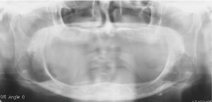
    Figure 8:  Preoperative panoramic radiography. Alveolar crest height is
severely reduced in both molar regions.
A surgical template was fabricated using computer-aided design/ computer-aided manufacturing (CAD/CAM), and two implants (Osseo speed TX; Dentsply IH AB) were inserted by flapless operation (Figure 9). Locator attachments (Creator Overdenture attachment system; KJ Meditech) were connected to the fixtures 3 months after implant placement. After loading of the implants, the implants and denture have remained stable, with no progressive marginal loss observed around the implants.
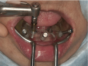
    Figure 9:  Clinical view of implant placement using non-flapped computerguided
surgery.
Evaluation of Masticatory Function
In both cases, the outcomes of prostheses were evaluated using chair side scoring to measure masticatory function [9]. Scoring was performed using the conventional complete dentures before implant placement, and using the two-IOD at 1 year postoperatively. The results showed substantial increases in evaluation scores for chewing function in both patients at 1 year postoperatively (Table 1).
    Chewing function score


    With conventional complete dentures
      before implant placement
    With two-IOD
      at 1 year postoperatively


    Case    1
    15
    65


    Case    2
    30
    75
Table 1: Laboratory values at hospital admission.
Discussion
In the past several decades, numerous studies have shown clinical advantages of IOD for the completely edentulous jaw. In our two cases, although consideration of severe bone resorption in the jaw and multiple systemic medical diseases was required, two-IOD treatment using Locator attachments is minimally invasive and is thus considered available restoration for elderly patients with various local and systemic disorders. Evaluation of masticatory function revealed significant improvements in oral function with two-IOD treatment as compared with the conventional complete dentures. Moreover, the combination of IOD treatment and computer-guided surgery could provide more accurate and safer management during implant insertion [10]. These tools might expand the indications for IOD treatment in various clinical cases.
With unsplinted attachments, although ball anchors have generally been used for the attachment of two-IOD for several decades, this material need as relatively large space in the denture, potentially leading to an excessive occlusal vertical dimension and over-countered prostheses to accommodate the attachments into the denture [8,11]. Conversely, fracture of the denture base around the attachments of the IOD is a common problem in prosthodontic practice. This fracture is generally associated with concentration of stress in anterior sites of the prostheses over the implants. Insufficient acrylic overlaying around the attachment may lead to denture base deformation and could result in fracture of the prosthesis [11]. To address this problem, the Locator attachment system uses a lower height than a ball anchor. These attachments are resilient [12] and self-aligning, provide dual retention, and area available in different colors with different retention values [13]. During the connection of the attachment, the Locator attachment system has a space of 0.2mm between the abutment top and nylon patrix for the retention disk. This allows vertical resilience and 8° hinging in any direction [8,14]. Elsyad et al. [11], using an acrylic model covered with resilient silicone mucosal simulation, studied deformation of the mandibular denture base with Locator and ball attachments for two-IOD. That study found that during bilateral load application to the prosthesis, Locator attachments for the IOD were associated with less deformation of the mandibular denture base over the implants compared to ball attachments. The Locator system recorded higher compressible strains and provided excellent settlement of the denture base without fulcrum formation. This mechanical condition without fulcrum formation could thus be considered to behave like the denture base of conventional mandibular complete dentures, and result in good prognosis for both implants and prostheses, and might contribute to long-term stability of the IOD. In general, since Asian patients often show lower interacts distance or denture height, a low-profile attachment might be more recommendable. Thus, Locator system can expected as an alternative to conventional ball attachments.
Conclusion
Two-IOD using Locator attachments appear highly applicable to various clinical situations. This method is considered to offer viable restoration for patients with local and systemic disorders.
References
- Mack F, Schwahn C, Feine JS, Mundt T, Bernhardt O, John U, et al. The Impact of Tooth Loss on General Health Related to Quality of Life Among Elderly Pomeranians: Results from the Study of Health in Pomerania (SHIP-O). Int J Prosthodent. 2005; 18: 414-419.
- Feine JS, Carlsson GE, Awad MA, Chehade A, Duncan WJ, Gizani S, et al. The McGill Consensus Statement on Overdentures. Montreal, Quebec, Canada. May 24-25, 2002. Int J Prosthodont. 2002; 15: 413-414.
- Attard NJ, Zarb GA. Long-term Treatment Outcomes in Edentulous Patients with Implant Overdentures: the Toronto study. Int J Prosthodont. 2004; 17: 425-433.
- Moberg LE, Kondell PA, Sagulin GB, Bolin A, Heimdahl A, Gynther GW. Branemark System and ITI Dental Implants System for Treatment of Mandibular Edentulism. A Comparative Randomized Study: 3-year Follow-up. Clin Oral Implants Res. 2001; 12: 450-461.
- Bernhart G, Koob A, Schmitter M, Gabbert O, Stober T, Rammelsberg P. Clinical Success of Implant-supported and Tooth-implant-supported Double Crown-retained Dentures. Clin Oral Investig. 2012; 16: 1031-1037.
- MacEntee MI, Walton JN, Glick N. A Clinical Trial of Patient Satisfaction and Prosthodontic Needs with Ball and Bar Attachments for Implant-retained Complete Overdentures: Three-year Results. J Prosthet Dent. 2005; 93: 28-37.
- Alsabeeha NH, Payne AG, Swain MV. Attachment Systems for Mandibular Two-implants Overdentures: A Review of In Vitro Investigations on Retention and Wear Features. Int J Prosthodont. 2008; 22: 429-440.
- Lee CK, Agar JR. Surgical and Prosthetic Planning for a Two-implant-retained Mandibular Overdenture: A Clinical Report. J Prosthet Dent. 2006; 95: 102-105.
- Sato Y, Minagi S, Akagawa Y, Nagasawa T. An Evaluation of Chewing Function of Complete Denture Wearers. J Prosthet Dent. 1989; 62: 50-53.
- Gallardo R, Natali Y, Silva-Olivio IRT, Mukai E, Morimoto S, Sesma N, et al. Accuracy Comparison of Guided Surgery for Dental Implants According to the Tissue of Support: a Systematic Review and Meta-analysis. Clin Oral Implants Res. 2016 [Epub ahead of print].
- ELsyad MA, Errabti HM, Mustafa AZ. Mandibular Denture Base Deformation with Locator and Ball Attachments of Implant-retained Overdentures. J Prosthodont. 2015; 00: 1-9.
- Nguyen CT, MasriR, Driscoll CF, Romberg E. The Effect of Denture Cleansing Solutions on the Retention of Pink Locator Attachments: an In Vitro Study. J Prosthodont. 2010; 19: 226-230.
- Trakas T, Michalakis K, Kang K, Hirayama H. Attachment Systems for Implant Retained Overdentures: a Literature Review. Implant Dent.2006; 15: 24-34.
- Chikunov I, Doan P, Vahidi F. Implant-retained Partial Overdenture with Resilient Attachments. J Prosthodont. 2008; 17: 141-148.