Case Report
J Dent App. 2016; 3(2): 325-327.
A Case of Nasopalatine Dust Cyst: Presentation, Diagnosis and Management
Nilesh K1*, Vande AV2, Suresh KV3, Pramod RC4, Suryavanshi P1 and Kadam NA5
1Department of Oral & Maxillofacial Surgery, School of Dental Sciences, KIMSDU, Karad, Maharashtra, India
2Department of Prosthodontics, School of Dental Sciences, KIMSDU, Karad, Maharashtra, India
3Faculty of Dentistry, SEGi University, Selangor, Malaysia
4Department of Oral Pathology, College of Dental Sciences, Davangere, Karnataka, India
5Department of Endodontics, School of Dental Sciences, KIMSDU, Karad, Maharashtra, India
*Corresponding author: Kumar Nilesh, Department of Oral & Maxillofacial Surgery, School of Dental Sciences, Krishna Hospital, Karad, Satara, India
Received: September 20, 2016; Accepted: October 17, 2016; Published: October 19, 2016
Abstract
Nasopalatine duct cyst is a non-odontogenic developmental cyst typically located in the maxillary midline between the tooth roots of central incisors. Unlike radicular cyst which is also associated with tooth roots, nasopalatine duct cysts are infrequent and can often be misdiagnosed as periapical lesion or cyst. This paper reports a case of nasopalatine duct cyst in a 57 year old male patient. Clinical presentation, radiological appearance, microscopic features and management of the lesion is presented.
Keywords: Non-odontogenic; Developmental Cyst; Maxilla; Periapical lesion
Introduction
Nasopalatine duct cyst (NPDC) was first described by Meyer in 1914, who wrongly identified it as a paranasal sinus. In the past, NPDC, was termed as anterior palatine cyst or incisor duct cyst and was regarded as fissural cysts formed between the two maxillae. World Health Organization in 1998 classified these lesions as nonodontogenic developmental cyst along with naso-alveolar and naso-labial cyst. Although they are most common non-odontogenic jaw cysts, it accounts for only 1% of all maxillary cysts [1]. Unlike radicular or dentigerous cysts which are frequently seen in clinical practice, NPDC are less commonly encountered.
NPDC is commonly seen in 4th and 5th decade of life with slight male predilection. Clinically it may be asymptomatic and get discovered during routine radiographic examination. Larger lesion presents as a swelling over anterior maxilla, located between the tooth roots of maxillary central incisors. As the presentation of NPDC mimics common periapical lesion like radicular cyst, it is not uncommon to see evidence of endodontic treatment of associated teeth due to previous misdiagnosis of the pathology.
Case Presentation
A fifty seven year old male patient was referred by private dental practitioner to Oral surgery clinic for opinion and management of a periapical lesion, non-responsive to endodontic therapy. The patient had chief complaint of swelling in anterior part of upper jaw since six months. There was no history of trauma. Patient did not have any underlying medical disorder. Intraoral examination revealed a welldefined oval swelling measuring about 1 X 2 cm, localized over apical region of maxillary incisors and obliterating labial vestibule (Figure 1A). The swelling was soft and tender on palpation. On electric pulp testing the maxillary incisor teeth were non-responsive, which was attributed to the previous endodontic treatment.

Figure 1: Overall survival, autologous stem cell transplant (ASCT) versus no ASCT (p=0.12).
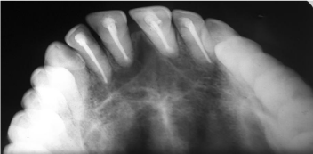
Radiographs available from patient’s record showed a welldefined, corticated heart shaped radiolucency measuring about 1.5 X 2 cm. between the roots of maxillary incisors, with lateral displacement of roots of maxillary central incisors. There was no evidence of root resorption (Figure 1B and 1C). Based on clinical and radiographic findings the intraosseous periapical lesion was provisionally diagnosed as nasopalatine duct cyst. A differential diagnosis of radicular cyst, central giant cell granuloma and lateral periodontal cyst was given.
Treatment plan included completion of root canal therapy of maxillary incisors followed by enucleation of the lesion under local an aesthesia. Bilateral infraorbital and nasopalatine nerve blocks were given. Full thickness trapezoidal.
Mucoperiosteal flap was raised from 13 to 23. A bony window was created in the midline between the apices of maxillary central incisors (Figure 2A). The cyst lining was enucleated and submitted for histopathologic examination. Root ends of the teeth were seen within the cyst cavity and were resected to achieve complete removal of the lining from behind the tooth root and undercuts. Resected root ends were prepared to receive MTA as retrograde root canal filling and to achieve a periapical seal. Hydroxyapatite bone graft was placed in the bony defect (Figure 2B). Heamostasis was achieved and closure done with 3-0 vicryl resorbable sutures (Figure 2C). Periodontal pack was applied over labial and palatal and maintained for a week. Patient was prescribed antibiotics, analgesics and mouthwash in postoperative period.

Figure 2: Overall survival, autologous stem cell transplant (ASCT) versus no ASCT (p=0.12).
Microscopic examination of the specimen revealed cystic lining of non- keratinized statified squamous epithelium of variable thickness. The underlying connective tissue showed dense chronic inflammatory cell infiltrate and numerous blood vessels. In certain areas presence of bony trabeculae and nerve bundles was noted (Figure 3). On basis of histopathology findings final diagnosis of nasopalatine duct cyst was established. Patient was kept on regular follow-up and showed uneventful healing. At six month review, the patient was disease free with no clinical signs of recurrence. Radiograph taken at this stage showed bone fill in the apical osseous defect (Figure 4).

Figure 3: Overall survival, autologous stem cell transplant (ASCT) versus no ASCT (p=0.12).

Figure 4: Overall survival, autologous stem cell transplant (ASCT) versus no ASCT (p=0.12).
Discussion
Nasopalatine duct cyst (NPDC) in a developmental nonodontogenic cyst which arises from epithelial remnants in the nasopalatine duct. The pathology is unique in that it develops only at single location, which is the maxillary midline between the roots of central incisors. Although they are commonly seen in adult patients with slight male predilection, it can be seen in any age group [2]. They are usually asymptomatic and get discovered during routine radiography. Larger lesion may present as swelling, commonly over anterior palate. Rarely the cyst may present as localized swelling over the labial mucosa or an extensive lesion involving the anterior palate and labial mucosa. Although infrequent, neurological symptoms including numbness or burning sensation over anterior palate may be experienced due to pressure on the nasopalatine nerve. It is typically seen associated with roots of maxillary central incisors. Although the root apex may be commonly involved, the teeth remain vital. NPDC must be differentiated with common periapical lesion like a radicular cyst, present in anterior maxilla associated with root apex of maxillary central incisor. Radiclar cyst unlike NPDC is commonly associated with non-vital teeth with positive history of trauma to anterior teeth or chronic pulpitis. Root displacement and resorption are rare findings in NPDC. In present case the lesion was localized over the labial vestibule in midline. The maxillary incisor teeth were nonvital due to previous endodontic therapy, rather than due to the lesion itself. The midline lesion caused slight displacement of roots of maxillary central incisors.
Intraoral periapical radiograph, maxillary occlusal view and orthopantamogram are used to radiographically evaluate the lesion. NPDC appear as a well defined, round or oval radiolucency located between the roots of maxillary central incisors with sclerotic border. Superimposition of anterior nasal spine gives the lesion typical heart shaped radiolucency. A large incisive canal opening may often be misdiagnosed as a NPDC. To avoid the confusion, 6 mm is considered as the upper limit for incisive canal opening and radiolucencies larger than 6 mm is regarded as potentially pathologic and should be investigated further [3]. Radiologically it is important to differentiate NPDC from other radiolucent lesions in maxillary midline, like lateral radicular cyst or lateral periodontal cyst arising from mesial root surface of maxillary central incisors. The radiolucency of lateral radicular cyst is continuous with lamina dura of the involved nonvital tooth, whereas lateral periodontal cyst is a developmental cyst associated with a vital tooth and the lamina dura of the tooth root is often destroyed. Clinical and radiological similarity of NPDC with periapical lesions like radicular cyst may lead to its misdiagnosis. This could have been the reason why the present case was possibly misdiagnosed as a periapical lesion and was being unsuccessfully managed by endodontic treatment when the patient was referred to us.
Histologically NPDC lesion is characterized by epithelial lining surrounded by connective tissue wall with varying degree of inflammation and presence of nerve bundles, mucous glands and adipose tissue. The epithelial lining of the cyst is variable ranging from squamous, columnar, cuboidal or combination of these epitheliums. The nature of the lining depends on proximity of the lesion to oral or nasal cavity, with those arising in the superior part of duct having pseudostratified squamous epithelium and those closer to oral cavity having stratified squamous epithelium [4].
Treatment of NPDC is surgical excision. The lesion can be approached from palatal or labial aspect depending on its location.
In the present case the lesion was localized over labial vestibule and was in close proximity to the root apex of maxillary incisors. After completion of endodontic treatment, the lesion was surgically approached from labial aspect. The periapical defect was grafted with bone graft for better bone healing. Some authors have proposed marsuplization for management of larger lesion [5]. The nasopalatine neurovascular bundle may be inadvertently severed during surgery giving rise to profuse bleeding, which can be managed by pressure packing, electrocautery or pizosurgery. Recurrence after surgical excision is very rare. Follow-up of the present case showed no clinical signs of recurrence and bone fill in the surgical defect at 6 months.
Conclusion
Although NPDC is most common non-odontogenic developmental cyst, they are rare pathology accounting for less than 1% of all maxillary cysts. Its clinical and radiological presentation can mimic common periapical lesion like radicular cyst, leading to its misdiagnosis. Treatment of choice is simple surgical excision and definitive diagnosis is established only after histological evaluation of the excised lining. Although recurrence is unlikely, follow-up is recommended to ensure resolution of the pathology.
References
- Swanson KS, Kaugars GE, Gunsolley JC. Nasopalatine duct cyst: an analysis of 334 cases. J Oral Maxillofac Surg. 1991; 49: 268-271.
- Robertson H, Palacios E. Nasopalatine duct cyst. Ear Nose Throat J. 2004; 83: 313.
- Francoli JS, Marques NA, Aytis LB, Escoda CG. Nasopalatine duct: report of 22 cases and review of literature. Med Oral Patol Oral Cir Bucal. 2008: 13: E438–E443.
- Cecchetti F, Ottria L, Bartuli F, Bramanti NE, Arcuri C. Prevalence, distribution, and differential diagnosis of nasopalatine duct cysts. Oral Implantol. 2012; 5: 47–53.
- Curtin HD, Wolf P, Gallia L, May M. Unusually large nasopalatine cyst: CT findings. J Comput Assist Tomogr. 1984; 8: 139–142.