
Special Article – Pedodontics
J Dent App. 2016; 3(2): 328-329.
Orthodontic Approach for Double Teeth
Jairath R¹, Chaudhry A²*, Chaudhary G³ and Bector K4
¹Department of Orthodontics, Christian Dental College, CMC, Ludhiana-141010, Punjab, India
¹Senior lecturer, Department of Orthodontics, Baba Jaswant Singh Dental College, Hospital and Research Institute, Chandigarh Road, Ludhiana-141010, Punjab, India
*Corresponding author: Anshul Chaudhry, Senior lecturer, Department Of Orthodontics, Christian Dental College, MC, Ludhiana-141010, Punjab, India
Received: October 06, 2016; Accepted: October 21, 2016; Published: October 24, 2016
Abstract
Odontogenic anomalies of teeth can be encountered frequently in dental practice. Double teeth or fused teeth not only create esthetic problems, but also cause malalignment and malocclusion by creating tooth size discrepancy. A case of bilateral maxillary permanent double teeth of the central incisors is described in this article
Keywords: Central incisor; Fusion; Orthodontic treatment
Introduction
Anomalies in the size and shape of teeth arise from abnormalities in the differentiation of tooth germs such as gemination, fusion, concrescence, double tooth. Fusion is a union of two or more separately developing tooth buds at dentinal level, presenting one single large tooth structure. Gemination is a developmental anomaly of form which is recognized as an attempt by a single tooth germ to divide resulting in a large single tooth with a bifid crown and usually a common root and root canal in which the tooth count is normal.
The etiology of double tooth is unknown, although some cases can be hereditary, evolution, trauma and environmental [1]. The unaesthetic appearance of double teeth presents a great challenge for dentist. By not treating the condition, complications may arise including caries, gingival problems and crowding. The extraction of these teeth would necessitate prosthetic replacement at a very early age. Reshaping and restoration usually failed to achieve the desired results in the most cases. Careful evaluation of a case will guide the clinician in treatment planning and in some instances no treatment may be carried out. This article represents the case of orthodontic treatment of fusion of permanent maxillary central incisors with supernumerary tooth.
Case Presentation
A 15 year old boy reported to an orthodontic clinic with chief complaint of presence of two large front teeth. There was no significant medical history. The teeth were asymptomatic. The right central incisor was in cross bite (Figure 1 & 2). The periapical view of upper central incisors showed crowns fused with supernumerary tooth (Figure 3 & 4). Upper right central incisor had two roots and two root canals, upper left central incisor had one root and one root canal.
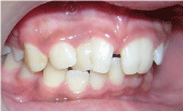
Figure 1: Fused 11 with supernumerary tooth.
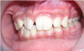
Figure 2: Fused 21 with supernumerary tooth.
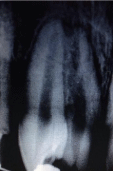
Figure 3: Preoperative intraoral Radiograph for 11.
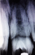
Figure 4: Preoperative intraoral Radiograph for 21.
Before initiation of treatment the treatment options were discussed with parents, they were mainly concerned with esthetics and preferred the option of retaining fused incisors. Pretorqued Roth brackets were used. A posterior mandibular bite-plate was placed to correct the cross bite in relation to 11. Following the active orthodontic treatment, a Hawley retainer was used for stability and retention. The final result was improvement in occlusion and in the appearance of teeth (Figure 5 & 6).
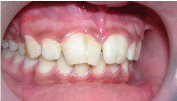
Figure 5: Post operative image from right lateral view.
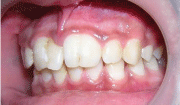
Figure 6: Post operative image from left lateral view.
Discussion
Bilateral double teeth are less common in the permanent dentition than the primary dentition [2]. Also, bilateral double tooth in the permanent dentition has been found more frequently in the maxilla than in the mandible [3]. Further, 100% of the permanent bilateral fusion cases in the maxilla involve the central incisors [4], 83% of these involve a supernumerary tooth, the same as in this case. Problems associated with double tooth include poor esthetics, crowding, abnormal eruption, residual periodontal defects, dental caries, over jet problems and difficulties in management. Although fusion is described in the literature as a developmental occurrence, its etiology is uncertain [4]. The morphology of fused teeth varies, and complex forms with separated or fused coronal pulp chambers are present. Even separated chambers can meet in the radicular area or can remain separated [5].
Conclusion
Double teeth are not usual condition but they are important dental anomalies. Early diagnosis of anomaly has considerable importance and it should be followed by careful clinical and radiographical observation that allows the dentist to plan the treatment at proper time. The minimal intervention technique should be advocated for double teeth.
References
- Brook AH, Winter GB. Double teeth. A retrospective study of ‘geminated’ and ‘fused’ teeth in children. Br Dent J. 1970; 129: 123-130.
- Grahnan H and Granath LE. Numerical variations in primary dentition and their correlation with the permanent dentition. Odontol Revy. 1961; 12: 341- 357.
- Shafer WG, Hine MK, Levy BM. A Textbook of oral pathology, 4th ed. Philadelphia: W.B. Saunders Co. 1983; 30-40.
- Mader CL. Fusion of teeth. J Am Dent Assoc. 1979; 98: 62-64.
- Marechaux SC. The treatment of fusion of a maxillary central incisor and a supernumerary: report of a case. J Dent Child. 1984; 51: 196–199.