
Special Article - Pediatric Dentistry
J Dent App. 2016; 3(3): 330-332.
Diagnosis and Management of Pre-Eruptive Intra- Coronal Resorption (PICRL) in a Primary Molar Tooth: A Case Report
Arwa G and Zilberman U*
Pediatric Dentistry Unit, Barzilai Medical University Center, Ashkelon, Israel
*Corresponding author: Zilberman Uri, Head of the Pediatric Dentistry Unit, Barzilai Medical University Center, 2nd Hahistadrut st, 78012, Ashkelon, Israel
Received: September 29, 2016; Accepted: October 24, 2016; Published: October 26, 2016
Abstract
Hidden caries (?) is a dentinal lesion beneath the dentino-enamel junction, visible on radiographs. A single report described this lesion in primary dentition. This case report describes a case of pre-eruptive intra-coronal resorption lesion (PICRL) in a mandibular second primary molar that caused pulp necrosis, misdiagnosed as malignant swelling. A three year old girl was referred to department of pediatric dentistry at Barzilai medical center with a chief complaint of pain and extra oral swelling on the right side of the mandible for the last three months. She was earlier referred to the surgical department for biopsy of the lesion. Radiographic and CT scan examination showed a peri-apical lesion with buccal plate resorption and radiolucency beneath the enamel on the mesial part of tooth 85. The tooth was extracted and follow-up of two years showed normal development of tooth 45. The main problem is early detection and treatment, since the outer surface of enamel may appear intact on tactile examination.
Keywords: Pre-eruptive intra-coronal resorption; Primary molar; Submandibular swelling
Introduction
Hidden caries is a dentinal lesion beneath the dentinoenamel junction, visible on radiographs. It is also known as preeruptive intracoronal resorption (PICR), pre eruptive intracoronal radiolucent lesion (PICRL) or pre eruptive caries. The prevalence of PICR in permanent dentition is 2% - 6%, depending on the tooth and radiographic technique. When bite-wing radiographs were used, the prevalence is 4% for the mandibular first molar, 2% for the mandibular first premolar, and 1% for the maxillary first molar, maxillary first premolar, and mandibular second molar [1]. When panoramic radiographs were used, the prevalence is 4% for maxillary first molars, and 3% for mandibular first molars. Usually, a single tooth is affected. However, cases of multiple PICRLs in individual subjects have also been reported [2]. A single report of PICRLs in primary teeth has been published [3]. Nearly half of the lesions extend to more than two thirds of the dentin [4]. No association was found between PICR and gender, race, medical conditions, systemic factors, or fluoride supplementation [1,4,5].
The etiology of PICRL remains a controversy. Suggested causes include apical inflammation of primary molars (relevant only to PICRL in premolars) and dental caries. The most likely hypothesis is that the defects are acquired as a result of coronal resorption. According to Seow [5], local factors play an important role in the etiology. There’s a significantly high association between ectopically positioned teeth and PICRL, which suggests that an ectopic position is a trigger factor. Pressure resulting from an abnormal position may induce sufficient local damage to the tooth’s protective covering causing resorptive cells to enter through the enamel. Loose of the integrity of the reduced enamel epithelium may allow osteoclasts, multinucleated giant cells, and chronic inflammatory cells to enter the tooth and initiate resorption of dentin [5].
This report describes a case of PICRL in a mandibular second primary molar and the subsequent treatment.
Case Presentation
A three year old girl was referred to department of pediatric dentistry at Barzilai medical center, with a chief complaint of pain and extra oral swelling on the right side of the mandible, for the last three months. Her medical history was not remarkable. She attended several private pediatric dentists for diagnosis, who prescribed antibiotic therapy. The swelling regressed and reappeared. Latter she was referred for a panoramic x-ray, misdiagnosed as malignant swelling, and scheduled for biopsy at oral surgery department. Before the biopsy she was referred for second opinion to the pediatric dentistry department.
Extra oral examination revealed swelling on the right side of the mandible which was diffuse, tender and warm to palpation. The overlying skin appeared flushed. Intraoral examination revealed intact primary dentition with buccal cortical plate expansion on the right side of the mandible (Figure 1).
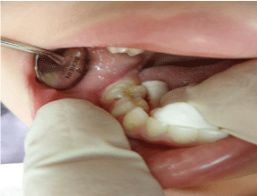
    Figure 1:  Intraoral examination at the first visit.
Note: Intact primary dentition with buccal cortical plate expansion on the right
side of the mandible.
On panoramic radiograph, tooth 85 had a peri-apical radiolucency (Figure 2). CT scan revealed bone resorption of the buccal cortical plate of tooth 85 with the MB root being in contact with the oral soft tissues and a radiolucent area in the crown extending from the occlusal surface into the dentin under the MB cusp (Figure 3 and 4). Pre-eruptive intra-coronal resorption was diagnosed with carious involvement, and the decision of tooth extraction was made.
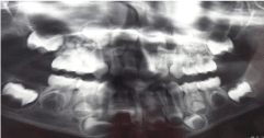
    Figure 2:  Panoramic radiograph.
Note: Periapical radiolucency at tooth 85.
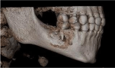
    Figure 3:  CT scan of right lower area.
Note: Extensive bone resorption of the buccal cortical plate of tooth 85 with
the MB root being in contact with the oral soft tissues.
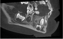
    Figure 4:  CT scan (cont).
Note: A radiolucent area in the crown extending from the occlusal surface
into the dentin under the MB cusp.
Local anesthesia was applied after sedation with nitrous-oxide and tooth 85 was extracted. The exterior opening of the hidden caries was located using a #8 file that entered from the mesial central fissure and reached the pulp horn (Figure 5).
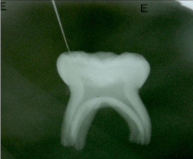
    Figure 5:  X-ray of tooth 85 after extraction.
Note: The exterior opening of the hidden caries was located using a #8 file
that entered from the mesial central fissure reaching the pulp horn.
Follow-up examination 1 week later showed soft tissue healing. Six months later, no clinical symptoms or swelling were observed (Figure 6 and 7). Two years later, radiographic findings showed normal development of the succedaneous tooth (Figure 8).
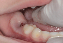
    Figure 6:  Follow-up examination after 1 week.
Note: Proper soft tissue healing.
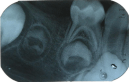
Figure 7: Periapical radiograph, six months after the extraction.
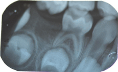
    Figure 8:  Periapical radiograph, two years later.
Note: Normal development of the succedaneous tooth.
Discussion
This case demonstrated a mis-diagnosed pre-eruptive intracoronal resorption of a mandibular second primary molar, as malignant swelling.
The purpose of this report is to increase the clinician’s awareness and clinical surveillance regarding diagnosis of PICRL, and to avoid further misdiagnosis (artifacts\malignancy). The management of PICRL in permanent dentition varies and may consist of immediate surgical exposure [5] with curettage of the defect or to wait for tooth eruption to achieve occlusal access for restoration of the defect [6]. In this case of a primary molar, extraction was the treatment of choice due to large periapical lesion reaching the underlying permanent tooth follicle and very young age.
Clinical and histological evidence from several case reports have suggested that these lesions are likely to be resorptive in nature [2- 4,7,8]. Although the etiology and factors associated with the initiation of resorption remain unknown, resorptive cells originating from the surrounding bone are thought to enter the dentin through a break in the dental follicle and enamel or cementum [3,9].
According to Kraus and Jordan [10], calcification may begin as early as eighteen weeks on the tip of the mesiobuccal cusp while the other cusps calcify later (mesiolingual-twenty three weeks, distobuccal-twenty six weeks, distal or distolingual – twenty eight weeks). At twenty eight weeks all five centers of calcification are present, and only then the intercuspal coalescence occurs, and varies with respect to location. Union between the mesiobuccal and distobuccal cusps may occur via their distal and mesial cusp ridges respectively (at thirty weeks), or the mesial marginal ridge may be the first to calcify (at thirty weeks) thereby connecting indirectly, or in a roundabout way the two mesial cusps. Whichever union occurs first, it is followed by the alternate coalescence to form a calcified ring uniting distobuccal, mesiobuccal and mesiolingual cusps (at thirty two weeks). Coalescence is complete by thirty six weeks and a small area of un-calcified tissue remains in the middle of the occlusal surface on the distal half of the crown. At thirty six week mandibular second molar crown features sharp conical cusps, angular ridges, and a relatively smooth occlusal surface, indicating that considerable deposition of enamel is yet to occur before the completion of calcification.
Based on this evidence hidden caries may be due to a faulty union or imperfection at the time of coalescence of the centers of calcification. This suggests that occlusal surface defects on molar crowns are intimately associated with the early calcification pattern peculiar to the individual teeth.
Conclusion
Increased awareness of this entity may improve diagnosis, allow early treatment and avoid mis-diagnosis.
References
- Seow WK. Pre-eruptive intracoronal resorption as an entity of occult caries. Pediatr Dent. 2000; 22: 370-376.
- Seow WK. Multiple pre-eruptive intracoronal radiolucent lesions in the permanent dentition: case report. Pediatr Dent. 1998; 20: 195-198.
- Seow WK, Hackley D. Pre-eruptive resorption of dentin in the primary and permanent dentitions: case reports and literature review. Ped Dent. 1996; 18: 67-71.
- Seow WK, Lu PC, Mcallan LH. Prevalence of preeruptive intracoronal dentin defects from panoramic radiographs. Pediatr Dent. 1999; 21: 332-339.
- Seow WK, Wan A, Mcallan LH. The prevalence of pre-eruptive dentin radiolucencies in the permanent dentition. Pediatr Dent. 1999; 21: 26-33.
- Holan G, Eidelman E, Mass E. Pre-eruptive coronal resorption of permanent teeth: Report of three cases and their treatments. Pediatr Dent. 1994; 16: 373-377.
- Rankow H, Croll TP, Miller AS. Pre-eruptive idiopathic coronal resorption of permanent teeth in children. J Endodont. 1986; 12: 36-39.
- Blackwood HJ. Resorption of enamel and dentine in the unerupted tooth. Oral Surg. 1958; 11: 79-85.
- Grundy GE, Pyle RJ, Adkins KF. Intra-coronal resorption of unerupted molars. Aust Dent J. 1984; 29: 175-179.
- Kraus BS, Jordan RE. Mandibular Second Primary Molar. In Berthram S Kraus and Ronald E Jordan (eds).The Human Dentition Before Birth. Lea & Febiger, Philadelphia. 1965: 54-68.