
Research Article
J Dent App. 2016; 3(3): 333-336.
Evaluation of Microleakage of Different Intraorifice Barrier Materials in Endodontically Treated Teeth
Özyürek T1*, Özsezer Demiryürek E1, Demiroğlu M2 and Sari ME3
¹Ondokuz Mayis University, Faculty of Dentistry, Department of Endodontics, Samsun, Turkey
²Staff Endodontist, Oral and Dental Health Hospital, Samsun, Turkey
³Ondokuz Mayis University, Faculty of Dentistry, Department of Pedodontics, Samsun, Turkey
*Corresponding author: Taha Özyürek, Faculty of Dentistry, Department of Endodontics, Ondokuz Mayis University, ID: orcid.org/0000-0003-3299-3361, Samsun, Turkey
Received: September 29, 2016; Accepted: November 02, 2016; Published: November 04, 2016
Abstract
Aim: The aim of this study was to compare the sealing ability of 4 different intraorifice barrier materials (MTA Angelus, Filtek Ultimate light-cured flowable composite resin, Filtek Z250 light-cured composite resin, SDR light-cured flowable bulk-fill composite resin)using the dye penetration method.
Materials and Methods: One hundred forty single-rooted teeth were obturated using warm vertical compaction technique. The teeth were randomly divided into four groups of 30 teeth each and positive and negative control group of 10. The access openings were filled with one of the tested intraorifice barrier materials in four groups. The sealing ability of the test materials was evaluated by dye penetration method.
Results: MTA Angelus group showed the lowest mean microleakage value and the flowable composite resin group had the highest microleakage value among the experimental groups (p = 0.001).
Conclusion: Within the limits of present study, MTA Angelus and SDR showed a better leakage resistance than the flowable composite resin and composite resin.
Keywords: Smart Dentin Replacement (SDR); Mineral Trioxide Aggregate (MTA); Dye Penetration; Leakage; Endodontics
Introduction
Microorganisms and their products are one of the main causes of periapical inflammation. Thus, root canal treatment aims at removing microorganisms from the root canals and preventing being re-infected [1,2]. Coronal leakage has an important place among the causes of failure following the completion of root canal treatment [3]. Ray and Trope [4] reported that the quality of coronal restoration was more important than the quality of root canal filling in protecting the periapical heath.
Studies have shown that gutta-percha and root canal sealer could not resist leakage for a long time when they contact with the oral flora all by themselves without any protective intraorifice barrier material [5]. Swanson and Madison [6] stated that contamination occurred in such a short time as 3 days when there is no coronal sealing.
Among the alternative methods suggested are placing an intraorifice barrier material on canal orifice by removing 3 or 4 mm part of the gutta-percha or canal sealer in order to prevent oral fluids and microorganisms from entering the root canals [6], or sealing the pulp chamber floor with a restorative material [7]. The studies have shown that sealing the pulp chamber floor with adhesive systems using intraorifice barrier materials after the root canal treatment constitutes a second defense line against bacterial leakage [7,8]. Different materials such as amalgam, Cavit, glass ionomer cement, composite resin, Mineral Trioxide Aggregate (MTA), and Intermediate Restorative Material (IRM) for this purpose [9,10].
Although there are many studies comparing the efficiencies of intraorifice barrier materials, there is no global consensus on how to use which material [9,10].
While our literature review resulted in finding no studies investigating the resistance of the light-cured bulk-fill flowable composite material (Surefil SDR; Dentsply Caulk, Milford, DE, USA) against leakage as an intraorifice barrier material, we found that there were very few studies on the coronal sealing of MTA. Therefore the purpose of this in vitro study was to compare the sealing ability of 4 different intraorifice barrier materials (MTA Angelus, Filtek Ultimate light-cured flowable composite resin, Filtek Z250 light-cured composite resin, Smart Dentin Replacement light-cured flowable bulk-fill composite resin) in extracted human teeth using the dye penetration method. The null hypothesis of our study was that there would be no difference between the dye leakage values of the tested restorative materials.
Materials and Methods
After ethic committee approval, 140 extracted human maxillary central incisors were used in this in vitro study. After the access cavity preparation, the pulp tissue was removed. The working length was determined by measuring the length of a #10 K-file (Dentsply, Maillefer, Switzerland) just visible at the apical foramen. The canals were instrumented up to 40.06 apical diameters with ProTaper NEXT (Dentsply, Maillefer, Switzerland) nickel titanium files using X1, X2, X3 and X4 files respectively. After each file 2ml 5.25% NaOCl was used for irrigation. To eliminate the smear layer in the final irrigation 2ml 17% EDTA for 3 minutes and 2ml 5.25% NaOCl were used respectively.
After the instrumentation all canals were dried with paper points (DiaDent Group International Inc., Cheungju, Korea). AH Plus (Dentsply De-Trey, Konstanz, Germany) was mixed according to the manufacturer’s instructions, and ProTaper X4 gutta-percha cones (Dentsply, Maillefer, Switzerland) were coated with sealer and placed into the root canal to the working length. Gutta-percha was then down-packed with a medium size plugger (Calamus Dual 3D Obturation System; Dentsply, Maillefer). Gutta-percha at the apical level was condensed using hand pluggers (Buchanan; SybronEndo, Orange, CA, USA). A backfill procedure was performed using the extruder hand-piece of the Calamus Dual 3D Obturation System. After completion of the filling procedures the teeth were sectioned just apical to the cement-enamel junction with a low-speed diamond saw.
The roots were randomly divided to four experimental groups with 30 samples each; 20 roots were served as control (10 teeth as positive control and 10 teeth as negative control). Coronal cavity was prepared by removing gutta-percha with System B (SybronEndo, Orange, CA, USA) to the experimental depth of 3mm. The depth was verified with a periodontal probe. The coronal 3 mm was rinsed with alcohol and distilled water respectively and dried with an air stream.
The first group (n: 30) received a 3mm barrier of MTA Angelus (Angelus, Londrina, PR, Brazil). The second group (n: 30) received light-cured flowable composite resin (Filtek Ultimate; 3M-ESPE, St. Paul, MN, USA). The third (n: 30) and fourth group (n: 30) were sealed with Smart Dentin Replacement light-cured flowable bulk-fill (SDR); and light-cured composite resin (Filtek Z250; 3M-ESPE, St. Paul, MN, USA) respectively. MTA Angelus was mixed and handled according to manufacturer’s instruction. Before usage of the other materials entire cavity surface treated with 37% phosphoric acid (3M ESPE) for 15s, rinse with water for 10s, and gentle dried with cotton pellets. A thin layer of bonding agent (Adper Single Bond 2; 3M-ESPE, St. Paul, MN, USA) and gentle air stream was applied and light cured (Elipar S10; 3M-ESPE, St. Paul, MN, USA) for 20s.
After placement of the test materials into the cavities, the samples were stored in 100% humidity at 37°C for one week. The samples were thermocycle for 100 cycles in distilled water at 5°C/55°C, with a dwell time of 4 hours in each bath. After thermocycling, the surfaces of specimens were dried and coated using nail varnish expect 1mm around the coronal filling cavity side. The samples in the experimental groups and positive control group were coated with two layers of nail varnish except for 1mm around the tooth-restoration interface. The positive control group consisted of 10 teeth obturated in the same manner as the experimental teeth without a coronal barrier.
The negative control group consisted of 10 matching obturated teeth without coronal barrier, but with crowns and roots covered completely with nail varnish and sticky wax. Then the samples were immersed in 1% Pelikan ink (Pelikan, Hannover, Germany) for 10 days. After 10 days all samples were immersed in 1% methylene blue dye and centrifuged at 30g for 5min. The samples were then washed under tap water for 1 hour and air-dried. Mid-sagittal cutting was performed using a diamond disc without water-cooling to prevent dye removal (Figure 1).
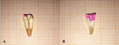
    Figure 1:  Mid-sagittal cutting was performed using a diamond disc without
water-cooling to prevent dye removal.
To measure the length of dye penetration, digital images of sample’s cross-sections were taken with a digital camera (Canon EOS 500D, Japan). The maximum lengths of penetrations, which occurred between the filling materials and the dentinal walls, were measured in the longitudinal linear direction.
Data were analyzed by Kolmogorov-Smirnov test to indicate normal distribution. As data had normal distribution one-way ANOVA was utilized for comparison followed by a Turkey’s Post Hoc test. Statistical significance was defined at P<0.05.
Result
All the experimental samples were showed microleakage at the interface of coronal barrier material and dentin wall. The mean leakage values, standard deviations and standard errors are shown in Table 1.
![]()
    Group
    N
    Mean
    Standard    Deviation
    Standard    Error
    P-value


    MTA    Angelus
    30
    2.498a
    0.816
    0.149


    SDR
    30
    2.984b
    0.692
    0.126


    Composite    Resin
    30
    3.107b
    0.973
    0.178
    0.05


    Flowable    Composite Resin
    30
    3.98c
    0.909
    0.166


    Positive    Control
    10
    12.08d
    1.545
    0.208


    a, b,    c, d : Means that do not share same superscript    letter are significantly different.
Table 1: Mean and standard deviation of microleakage of tested coronal barrier materials (in mm).
MTA Angelus group showed the lowest mean microleakage value (2.498) and the flowable composite group had the highest microleakage value (3.98) among the experimental groups. The negative control group showed no dye penetration but the all teeth in positive control group showed microleakage. Comparing the mean microleakage values of all groups showed statistically significant difference among all the experimental groups except between composite resin and SDR (P = 0.001).
Discussion
Achieving coronal sealing is highly crucial for a successful root canal treatment. Thus, the quality of coronal restoration plays a critical role in endodontic success [3]. If the coronal restoration has been performed insufficiently or improperly, a quality coronal barrier material to be placed under the coronal restoration may reduce bacterial penetration [11].
In order to investigate in vitro the resistance of dental materials to microleakage, a variety of methods has been used such as bacterial leakage, dye leakage, electrochemical method, fluid filtration method, radioisotope labeling and scanning electron microscope [12]. Since it is cheap, easy to use and capable of high staining, dye penetration method is a frequently used method in microleakage studies [13,14]. Furthermore, its molecular weight is lower than those of the bacterial toxins and it has a similar leakage value to butyric acid, a bacterial product [15]. Besides these advantages, it has also some disadvantages such as dissolving during demineralization and failure to observe the maximum leakage point in some cases [16]. It was also found that applying or not applying vacuum to the samples before dye penetration did not affect the study results [17,18]. For these purposes, dye penetration method was used in the present study while no vacuum was applied to the samples before immersing to the dye.
Even though many studies have pointed out the importance of using the coronal barrier after root canal treatment, there are different views on which one is the ideal intraorifice barrier material. However, it was determined that the thickness of the material to be used should be within the range of 3 to 4 mm [19-21]. In compliance with the previous studies, the present study has also determined the thickness of the coronal barrier materials to be 3mm.
Thermocycling was used to simulate the stress conditions restorative materials may be exposed to under normal clinical conditions [22]. Therefore, thermocycling within the range of 5°C and 55°C, which are the temperatures that can be observed under normal oral conditions, was applied to the samples before dye penetration [23].
In their study evaluating the efficacy of vertical or horizontal sectioning, decalcification and clearing methods used in the dye leakage studies, Wu and Wesselink reported that higher dye penetration values were obtained from the samples with vertical sectioning [24]. Thus, in the present study vertical sectioning method were used to evaluate the dye penetration.
The conventional root canal filling materials, gutta-percha and root canal sealer, provide minimal resistance to bacterial leakage [21,25]. This finding is supported by the results of our study in which the samples in the positive control group showed the statistically highest microleakage values.
In our study, the flowable composite resin group had statistically higher leakage value than the composite resin and SDR groups. Composite resins are bound to the dental tissue through adhesives, and accordingly, if the adhesives used fail to resist polymerization stresses, it is inevitable that there will be micro-gaps between the tooth and composite and the restoration will leak [26]. The amount of filler contained by the composite resins plays an important role in polymerization shrinkage. It may be concluded that the flowable composite resin used in the present study is exposed to a higher level of polymerization shrinkage and thus leaked more as it contains a lower amount of filler than the composite resin and SDR. Furthermore, the effective binding of adhesive systems to the dentin depends on the structure of the collagen-rich predentin and the number and permeability of the dentinal tubules [27]. NaOCl administered in endodontic treatment may irreversibly impair the physical structure of the dentin [27,28]. Furthermore, NaOCl dissolves into sodium chloride and oxygen. Oxygen-induced chemical reactions severely inhibit the polymerizations of the adhesive systems [28]. Moreover, when an adhesive material is placed on gutta-percha in a singlerooted tooth, more than half of the bond surface will be constituted by gutta-percha. A study showed that some acetone-based adhesive systems cannot be polymerized well due to some substances produced by the contents of gutta-percha [29]. Therefore, the resin should be able to polymerize well on gutta-percha. Since the adhesive system used in the present study is acetone-based as well, a polymerization problem on gutta-percha might have occurred. Similar to the results of our study, Yavariniet al. [30] and Divyaet al. [31] reported in their leakage studies using dye penetration method that composite resin leaked more than MTA. On the contrary, in their leakage study using dye penetration method, Jenkins et al. [11] reported that composite resin leaked less than Cavit and MTA. A separate leakage study using glucose penetration method found that Cavit, composite resin and MTA had comparable leakage values [25]. Another study using dye penetration method reported that composite resin and MTA had similar leakage values [32]. The different results obtained in these studies may have been caused by the use of different material contents and study methodologies.
The results of present study showed that MTA Angelus group had statistically lower leakage values than the other groups. This finding is supported by many previous studies [33-35]. In their in vitro study with MTA, Torabinejad et al. [36] reported that MTA had a superior sealing ability. They expressed that it was due to the fact that MTA, which is hydrophilic, absorbed water during setting and expanded which granted it with a good marginal adaptation.
Conclusion
Within the limits of present dye penetration study, MTA Angelus and SDR showed a better leakage resistance than the flowable composite resin and composite resin. However, further in vivo studies are needed to determine the performance of these materials in clinical use.
References
- Byström A, Sunvqvist G. The antibacterial action of sodium hypochlorite and EDTA in 60 cases of endodontic therapy. Int Endod J. 1985; 18: 35-40.
- Madison S, Wilcox LR. An evaluation of coronal microleakage in endodontically treated teeth. Part III. In vivo study. J Endod. 1988; 14: 455-458.
- Saunders W, Saunders E. Coronal leakage as a cause of failure in root-canal therapy: a review. Dent Traumatol. 1994; 10: 105-108.
- Ray H, Trope M. Periapical status of endodontically treated teeth in relation to the technical quality of the root filling and the coronal restoration. Int Endod J. 1995; 28: 12-18.
- Khayat A, Lee S-J, Torabinejad M. Human saliva penetration of coronally unsealed obturated root canals. J Endod. 1993; 19: 458-461.
- Swanson K, Madison S. An evaluation of coronal microleakage in endodontically treated teeth. Part I. Time periods. J Endod. 1987; 13: 56-59.
- Carman JE, Wallace JA. An in vitro comparison of microleakage of restorative materials in the pulp chambers of human molar teeth. J Endod. 1994; 20: 571-575.
- Roghanizad N, Jones JJ. Evaluation of coronal microleakage after endodontic treatment. J Endod. 1996; 22: 471-473.
- Canoglu E, Gulsahi K, Sahin C, Altundasar E, Cehreli ZC. Effect of bleaching agents on sealing properties of different intraorifice barriers and root filling materials. Med Oral Patol Oral Cir Bucal. 2012; 17: e710-715.
- Galvan RR, West LA, Liewehr FR, Pashley DH. Coronal microleakage of five materials used to create an intracoronal seal in endodontically treated teeth. J Endod. 2002; 28: 59-61.
- Jenkins S, Kulild J, Williams K, Lyons W, Lee C. Sealing ability of three materials in the orifice of root canal systems obturated with gutta-percha. J Endod. 2006; 32: 225-227.
- Alani AH, Toh CG. Detection of microleakage around dental restorations: A review. Oper Dent. 1997; 22: 173-185.
- Camps J, Pashley D. Reliability of the dye penetration studies. J Endod. 2003; 29: 592-594.
- Kontakiotis E, Georgopoulou M, Morfis A. Dye penetration in dry and water-filled gaps along root fillings. Int Endod J. 2001; 34: 133-136.
- Kersten H, Moorer W. Particles and molecules in endodontic leakage. Int Endod J. 1989; 22: 118-124.
- Ahlberg K, Assavanop P, Tay W. A comparison of the apical dye penetration patterns shown by methylene blue and India ink in root-filled teeth. Int Endod J. 1995; 28: 30-34.
- Masters J, Higa R, Torabinejad M. Effects of vacuuming on dye penetration patterns in root canals and glass tubes. J Endod. 1995; 21: 332-334.
- Roda R, Gutmann J. Reliability of reduced air pressure methods used to assess the apical seal. Int Endod J. 1995; 28: 154-162.
- Pisano DM, DiFiore PM, McClanahan SB, Lautenschlager EP, Duncan JL. Intraorifice sealing of gutta-percha obturated root canals to prevent coronal microleakage. J Endod. 1998; 24: 659-662.
- Sauáia TS, Gomes BP, Pinheiro ET, Zaia AA, Ferraz CC, Souza-Filho FJ. Microleakage evaluation of intraorifice sealing materials in endodontically treated teeth. Oral Surg Oral Med Oral Pathol Oral Radiol Endod. 2006; 102: 242-246.
- Wolcott JF, Hicks ML, Himel VT. Evaluation of pigmented intraorifice barriers in endodontically treated teeth. J Endod. 1999; 25: 589-592.
- Kidd EA. Microleakage: a review. J Dent. 1976; 4: 199-206.
- Ziskind D, Avivi-Arber L, Haramati O, Hirschfeld Z. Amalgam alternatives--micro-leakage evaluation of clinical procedures. Part I: direct composite/composite inlay/ceramic inlay. J Oral Rehabil. 1998; 25: 443-447.
- Wu MK, Wesselink P. Endodontic leakage studies reconsidered. Part I. Methodology, application and relevance. Int Endod J. 1993; 26: 37-43.
- Bailón-Sánchez M-E, González-Castillo S, González-Rodríguez M-P, Poyatos-Martinez R, Ferrer-Luque C-M. Intraorifice sealing ability of different materials in endodontically treated teeth. Med Oral Patol Oral Cir Bucal. 2011; 16: e105-109.
- Banomyong D, Palamara JE, Messer HH, Burrow MF. Sealing ability of occlusal resin composite restoration using four restorative procedures. Eur J Oral Sci. 2008; 116: 571-578.
- Morris MD, Lee K-W, Agee KA, Bouillaguet S, Pashley DH. Effects of sodium hypochlorite and RC-prep on bond strengths of resin cement to endodontic surfaces. J Endod. 2001; 27: 753-757.
- Lai S, Mak Y, Cheung G, Osorio R, Toledano M, Carvalho R, et al. Reversal of compromised bonding to oxidized etched dentin. J Dent Res. 2001; 80: 1919-1924.
- Belli S, Zhang Y, Pereira PN, Pashley DH. Adhesive sealing of the pulp chamber. J Endod. 2001; 27: 521-526.
- Yavari H, Samiei M, Eskandarinezhad M, Shahi S, Aghazadeh M, Pasvey Y. An In vitro Comparison of Coronal Microleakage of Three Orifice Barriers Filling Materials. Iran Endodo J. 2012; 7: 156-160.
- Divya K, Satish G, Srinivasa T, Reddy V, Umashankar K, Rao BM. Comparative evaluation of sealing ability of four different restorative materials used as coronal sealants: an in vitro study. J Int Oral Health. 2014; 6: 12-17.
- Lee KS, Kim JS, Lee DY, Kim RJY, Shin JH. In vitro microleakage of six different dental materials as intraorifice barriers in endodontically treated teeth. Dent Mater J. 2015; 4: 425-431.
- Malik G, Bogra P, Singh S, Samra RK. Comparative evaluation of intracanal sealing ability of mineral trioxide aggregate and glass ionomer cement: An in vitro study. J Conserv Dent. 2013; 16: 540-545.
- Barrieshi-Nusair K, Hammad H. Intracoronal sealing comparison of mineral trioxide aggregate and glass ionomer. Quintessence Int. 2004; 36: 539-545.
- John AD, Webb TD, Imamura G, Goodell GG. Fluid flow evaluation of Fuji Triage and gray and white ProRoot mineral trioxide aggregate intraorifice barriers. J Endod. 2008; 34: 830-832.
- Torabinejad M, Smith PW, Kettering JD, Ford TRP. Comparative investigation of marginal adaptation of mineral trioxide aggregate and other commonly used root-end filling materials. J Endod. 1995; 21: 295-299.