Review Article
J Dent App. 2016; 3(4): 365-369.
Piezosurgery: A Boon to Dentistry
Firoz Babu P¹, Polepalle T², Loya M², Mani Deepthi CH³ and Nayyar AS4*
¹Department of Orthodontics and Dentofacial Orthopedics, Rama Dental College and Hospital, Kanpur, Uttar Pradesh, India
²Department of Periodontics and Implantology, Sibar Institute of Dental Sciences, Guntur, Andhra Pradesh, India
³Department of Oral Pathology and Microbiology, Sibar Institute of Dental Sciences, Guntur, Andhra Pradesh, India
4Department of Oral Medicine and Radiology, Saraswati- Dhanwantari Dental College and Hospital and Post- Graduate Research Institute, Parbhani, Maharashtra, India
*Corresponding author: Abhishek Singh Nayyar, 44, Behind Singla Nursing Home, New Friends’ Colony, Zodel Town, Panipat-132 103, Haryana, India
Received: December 06, 2016; Accepted: December 29, 2016; Published: December 30, 2016
Abstract
Dentistry had undergone lots of advancement and has seen lots of changing concepts over the past few decades and one such novel innovation is Piezosurgery. Piezoelectric bone surgery, otherwise, known as Piezosurgery is a new technique that was introduced by Professor Vercellotti in the year 1988 to overcome the limitations of the conventional scalpel blade surgical procedures in oral bone surgery by modifying and improving the conventional ultrasound technology. It is a promising, meticulous and soft tissue sparing technique for bone cutting based on low frequency ultrasonic micro-vibrations. The absence of macro-vibrations makes the instrument more manageable and allows greater intra-operative control with a significant increase in the cutting safety in the more difficult anatomical cutting zones. The present review outweighs Piezosurgery over the conventional oral surgical procedures and emphasizes on its mechanism of action, instruments, biologic effects, advantages and limitations and its varied applications in the field of oral surgery and dentistry per se.
Keywords: Piezosurgery; Piezoelectric device; Cavitation
Introduction
Over the past few decades, there has been rapid development in various dental surgical techniques evolving a world of painless dentistry. Traditionally, osseous surgery was performed using hand instruments and various rotary instruments with different burs which required external copious irrigation due to production of heat using these instruments. Besides heat, considerable pressure was, also, exerted in osseous surgeries and had limitations in case of fractured and/or, brittle bones [1]. To overcome the above limitations, a novel surgical technique was introduced based on ultrasonic microvibrations for precise and selective bone cutting with an added advantage of sparing the surrounding soft tissues [2]. This new alternative method, introduced in dentistry for bone related surgical procedures, was termed as Piezosurgery.
What is Piezosurgery? Piezosurgery is relatively a new technique introduced by Professor Vercellotti in the year 1988 which offers advantages and overcomes the limitations of traditional instrumentation in oral bone surgeries by modifying and improving the conventional ultrasound technology. The basic principle of piezoelectricity discovered by Jacques and Pierre Curie in late 19th century for bone cutting was based on ultrasonic micro-vibrations [3]. These micro-vibrations were created by the piezoelectric effect where certain ceramics and crystals deformed when an electric current was passed across them resulting in oscillations of ultrasonic frequency [4]. The current device consists of a novel piezoelectric ultrasonic transducer powered by an ultrasonic generator capable of driving a range of resonant cutting inserts, a hand piece and a footswitch connected to the main unit, which supplies power and has holders for the hand piece and irrigant fluids, besides a peristaltic pump for cooling with a jet of solution that discharges from the inserts and also, helps in removing the debris from the cutting area. The device has a control panel with a digital display to set the power and frequency modulation. The device, also, has several autoclavable tooltips known as inserts which are titanium and/or, diamond coated in various grades and which move by micro-vibrations created in the piezoelectric hand piece [5]. The device is widely used in dentistry serving various applications including root planning, removal of supra-and sub-gingival deposits and stains from teeth, crown lengthening procedures, a traumatic tooth extractions, ridge augmentation procedures, sinus floor elevation procedures, bone graft harvesting, lateralization of inferior alveolar nerve, implant surgeries, and ridge expansion procedures etc [6,7].
History
The term piezo originates from the Greek word pieze in which means to press tight and/or, squeeze. In the year 1880, piezoelectricity was first discovered by Jacques and Pierre Curie who found that applying pressure on various crystals, ceramics and bone, created electricity. This piezoelectric effect is based on the principle of physical interactions and the phenomena of basic electric and mechanical dimensions such as electric field strength, polarization and tension and extension in crystalline field which states that deformation in crystals on passing of electric current results in oscillations of ultrasonic frequency. The vibrations obtained are amplified and transferred to a vibration tip which, when applied with slight pressure on bone tissue, results in cavitation phenomena that is a cutting effect, exclusively on the mineralized tissues [4]. Gabriel Lippmann, in the year 1881, found the converse piezoelectric effect which was, further, investigated by different scientists. In the year 1953, Catuna MC developed ultrasonic drill for cavity preparation on human teeth and published an article on the effects of ultrasound on hard tissues [8]. In the year 1957, Richman MJ was the first to report the surgical use of an ultrasonic chisel without slurry to remove bone and resect roots in apicoectomies [9]. In the year 1960, Mazarow HB reported evidence of an ultrasonic scalpel-like blade to directly cut osseous tissues [10]. In the year 1961, Mcfall TA et al evaluated distinction of healing by comparison of rotating instruments and oscillating scalpel blades and found a slow healing with no severe complications reported by use of these scalpel blades [11]. In the year 1980, Horton JE et al stated that bone regeneration was better using ultrasonic devices [12]. A year later, he evaluated the clinical use of ultrasonic instrumentation in the surgical removal of bone and observed the removal of the mineralized tissue with an ease and efficiency with good acceptance by patient without any complications [13]. In the year 1998, Torrella F et al stated better use of ultrasonic generators compared to magnetostrictive devices due to their greater efficiency of cutting bone with less tissue destruction [14]. In the year 1999, Vercellotti T invented Piezoelectric bone surgery in collaboration with Mectron Spa and published about this topic in the year 2000 [15]. Subsequently, in the year 2001, Piezosurgery® was introduced. In 2005, the US Food and Drug Administration extended the use of ultrasonics in dentistry to include bone surgeries [16].
Mechanism of Action
Piezoelectric ultrasonic frequency is created by driving an electric current from a generator over piezoceramic rings leading to their deformation. In dental applications, the ultrasonic frequency usually ranges from 24-36 kHz with a capability of cutting mineralized tissues. Thus, the resulting movement from the deformation of piezoceramic rings sets-up a vibration in the transducer which creates the ultrasound output. These waves transmitted to the hand piece tips, also, known as inserts, leads to longitudinal movements resulting in cutting of the osseous tissues by microscopic shattering of bone [17]. The transducer is a very important part of instrument system as it incorporates a piezoelectric element which converts electrical signals into mechanical vibrations and mechanical vibrations into electrical signals.
Cavitation
It is the micro-boiling phenomenon occurring in liquids on any solid-liquid interface vibrating to an intermediate frequency corresponding to a rupture of the molecular cohesion in liquids and the appearance of zones of depressions, craters or, troughs, that fillup with vapor until they form bubbles about to implode. In case of detartrating tools, cavitation occurs when the water spray contacts the inserts vibrating to intermediate frequencies [18].
Piezoelectric Device
It consists of a hand piece and a foot-switch that are connected to the main unit which supplies power and has holder for hand piece and irrigant fluids. The main unit comprises of a platform with a control panel with a digital display and a keypad. A frequency of 25- 29 kHz is present with a series of inserts of different forms with a linear vibration range from 60-200μm. The power of the commonly used devices is 5W [6]. From a clinical point of view, this unit offers three different power levels:
- Low mode: For orthodontic surgeries; apico-endo-canal cleaning procedures;
- High mode: For cleaning and smoothing the radicular surfaces; and
- Boosted mode: For bone surgeries and for performing osteotomy and osteoplasty procedures.
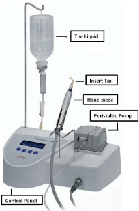
Parts of instrument (Figure 1):

Figure 1: Piezosurgery unit.
- Control panel: The piezosurgery unit is solely controlled by means of an interactive keyboard which has two basic.
- Programs: Bone and Root.
In Bone program, it is possible to adapt the power to any four levels depending upon the bone quality; while
In Root program, the power can be set to either Perio and/or, Endo mode.
With this system, any interference present in the unit, hand piece and/or, electronics, are recognized and highlighted on the display.
Surgical Tray
It contains all the components necessary for the various surgical procedures utilizing this unit.
- Dynamometric wrench: It is an added instrument used to tighten insert tips into the hand piece and applies a pre-defined force to obtain the energy transmission.
- Hand piece: Each piezosurgery unit comes with two hand pieces and is permanently connected to hand piece cord.
- The Peristaltic pump: It contains irrigant fluids which are discharged from the inserts with an adjustable flow of 0-60ml/min for the cooling and removal of the detritus from the cutting area. For cooling effect, the solution is refrigerated at 4 degree Celsius and the quantity of liquid might be adjusted using + and – buttons.
- The Liquid: It is drawn from the bottle which hangs from the rod provided and passes into the hand piece through a cord.
Insert tips: Various insert tips are available and can be classified as with:
- Titanium Nitride coated: They are particularly effective for osteoplasty procedures and/or, for harvesting of bone chips as they have maximum cutting efficiency, avoid corrosion and have increased working lives; and
- Diamond coated: They are used in case of thin bone osteotomy procedures and/or, in case of proximity to anatomic structures; histologically, are more traumatic than cutting inserts, but much safer. They are, further, classified as:
- Sharp Insert tips: For osteoplasty procedures and/or, for harvesting of bone chips as they have maximum cutting efficiency;
- Smooth Insert tips: For precise and controlled cutting on bone structures. They are used in osteotomy procedures when it is necessary to prepare difficult and delicate structures like those preparing for sinus window and/or, access to nerve; and
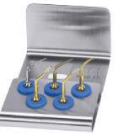
- Blunt Insert tips: For preparing soft tissues, e.g., elevating Schneider’s membrane and/or, lateralization of inferior alveolar nerve. In periodontics, these tips are used for root planning (Figure 2).
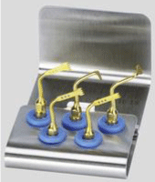
- Gold: Insert tips which are specifically used for surgical procedures related to bone. This gold color is obtained due to titanium nitride to improve the surface hardness for longer working life (Figure 3); and
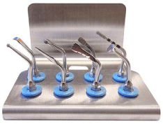
- Steel: Insert tips which are specifically used for soft tissue procedures and/or, delicate structures such as roots of teeth (Figure 4) [18,19].
- Orthodontic applications including corticotomies, exposure of impacted canines, etc.;
- Oral surgical procedures including TMJ ankylosis, nerve mobilization and/or, transposition, lateralization of inferior alveolar nerve, atraumatic tooth extractions, cyst enucleations;
- Periodontology procedures including root planning, removal of supra-and sub-gingival deposits and stains from teeth, periodontal pocket lavage, crown lengthening procedures, soft tissue debridement, resective surgeries and various regenerative surgeries for obtaining autogenous grafts from the donor sites;
- Implantology procedures for harvesting bone grafts, osteotomy procedures, distraction osteogenesis followed by implant placements, for retrieval of blade implants, in ridge expansion procedures, maxillary sinus elevations, drilling holes in bone for implant placements and for insertion of implants etc.
- Optimal vibration frequency for the mineralized tissues required during cutting with maintenance of relatively clean surgical field avoiding excessive temperatures;
- Allowing highly precise and safe cutting of hard tissues while sparing the adjacent soft tissue and nerve making it superior to conventional rotary instruments;
- Strength required to make cuts is far less compared to that with drills and/or, oscillating saws making surgical control with piezoelectric instruments maximum;
- Offering direct visibility during procedures minimizing risk to adjacent oral soft tissues;
- Being 3-times more powerful than the conventional ultrasonic units which makes it possible to cut highly mineralized tissues;
- Minimal intra-operative bleeding with rapid post-operative wound healing;
- Using piezosurgery tips, the harvested bone can be modified and shaped to fit accurately to the recipient sites;
- Cavitation effect created by interaction between the irrigant fluids and the oscillating tips makes surgical site clean during the osteotomy procedures; and
- Possibility to adapt the frequency of the vibrations of piezoelectric ceramics producing an efficacious and efficient cutting action on the bone of different qualities.
- Adequate dexterity and gentle touch is required for these type of procedures with a different learning curve;
- With increase in the working pressure above a certain limit, impedes the vibrations of inserts, transforming the energy into heat; thus, the most effective way to use piezoelectric instruments is to use them at higher speed and a lower pressure;
- Increase in operative time as compared to conventional cutting instruments;
- Difficulties encountered in deeper osteotomy sites due to lack of inserts of appropriate length and thickness;
- Inserts get worn away very rapidly and hence, are recommended not to go beyond ten little uses in bone surgeries as they have a probability to break and/or, cause damage to the tissues by uncontrollable heat; and
- Not as cost-effective as against the conventional scalpel blade surgical procedures.
- Rashad A, Kaiser A, Prochnow N, Schmitz I, Hoffmann E, Haurer P. Heat Production during different Ultrasonic and conventional osteotomy preparation for dental Implants. Clin Oral Implant Res. 2011; 22: 1361-1365.
- Chopra P, Chopra P. Piezosurgery and its applications in Periodontology and Implantology. Int J Contemp Dent. 2011; 2: 16-24.
- Pavlikova G, Foltan R, Horka M, Hanzelka T, Brounska H, Sedy J. Piezosurgery in oral and maxillofacial surgery. Int J Oral Maxillofac Surg. 2011; 40: 451-457.
- Labanca M, Azzola F, Vinci R, Rodella LF. Piezoelectric surgery: Twenty years of use. Br J Oral Maxillofac Surg. 2008; 46: 265-269.
- Georg E, Klein J, Bank J, Hassfeld S. Piezosurgery: An ultrasound device for cutting bone and its use and limitations in maxillofacial surgery. Br J Oral Maxillofac Surg. 2004; 42: 451-453.
- Schlee M. Piezosurgery: A precise and safe new oral surgery technique. Aust Dent Pract. 2009; 2009: 144-148.
- Nalbandian S. Piezosurgery techniques in implant dentistry. Aust Dent Pract. 2011; 116-126.
- Catuna MC. Sonic Surgery. Ann Dent. 1953; 12: 100-129.
- Richman MJ. The use of ultrasonics in root canal therapy and root resection. Med Dent J. 1957; 12: 12-18.
- Mazarow HB. Bone repair after experimentally produced defects. J Oral Surg. 1960; 18: 107-115.
- Mcfall TA, Yamane GM, Burnett GW. Comparison of cutting effect on bone of ultrasonic cutting device and rotary burs. J Oral Surg. 1961; 19: 200-209.
- Horton JE, Tarpley TM, Wood LD. The healing of surgical defects in alveolar bone produced with ultrasonic instrumentation, chisel and rotary bur. Oral Surg. 1975; 39: 536-546.
- Horton JE, Tarpley TM Jr, Jacoway JR, Chapel Hill NC. Clinical applications of ultrasonic instrumentation in the surgical removal of bone. Oral Surg Oral Med Oral Pathol. 1981; 51: 236-242.
- Torrella F, Pitarch J, Cabanes G, Aniuta E. Ultrasonic osteotomy for surgical approach of maxillary sinus: A technical note. Int J Oral Maxillofacial Implants. 1998; 13: 697-700.
- Vercellotti T. Piezoelectric surgery in implantology: A case report- A new piezoelectric ridge expansion technique. Int J Perio Rest Dent. 2000; 20: 358-365.
- Thomas J. piezoelectric ultrasonic bone surgery: Benefits for the interdisciplinary team and patients. Dent India. 2008; 2: 20-24.
- Leclercq P, Zenati C, Amr S, Dohan DM. Ultrasonic bone cut part 1: State-of-art technologies and common applications. J Oral Maxillofac Surg. 2008; 66: 177-182.
- Piezosurgery: The new dimension in bone surgery. Mectron medical technologies.
- Lands CA, Stubinger S, Laudemann K, Rieger J, Sader R. Bone harvesting at the anterior iliac crest using piezo-osteotomy versus conventional open harvesting: A pilot study. Oral Surg Oral Med Oral Pathol Oral Radio Endod. 2008; 105: 19-28.
- Chiriac G, Herten M, Schwarz F, Rothamel D, Becker J. Autogenous bone chips: Influence of a new piezoelectric device (Piezosurgery®) on chips morphology, cell viability and differentiation. J clin Periodontol. 2005; 32: 994-999.
- Vercellotti T, Nevins ML, Kim DV, Nevins M, Wada K, Schenk RK, et al. Osseous response following respective therapy with a piezosurgery. Int J Perio Rest Dent. 2005; 25: 543-549.
- Crosetti E, Battiston B, Succo G. Piezosurgery In head and neck oncological and reconstructive surgery: Personal experience on 127 cases. Acta Otorhinolaryngol Ital. 2009; 29: 1-9.
- Schlee M, Steigmann M, Bratu E, Garg AK. Piezosurgery: Basics and possibilities. Implant Dent. 2006; 15: 334-340.
- Walsh LJ. Piezosurgery: An increasing role in dental hard tissue surgery. Aust Dent Pract. 2007; 18: 52-56.

Figure 2: Perio Kit.
Based on Insert tip color:

Figure 3: Gold insert tips.

Figure 4: Steel insert tips.
Biological Effects on bone cutting by piezoelectric devices: The effect on cortical and cancellous bone and the surface roughness produced by different osteotomy techniques have a strong biological effect on the bone tissue and its eventual healing [9]. For piezoelectric osteotomy, the comparatively low amplitude of the cutting tool as opposed to that of an oscillating saw yields more precise cutting in the clinical practice.19 Blood free surgical site is another advantage as it gives better intra-operative view. Localised overheating, cavitation effects, flow of cooling fluid by use of the piezoelectric tools might serve as possible explanations for the absence of blood at the osteotomy site [20]. In a study, rate of post-operative wound healing in a dog model following ostectomy and osteoplasty was the marker used to compare the efficacy of piezoelectric instruments with commonly used carbide burs and/or, diamond burs. The surgical sites treated by carbide burs and/or, diamond burs lost bone in comparison to the baseline measurements by the 14th post-operative day while the surgical sites treated by piezoelectric instruments revealed a gain in the bone level. By 28th post-operative day, the surgical sites treated by all three instruments demonstrated an increase in the bone level and regeneration of cementum and periodontal ligament. However, by the end of 56th post-operative day, the surgical sites treated by carbide burs and/or, diamond burs evidenced a loss of bone versus a bone gain as compared to the ones treated by piezoelectric instruments. Thus, it appears that Piezoelectric bone surgery provided more favorable osseous repair and remodeling than carbide burs and/or, diamond burs when surgical procedures were performed. Therefore, Piezoelectric bone surgery could be regarded as being efficacious for use in the various osseous surgical procedures [21].
Applications of Piezosurgery in the Medical Field
Cranial Osteoplasties;
ENT Surgeries, Neurosurgeries, Pediatric Surgeries and numerous Orthopedics procedures;
Rhinoplasty procedures; and
Otologic Surgeries.
Applications of Piezosurgery Pertaining to Dentistry
Advantages [22-24]
Limitations [17]
Conclusion
Piezosurgery is a relatively new surgical technique which can be used in a variety of surgical procedures to complement conventional oral surgical procedures and in few cases, replace conventional procedures. It has, also, been found to have additional advantages in terms of bone healing, minimal intra-operative bleeding etc. apart from seeming to be more efficient in the first phases of bone healing. Besides its limitations, when used with variable frequency and power, it serves as a platform for a range of applications in the various fields of dentistry making it a highly effective tool in the clinical practice.
References