
Case Report
J Dent App. 2017; 4(2): 392-395.
Complete Mouth Rehabilitation of a Young Adult with Amelogenesis Imperfecta and Skeletal Class II Malocclusion
Kulkarni RS¹*, Pawar RS², Pimpale SK³ and Chandorikar HM4
¹Associate Professor, Department of Prosthodontics, Nair Hospital Dental College, Mumbai, India
²Assistant Professor, Department of Prosthodontics, Nair Hospital Dental College, Mumbai, India
³Assistant Professor, Department of Periodontics, Nair Hospital Dental College, Mumbai, India
4Assistant Professor, Department of Orthodontics, Nair Hospital Dental College, Mumbai, India
*Corresponding author: Rahul S Kulkarni, Flat no. 703, B wing, Satsang – 2 Apartments, Poonam Sagar Complex, Miraroad (East), Thane, Maharashtra, India
Received: November 13, 2017; Accepted: December 05, 2017; Published: December 12, 2017
Abstract
Complete mouth rehabilitation of a patient with esthetically and functionally compromised dentition often requires a systematic approach. In these patients, functional, esthetic, biological and restorative goals must be clearly defined before starting the treatment, and their incorporation must be made in design of prosthesis for long term success of treatment and preservation of the stomatognathic system. This case report describes treatment of a patient with hypoplastic amelogenesis imperfecta having severe attrition of tooth structure, multiple missing posterior teeth, and convex profile with underlying skeletal Class II pattern.
Keywords: Fixed partial denture; Occlusal splints; Overdentures; Amelogenesis imperfecta
Case Presentation
Case history and intraoral examination
A 21-year-old female earlier diagnosed with hypoplastictype amelogenesis imperfecta (AI) was referred to Department of Prosthodontics for restorative treatment. Patient complained of unesthetic appearance of teeth, difficulty during mastication, and teeth sensitivity. Patient had non-contributory medical history. Maxillary first and mandibular first and second molars on both sides were extracted due to caries three years ago, but prosthetic treatment was not taken by patient after extractions. Careful intraoral examination revealed that she had severe attrition of teeth with brownish yellow discoloration of enamel (Figure 1 and 2). There was extrusion of maxillary first molars due to non replacement of extracted mandibular molars. Periodontal examination indicated a healthy periodontium. Lateral cephalogram indicated that patient had a skeletal Class II jaw relationship, convex facial profile, deficient chin and clockwise rotation of mandible. During assessment of occlusal vertical dimension (OVD) and interocclusal distance, the interocclusal space measured at the premolar region during physiological rest was 3mm. Patient had no facial asymmetry. Esthetic evaluation revealed incompetent lips and undesirable display of gingiva during speech and smiling (Figure 3). There were no signs or symptoms related to temporomandibular joint, such as pain, limited range of jaw opening, or clicking. Family history revealed that patient was born to nonconsanguineous parents without history of AI, and had two male siblings out of which one was affected by the condition.
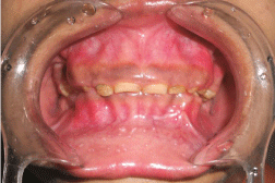
    Figure 1:  Preoperative frontal view in centric occlusion. Note severe attrition
of maxillary teeth and anterior deep bite.
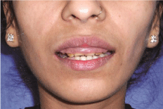
    Figure 2:  Preoperative extraoral view exhibits unaesthetic gingival display,
forwardly placed maxilla and incompetent lips.
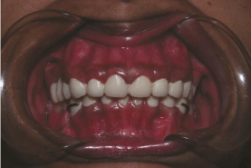
    Figure 3:  Crown lengthening surgery with osteotomy was performed to
reduce unesthetic gingival display and improve clinical crown height.
Cephalometric analysis
Considering the skeletal class II jaw relation and compensatory overgrowth of alveolar process due to attrition of teeth, lateral cephalometric radiograph was made. During radiography, the subject was in the natural head position (NHP), having teeth in maximum intercuspation and lips in repose. The film to source distance was 5 ft 2” and the distance between the film and patient’s mid-sagittal plane was 6” [1]. Tracings were done on 75μm lacquered polyester papers using a 0.3mm lead pencil, and linear and angular measurements were made to assess hard and soft tissue changes (Figure 4). Cephalometric readings at pre-treatment stage revealed severe skeletal Class II pattern [ANB 9°(normal range 2±2°)], severe mandibular retrognathism [SNB 69°(normal range 82±2°)], hyperdivergent jaw bases [mandibular plane angle 36°(normal range 28±3°)] with reduced chin prominence and profile convexity [facial angle 73°(normal range 89.5±2°)] [2]. These cephalometric findings, along with loss of vertical dimension due to attrition of teeth, added complexity to the un-aesthetic appearance.
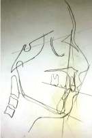
Figure 4: Preoperative cephalometric analysis.
Diagnosis and treatment planning
Diagnostic phase comprised of making radiographs, diagnostic impressions and casts, mounting on semi adjustable articulator, articulator programming, and developing the diagnostic wax-up. An interdisciplinary treatment approach was deemed necessary because of the complexity of this case, which included dental discoloration, attrition, multiple missing teeth, and excessive gingival display. Objective of the interdisciplinary treatment planning was to achieve the best restorative, esthetic, and functional outcomes. Consultation with other specialists such as orthodontist, periodontist and oral surgeon was carried out as a part of interdisciplinary treatment planning. Following treatment alternatives were presented to patient: A) Orthognathic surgical correction of skeletal class II relation, followed by prosthodontic treatment. B) Surgical repositioning of entire maxillary dentoalveolar segment to reduce unesthetic gingival display, followed by prosthodontic rehabilitation. C) Maxillary complete arch fixed orthodontic therapy, followed by crown lengthening surgery and prosthetic rehabilitation. D) Complete mouth rehabilitation using conventional fixed and removable dental prostheses. Patient declined options A and B due to apprehension for surgical procedure under general anesthesia, and option C due to long duration of treatment. Hence, complete mouth rehabilitation using conventional fixed and removable dental prostheses was planned, and informed written consent was obtained from the patient.
Fabrication of removable overlay splint
Overlay splint was fabricated by processing diagnostic wax up in acrylic resin (Figure 5). Modelling wax was added to the maxillary mock-up in form of palatal plate, and it was acrylized following standard laboratory technique. During packing, tooth colored resin was placed in areas of teeth, and clear resin in the palatal plate and flange areas. The splint obtained after processing was evaluated intraorally for adaptation, occlusion, retention and stability, and then delivered to the patient. Instructions were provided regarding maintenance and use of the splint. The patient was evaluated for 3 months to rule out presence of excessive signs of wear on the prosthesis, symptoms of temporomandibular disease, and muscle tenderness. The removable overlay splint was used to assess effect of restoring occlusal vertical dimension, esthetic evaluation, and as template during crown lengthening surgery.
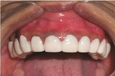
    Figure 5:  Removable overlay splint was used to assess effect of restoration
of occlusal vertical dimension, and as a template during crown lengthening
surgery.
Crown lengthening surgery
The reported case required deemed clinical crown exposure with regards to maxillary and mandibular anterior teeth. Clinical crown lengthening with osteotomy was the selected procedure to achieve proper aesthetics in the area (Figure 6) [3]. Removable overlay splint fabricated previously was used to estimate the amount of exposure desired in clinical crowns. Before the procedure, local anaesthesia was infiltrated in the surgical area. Splint was placed in desired position and markings with a sharp probe were done on the gingiva. After the markings were made, reverse bevel incisions were made with surgical BP blade from canine to canine in both the arches. Followed by reverse bevel, sulcular incisions were placed and the excised gingival mass was removed. After reflection, osteotomy of around 2 mm was done. Haemostasis was achieved and sutures were placed. Periodontal pack was placed for 7 days [4]. Post operative antibiotic and analgesics prescriptions were given. After 7 days, pack was removed and the area was irrigated with saline. On recall appointments, healing was satisfactory. By utilizing the apparently increased height of the teeth, improved resistance and retention forms were obtained in tooth preparations.
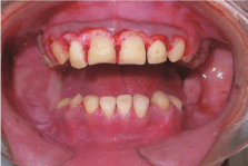
    Figure 6:  Crown lengthening surgery with osteotomy was performed to
reduce unesthetic gingival display and improve clinical crown height.
Tooth preparation, jaw relation and prosthetic procedures
Maxillary and mandibular tooth preparations were carried out for porcelain fused to metal (PFM) restorations following established clinical guidelines [5]. Minimal tooth reduction was performed on incisal and occlusal surfaces to avoid pulpal trauma and need for endodontic treatment. Gingival retraction was carried out in the maxillary and mandibular arches, impressions were recorded using polyvinyl siloxane impression material and working casts were obtained using die stone. The maxillary and the mandibular casts were mounted on semi adjustable articulator using average values of condylar and incisal guidance. The wax-up was developed for provisional restorations, which consisted of maxillary full arch and mandibular anterior heat polymerized poly (methyl methacrylate) fixed partial dentures (FPDs), and interim removable partial denture (RPD) for the mandibular posterior region (Figure 7). The provisional FPDs obtained after processing were evaluated intraorally for occlusion, margins, esthetics and then cemented. Interim RPD was subsequently delivered, and periodic recalls were scheduled for 3 months. The anterior guidance of the provisional restorations was recorded using a technique previously mentioned in the literature [6]. For fabrication of definitive prostheses, final tooth modification and gingival retraction were carried out in the maxillary and mandibular arches, final impressions were recorded, and working casts were obtained. Jaw relation procedures and articulator mounting were carried out. Individual ceramometal crowns were made for maxillary and mandibular anterior teeth, FPDs for maxillary posterior regions, and telescopic crown supported overdenture for mandibular posterior regions (Figure 8-10). The telescopic crowns were fabricated on mandibular first premolar on each side which was prepared to establish a single path of insertion. An occlusal convergence angle of 4 degrees was obtained in telescopic crowns by milling the surfaces with the aid of a milling system. The telescopic crowns were cemented to the premolars using resin cement under isolation. A new mandibular cast, obtained from a putty/wax impression was surveyed and the appropriate undercuts were blocked out. The cast was then duplicated with agar duplicating material and poured with investment material to obtain the refractory cast. Wax pattern of the overdenture framework was prepared and invested. Following casting, the metal framework was fitted to the master cast, and checked intraorally for passive fit, following which centric relation was recorded at predetermined vertical height. A try in of the waxed up denture was carried out, followed by processing and insertion of the telescopic overdenture. Recall evaluations were carried out at 3-month intervals for a period of 1 year.
    Figure 7:  Maxillary complete arch and mandibular anterior fixed provisional
restorations and mandibular posterior interim partial dentures.
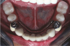
    Figure 8:  Mandibular ceramometal crowns and telescopic over dentures in
place.
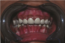
Figure 9: Frontal view of definitive restorations.
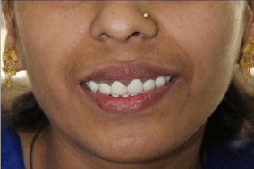
Figure 10: Post operative extraoral view.
Discussion
Complete mouth rehabilitation of a patient with esthetically and functionally compromised dentition often requires a systematic approach [3]. These patients commonly present with varying combinations of conditions such as loss of multiple teeth, pathologic migration and extrusion of teeth, gingival recession and overgrowth, diastema, inadequate pontic space, abrasion, attriton and unfavourable occlusal plane, to name a few. In these patients, functional, esthetic, biological and restorative goals must be clearly defined before the treatment, and their incorporation must be made in design of prosthesis for long term success of treatment and preservation of the stomatognathic system. Careful patient selection and systematic treatment planning are vital for successful result and patient satisfaction. This case report describes treatment for a young female patient with amelogenesis imperfecta, having severe attrition of tooth structure, multiple missing posterior teeth, and convex profile with underlying skeletal Class II pattern.
Lower facial height, facial profile, facial convexity angle, chin prominence, mandibular GoGn-SN angle have been assessed as important parameters in perception of facial esthetics as judged by researchers, clinicians and lay persons [7]. Studies have shown that balanced faces with straight profile and underlying skeletal Class I occlusion were found to be more esthetic in comparison to faces with increase in convexity (skeletal Class II) or increase in concavity (skeletal class III) [7]. Cephalometric analysis of the patient revealed skeletal mandibular retrognathism, prognathic maxilla, reduced chin prominence and clockwise rotation of mandible. Considering this, orthognathic surgical correction or surgical repositioning of alveolar process to reduce unesthetic gingival display was advisable prior to restorative treatment. However, patient was unwilling to undergo surgery. Orthodontic intrusion of extruded teeth was ruled out due to time constraints. Hence, conventional prosthodontic treatment in form of fixed and removable dental prostheses was delivered. Since there was un-esthetic gingival display, crown lengthening surgery (CLS) was performed [4]. Removable overlay splint was fabricated by acrylization of diagnostic mock up, and used as an appliance to evaluate response of patient to restoration of OVD, and during crown lengthening surgery. This was followed by fabrication of fixed provisional FPDs and interim partial denture, which was eventually, replaced by ceramometal FPDs and telescopic overdentures. Multiple papers have been published on management of AI, however, many of them failed to discuss a systematic and interdisciplinary approach towards the condition [8]. This case report uniquely describes cephalometric analysis, use of removable overlay splint, telescopic overdentures, and various treatment options during management of AI.
Patient was advised to follow periodic recall visits, use fluoride mouthwash, and brushing twice a day following proper technique to achieve long term favorable prognosis.
Conclusion
This case report describes a systematic approach used to accomplish complete-mouth rehabilitation of a patient affected by AI. A confluence of periodontal, orthodontic and prosthodontic disciplines during diagnosis and treatment planning, and meticulous prosthodontic management resulted in improved esthetics and enhanced masticatory efficiency. Satisfactory gingival health, esthetics, and function were observed at subsequent recall visits.
Acknowledgement
Authors are thankful to Dr. Rakesh K Kontham, Professor (Addl.), Department of Orthodontics, Nair Hospital Dental College, Mumbai, India for his positive inputs.
References
- Dahl BL, Carlsson GE, Ekfeldt A. Occlusal wear of teeth and restorative materials. A review of classi?cation, etiology, mechanisms of wear, and some aspects of restorative procedures. Acta Odontol Scand. 1993; 51: 299–311.
- Atit MB, Deshmukh SV, Rahalkar J, Subramanian V, Naik C, Darda M. Mean values of Steiner, Tweed, Ricketts and McNamara analysis in Maratha ethnic population: A cephalometric study. APOS Trends Orthod. 2013; 3: 137-151.
- Allen EP. Surgical crown lengthening for function and esthetics. Dent Clin North Am. 1993; 37: 163-179.
- Takei HH, Carranza FA, Do JH. The periodontal flap. In: Newman, Takei, Klokkevold, Carranza, editors. Carranza’s clinical periodontology, 12th ed., St Louis: Elsevier – Saunders. 2014: 586.e1-586.e2.
- Osensteil SF. Principles of tooth preparation. In: Rosensteil, Land, Fujimoto, editors. Contemporary fixed prosthodontics, 4th ed., St Louis: Mosby- Elsevier. 2006: 209-258.
- Pontons-Melo JC, Pizzatto E, Furuse AY, Mondelli J. A conservative approach for restoring anterior guidance: a case report. J Esthet Restor Dent. 2012; 24: 171-182.
- Orenstein NP, Bidra AS, Agar JR, Taylor TD, Uribe F, Little MD. Changes in lower facial height and facial esthetics with incremental increases in occlusal vertical dimension in dentate subjects. Int J Prosthodont. 2015: 28: 363-370.
- Dashash M, Yeung C, Jamous I, Blinkhorn A. Interventions for the restorative care of amelogenesis imperfecta in children and adolescents. Cochrane Database of Systematic Reviews. 2013; 6: CD007157.