
Research Article
J Dent App. 2021; 7(1): 451-455.
Two-Dimensional Intra-Oral Radiographs Compared to Three-Dimensional CBCT at Six-Month Post-Operative Evaluation of Secondary Bone-Grafting in Patients with Cleft Lip and Palate
Wiedel A-P1*, Svensson H2, Hellén-Halme K3, Ghaffari H4,5 and Becker M2
1Department of Oral and Maxillofacial Surgery, Skane University Hospital, Malmö, Sweden
2Department of Plastic and Reconstructive Surgery, Skane University Hospital and Department of Clinical Sciences in Malmö, Lund University, Malmö, Sweden
3Oral and Maxillofacial Radiology, Faculty of Odontology, Malmö University, Malmö, Sweden
4Department of Plastic and Reconstructive Surgery, Skane University Hospital Malmö, Sweden
5Department of Plastic Surgery, St George’s Hospital, London, Great Britain
*Corresponding author: Anna-Paulina Wiedel, Department of Oral and Maxillofacial Surgery, Jan Waldenströmsgata 18, Skane University Hospital, Malmö, 20502, Sweden
Received: April 30, 2021; Accepted: May 22, 2021; Published: May 29, 2021
Abstract
Background: The aim of this study was to investigate whether a complementary Cone-Beam Computed Tomography (CBCT) in patients with Cleft Lip and Palate (CLP) after alveolar bone-grafting to clefts gave substantial additional information, and particularly whether such new information had any implications for the further care of the patients.
Methods: Seventeen children, with complete CLP, 10 unilateral and seven bilateral clefts, in all 24 clefts, were evaluated six months after secondary alveolar bone-grafting with two-dimensional intra-oral radiographs complemented with CBCT. The mean age at bone-grafting was 8.8 years. Three different examiners evaluated the radiographic documentation.
Results: The mean pre-operative cleft width was 5.8mm. In 15 of the 24 clefts the same interpretation was made on both two-dimensional radiographs and CBCT. In the remaining nine clefts, CBCT added important information to the treatment decision.
Conclusions: For the evaluation six months post-operatively of the success of alveolar bone-grafting to clefts, the two-dimensional radiograph should be complemented with CBCT unless the two-dimensional radiograph without doubt reveals open residual cleft and clinical findings indicate graft failure.
Keywords: Alveolar cleft; Secondary bone-grafting; Orthodontic; Two- Dimensional radiographs; Three-dimensional radiographs; CBCT
Introduction
For alveolar crest repair in patients with clefts, Secondary Alveolar Bone-Grafting (SABG), which was first described by Boyne and Sands in 1972, has become the golden standard and is one of the mandatory surgical procedures [1,2]. Alveolar bone-grafting closes oro-nasal residual clefts, stabilizes the maxillary arch, provides bone support for cleft-adjacent teeth and facilitates orthodontic up righting of teeth [3].
To evaluate the result of alveolar bone-grafting radiographically, two-dimensional periapical or occlusal intra-oral radiographs are used widely [2,3]. One of the most common indexes for two-dimensional radiographs in assessing bone graft height is the Bergland index [2]. Lately the three-dimensional radiographs in the form of Cone-Beam Computed Tomography (CBCT) have become more widely used [4- 6]. Measurements using CBCT for evaluation of the results of alveolar cleft repair after alveolar bone-grafting have been suggested, namely the vertical height of the bone, the buccal-palatal thickness of the bone, and the nasal floor or nasal floor height difference [5,6]. Extensive bone resorption has been found in the buccal-palatal dimension of the alveolar portion of the transplant visible in CBCT analyses but is often underestimated in two-dimensional radiographs [7].
Two-dimensional intra-oral radiographs have constituted the clinical routine for many years at our cleft centre. However, we had a few patients in whom an evaluation also using CBCT was performed at the six-month post-operative evaluation. The reason for the extended investigation with CBCT was the uncertainty in the evaluation of the intra-oral radiographs according to the healing process. The aim of this study was therefore to investigate whether a complementary CBCT in these patients gave substantial additional information, and particularly whether such new information had any implications for the further care of the patients. A secondary aim was to evaluate other potential benefits of CBCT at this evaluation.
Material and Methods
Patients
Altogether, 24 clefts in 17 children, (14 boys, three girls) with complete CLP and a mean age of 8.8 years (range 7.9 -10.5 years) at secondary alveolar bone-grafting surgery were eligible for evaluation at the CLP care-centre, Skane University Hospital in Malmo, Sweden. Ten clefts were unilateral CLP, with nine left-sided and one right-sided. Seven clefts were bilateral CLP. Inclusion criteria was patients with CLP, bone grafted to the cleft area and with both twodimensional intra-oral radiograph and CBCT for evaluation of bone healing six months after bone grafting to the cleft area. The study was approved by the Regional Ethical Review Board, 017/593 in Sweden in accordance with the ethical standards of Helsinki Declaration. To obtain informed consent to participate, all patients and their parents were informed by post about the study. They were given the opportunity to get further information by telephone and to refrain from participation without any consequences for their future medical care. All patients and their parents chose to participate.
Surgical procedure
Secondary bone-grafting with muco-periosteal flaps was performed according to the technique described by Åbyholm [2]. Bilateral clefts were reconstructed in one procedure. The iliac crest was used as a donor site. All operations were performed by experienced surgeons (authors HS or MB or both).
Radiographic examination
A pre-operative two-dimensional intra-oral radiograph was taken the day before surgery to evaluate the cleft morphology regarding positions of teeth near the cleft area and the cleft width. The six- month post-operative two-dimensional intra-oral radiograph evaluated bone healing in the cleft area according to the Bergland index (Figure 1) [2]. The index group 1 had inter-alveolar bone at the amelocemental junction to the adjacent teeth; group 2 had a bone level of at least 75% of adjacent teeth; group 3 had a bone level less than 75% of the adjacent teeth; and group 4 had no bone-bridge over the cleft area. The complementary post-operative CBCT was used to measure the height of the bone graft (Figure 2), the thickness of the bone graft (Figure 3), and the nasal floor height difference (Figure 4). The height and thickness of the bone graft were measured at the thinnest point of the bone-bridge. In unilateral clefts, the nasal floor height was measured as the difference between the cleft side and the normal noncleft side. In bilateral cases, the nasal floor height was measured as the difference between the two sides. The two-dimensional radiographs and CBCTs were analyzed in the radiographic programmed, Sectra IDS7, by two of the authors (HG, KH-H), and one year later, the data was quality controlled by a third author (A-PW).
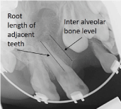
    Figure 1: Bergland index is based on inter-alveolar bone level divided by the
root length of adjacent teeth, times 100.
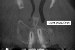
Figure 2: Height of bone graft.
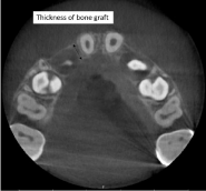
Figure 3: Thickness of bone graft.
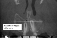
Figure 4: Nasal floor height difference.
Statistical analysis
Correlations coefficients were calculated for the interrelation between the cleft width and both the height of the bone graft and the thickness of the bone graft.
Results
Measurements
The results of the measurements are shown in (Table 1) and in (Figure 5). The pre-operative cleft width varied from 1.6 to 11 mm, with a mean of 5.8. When the six-month post-operative two-dimensional radiographs were taken, ten clefts had a Bergland index of 1; while for 11 clefts the index was 2; and for three clefts it was 3. The height of the bone graft evaluated at the six-month post-operative CBCT ranged from 0 to 15.4 mm with a mean value of 10.0. The thickness of the bone graft ranged from 0 to 13.8 mm, with a mean value of 6.7. In patients with unilateral CLP, two patients had no difference in nasal floor height whereas the nasal floor height was lower on the cleft side compared with the non-cleft side in eight patients. The difference ranged from 0.3 to 5.7 mm. Among the patients with bilateral CLP, two patients had equal nasal floor height whereas five patients had a difference ranging from 0.5 to 5.4 mm.
    Cleft    nr
    Cleft    width
    Bergland    index
    Height    of bone graft
    Thickness    of bone graft
    Nasal    floor height difference


    1 right
    2.8
    1
    11.7
    10.4
    1.5


    1 left
    4.2
    1
    10.2
    7.4


    2 right
    2.7
    1
    14.6
    8.8
    5.4


    2 left
    3.9
    2
    10.6
    9.8


    3
    2.3
    2
    12.9
    12
    0


    4
    8
    2
    9.1
    8
    5,7


    5
    7.9
    1
    13.6
    9.1
    3.3


    6
    9.1
    1
    12.9
    7
    0.3


    7
    5.4
    2
    8.3
    9
    1


    8
    4
    2
    8.4
    4.2
    0


    9 right
    6.5
    2
    12.7
    2
    3.2


    9 left
    7.5
    2
    10.8
    6.2


    10 right
    10.6
    3
    3.6
    1
    0


    10 left
    6.2
    2
    8.6
    3.6


    11 right
    2.7
    2
    13.2
    13.8
    0


    11 left
    11
    1
    11.6
    11


    12
    1.6
    1
    12.7
    5.8
    2.1


    13 right
    8.1
    1
    14.4
    2.9
    0.5


    13 left
    5
    2
    5.6
    8.4


    14
    8
    3
    4.8
    2.4
    5.4


    15
    6.3
    3
    0
    0
    1.5


    16 right
    2.4
    2
    15.4
    7.2
    1.3


    16 left
    6.6
    1
    2.8
    3.7


    17
    7.5
    1
    11.7
    8.4
    4.1
Table 1: The various finding in 17 patients. The cleft width (mm) was measured
in the pre-operative two-dimensional radiographs, and the Bergland index was
assessed in the post-operative two-dimensional radiographs. Other measures
(mm) refer to the post-operative CBCTs. In unilateral CLP, the non-cleft side
was used as the reference level for the nasal floor height difference, with lower
values for the grafted side in most instances. In bilateral CLP, the side with the
best vertical height was used as the reference level for the nasal floor height
difference, with lower values for the other side indicated in the table.
Interrelations between the pre-operative cleft width and the postoperative height of the bone graft had a correlation coefficient of -0.31 (SD: 4.0). The correlation coefficient for pre-operative cleft width and post-operative thickness of the bone graft was -0.38 (SD: 3.2) (Figure 5).
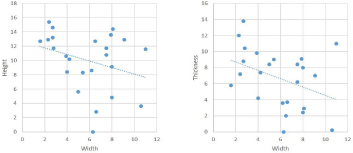
    Figure 5: Interrelations between pre-operative cleft width and post-operative
height of bone graft (left) and thickness of bone graft (right) with coefficients of
correlation of - 0.31 (SD: 4.0) and - 0.38 (SD: 3.2), respectively.
Discrepancies between two-dimensional intraoral radiographs and three-dimensional CBCT at six-month evaluation
In five clefts (cleft nr4, 7, 9 right, 13 right and left), evaluating whether or not the grafting was successful was difficult from the two-dimensional radiographs because adjacent teeth overlapped the former cleft area. This ambiguity also made it hard to decide whether further measures were indicated or not. However, in all five clefts, CBCT showed sufficient buccal or palatal bone with the overlapping teeth.
One cleft (cleft nr10 right) seemed to have a thin, almost complete cleft on two-dimensional radiographs and would be scheduled for complementary bone-grafting. However, on CBCT, a continuous bone bridge was found with buccal bone, although it was thin due to a palatal invagination. The decision was changed to no need for complementary bone-grafting and to await the eruption of adjacent teeth.
In one cleft (cleft nr14), the evaluation showed a bone level that was too low on two-dimensional radiographs, and the patient would have been sent for complementary grafting. However, in CBCT, more bone could be seen, and the decision was changed to expectance and to await the eruption of adjacent teeth.
In one cleft (cleft nr15), minor bone bridges could be seen in the two-dimensional radiographs covering the cleft area but not continuously supporting the entire height. We planned for expectance, but on CBCT, a complete cleft could clearly be seen, and the treatment decision was changed to make a complementary bonegrafting.
One cleft (cleft nr16 left) was considered successfully grafted on the two-dimensional radiographs, but on the CBCT, an inferior residual cleft was seen corresponding to the whole root height with buccal/palatal overlapping. Only a small and very superior bone bridge could be seen, and the treatment decision was changed to make a complementary bone-grafting.
To summarize these results, in 15 of the 24 clefts, the same interpretation was made on both two-dimensional radiographs and CBCT. In the remaining nine clefts, CBCT added important information regarding the treatment decision. In seven of the clefts, sufficient bone healing was noted. In two of them, CBCT pointed out the need for complementary bone-grafting.
Other findings on CBCT
CBCTs also gave information about the permanent canine position, for example, regarding impaction, root resorption, and the need for extraction of the primary canine. Such findings became important in the decision-making on future orthodontic treatment in eight of the clefts. In three clefts, buccal or palatal invagination gave important information for future orthodontic treatment considering the prospect of moving teeth across the former cleft area. In another three clefts, thin or missing bone could be seen at adjacent roots, which also influenced the post-operative orthodontic treatment. In one cleft, one adjacent tooth showed an unfortunate eruption angle, a distorted crown and a malformed root, and it was extracted without delay.
Discussion
Children with clefts are exposed to a three to five time higher cumulative radiation dose from dental radiography compared with children without clefts [8]. A major concern is based on the marked difference in the magnitude of radiation between an intra-oral radiograph and that of a CBCT investigation, even if the smallest volume, usually 4x4 cm, is used. Furthermore, the weighting factor table according to the International Commission on Radiological Protection for the different sensitivity of human tissue to ionisation radiation is defined for adults only. Roughly, the estimation is that one small three-dimensional CBCT equals about one hundred twodimensional radiographs when estimating an effective radiation dose [9]. According to ALARA (the As Low As Reasonably Achievable principle), a recommendation by the International Commission on Radiological Protection, one is obliged to use the radiographic investigation that gives the patients the lowest radiographic dose possible in order to answer the diagnostic question [10]. From the perspective of minimizing the total exposure of radiation in the general population, it seems justified to use CBCT only when it is meant to give information of significant clinical importance. Hence, CBCT is indicated when two-dimensional radiographs are perceived as giving insufficient information, and further treatment options are then based on this [11].
Before bone-grafting to the alveolar cleft, Wriedt et al. found no difference in orthodontic treatment planning when evaluating preoperative two-or three-dimensional radiographs [12]. This indicates that two-dimensional radiography is satisfactory for pre-operative documentation.
For the evaluation of the success of alveolar bone-grafting, postoperative CBCT has become more widely used. One reason is that the buccal-palatal dimension cannot be seen in two-dimensional radiographs, which is part of the reason CBCT is an interesting additional approach. The bone-support for cleft-adjacent teeth analyzed with two-dimensional radiographs might be both overestimated and underestimated by 20% in comparison with three-dimensional radiographs [4]. According to Feichtinger et al., bone resorption by 50% can be documented in the buccal-palatal dimension of the alveolar portion of the transplant 0 -1 year after the bone-grafting [7]. In three of the clefts in our series of patients, buccal or palatal invagination could be seen on CBCT, and this gave important information for orthodontic treatment when considering the possibility to move teeth across the former cleft area.
In this study, in five clefts (clefts nr4, 7, 9 right, 13 right and left) where no certain decision could be made about whether the grafting was successful or not because of teeth overlapping the bone bridge in the two-dimensional radiographs, CBCT radiographs revealed acceptable bone bridges buccal/palatal to the overlapping teeth and the future orthodontic treatment could be planned accordingly. Two clefts (cleft nr10 and 14) evaluated as not successful or doubtfully successful on two-dimensional radiographs proved to have enough bone support on CBCT, and in these cases, complementary bonegrafting could be avoided. In contrast, two clefts (cleft nr15 and 16) were perceived as successfully grafted on two-dimensional radiographs, but on CBCT, residual clefts could be seen. Buccalpalatal overlapping of bone margins was noted without continuous bone bridging of the total root height, and complementary bonegrafting was consequently performed. The risk would otherwise be that one might orthodontically move cleft-adjacent teeth into the open cleft area with loss of periodontal support.
Alveolar bone-grafting is advisable before eruption of the permanent canine. CBCT taken six-month post-operatively can be useful for determining the exact canine position after successful grafting when orthodontic leveling of the teeth may be of interest. Impacted canines seem to have a high prevalence in connection with clefts. A study by Enemark et al. reported a prevalence of 35% of canine impaction in connection with clefts compared with the general population, where the prevalence of impacted canines is around 1-2 % [3]. Oberoi et al. reported a prevalence of 12% impacted clefts near canines that required surgical exposure according to CBCT at one year after alveolar bone-grafting [13]. Furthermore, orthodontic assistance to reach occlusion is necessary in most cases.
Apart from the issue of impacted canines, CBCT can also provide other information of clinical relevance, for instance, regarding whether the primary canine with advantage should be extracted before awaiting the spontaneous eruption of the permanent canine. Also, other teeth near the cleft can be shown to have an abnormal route of eruption, thereby identifying treatment needs in order to prevent root resorption.
We found an overall negative correlation between pre-operative cleft width and the post-operative height of the bone graft as well as the thickness of the bone graft (Figure 5). Although variability is obvious and coefficients of correlation low, this finding indicates that it might be difficult to reach full graft dimensions if the cleft is wider. This reasoning is also strengthened by the observation that all but one patient (cleft nr13) with bilateral CLP had the higher values for height of the bone graft on the side with the narrowest cleft preoperation. Furthermore, the nasal floor height difference was 0 in two patients with unilateral CLP (cleft nr3 and 8), and both clefts were narrower than the mean cleft width. Nasal floor height difference is a relative measure of secondary importance, particularly in patients with bilateral CLP where a normal point of reference does not exist. From the orthodontic perspective, some degree of nasal floor height difference can be handled, if the height and thickness of the graft are within acceptable ranges. However, a notable nasal floor height difference may be important to consider when a later rhinoplasty is in prospect.
In this study, the results of alveolar bone-grafting were basically evaluated in CBCTs using the variables of the height of the bone graft, the thickness of the bone graft, and nasal floor height difference. These variables are those proposed by Soumalainen et al. [5]. However, alternative models for the interpretation of the CBCTs regarding the success of secondary bone-grafting exist. One such model is to give the variables of bone height, bone width and nasal floor categorical numbers, which thereafter are summarized to a score ranging from 1 to 10 [6]. This kind of score may seem attractive in concept, but is not always meaningful in clinical practice. In our experience, it is more useful to use the variables separately.
According to Feichtinger et al., there is initial bone loss, but there after the transplant remains almost constant in the following two years [7]. Two-dimensional radiographs might thus provide a sufficient image for the long-term follow-up of successfully grafted clefts where an adequate bone bridge has been confirmed six-month post-operatively with CBCT. For such for a long-term follow-up, another study found that conventional two-dimensional radiographs provide an adequate image for evaluating bone height with the Bergland index in the long term [14]. Although the Bergland index has its shortcomings in the six-month perspective, it is still a useful tool for categorizing findings in these long-term, two-dimensional radiographs.
Although we still use two-dimensional radiographs for the sixmonth post-operative evaluation of secondary bone-grafting, we now send patients more often for an extended radiographic evaluation with CBCT. The main reason is that there is a risk of both overestimating and underestimating the bone-grafting results with two-dimensional radiographs. However, if the two-dimensional radiograph shows an obvious residual cleft, supported by clinical findings of failure, then there is no reason to perform a CBCT. Assuming a success rate of about 85% [15, 16], this strategy will reduce the exposure to radiation by approximately 15% compared with using CBCT as a clinical routine for the post-operative evaluation of secondary bone-grafting in all cases. It should be stressed that any radiographic examination should always be indicated for each patient, and no radiographic investigation should be performed as a routine.
Conclusion
The study found that a two-dimensional intra-oral radiograph is the first step in determining the success of secondary alveolar bonegrafting six-months post-operatively, and is useful for comparison with two-dimensional radiographs taken before surgery.
However, for the evaluation of the success of alveolar bonegrafting six-months post-operatively when cases assessed with twodimensional intra-oral radiographs seem to show successful grafting, CBCT investigations were a useful tool since a three-dimensional image reveals possible overlapping of bone segments, indicating the need for supplementary grafting.
If the two-dimensional radiograph reveals without any doubt an open residual cleft, and clinical findings indicate graft failure, CBCT can be saved to reduce the exposure to radiation until evaluation after complementary alveolar bone-grafting is conducted.
After successful bone-grafting, CBCT can additionally provide useful information for future orthodontic treatment.
Acknowledgement
We are indebted to DDS Ingemar Swanholm, who was one of the initiators of this study.
The study was approved by the Regional Ethical Review Board, 2017/593 in Sweden in accordance with the ethical standards of the Helsinki Declaration.
References
- Boyne PJ, Sands NR. Secondary bone grafting of residual alveolar and palatal clefts. J Oral Surg. 1972; 30: 87-92.
- Bergland O, Semb G, Abyholm FE. Elimination of the residual alveolar cleft by secondary bone grafting and subsequent orthodontic treatment. Cleft Palate J. 1986; 23: 175-205.
- Enemark H, Krantz-Simonsen E, Schramm JE. Secondary bone grafting in unilateral cleft lip palate patients: indications and treatment procedure. Int J Oral Surg. 1985; 14: 2-10.
- Rosenstein SW, Long RE Jr, Dado DV, Vinson B, Alder ME. Comparison of 2-D calculations from periapical and occlusal radiographs versus 3-D calculations from CAT scans in determining bone support for cleft-adjacent teeth following early alveolar bone grafts. Cleft Palate Craniofac J. 1997; 34: 199-205.
- Suomalainen A, Åberg T, Rautio J, Hurmerinta K. Cone beam computed tomography in the assessment of alveolar bone grafting in children with unilateral cleft lip and palate. Eur J Orthod. 2014; 36: 603-11.
- Kamperos G, Theologie-Lygidakis N, Tsiklakis K, Iatrou I. A novel success scale for evaluating alveolar cleft repair using cone-beam computed tomography. J Craniomaxillofac Surg. 2020; 48: 391-398.
- Feichtinger M, Mossböck R, Kärcher H. Assessment of bone resorption after secondary alveolar bone grafting using three-dimensional computed tomography: a three-year study. Cleft Palate Craniofac J. 2007; 44: 142-148.
- Jacobs R, Pauwels R, Scarfe WC, De Cock C, Dula K, Willems G, et al. Pediatric cleft palate patients show a 3- to 5-fold increase in cumulative radiation exposure from dental radiology compared with an age- and gendermatched population: a retrospective cohort study. Clin Oral Investig. 2018; 22: 1783-1793.
- Pauwels R, Beinsberger J, Collaert B, Theodorakou C, Rogers J, Walker A, et al. SEDENTEXCT Project Consortium. Effective dose range for dental cone beam computed tomography scanners. Eur J Radiol. 2012; 81: 267-271.
- ICRP. The 2007 recommendations of the International Commission on Radiological Protection. ICRP publication 103. Ann ICRP 2007; 37.
- De Grauwe A, Ayaz I, Shujaat S, Dimitrov S, Gbadegbegnon L, Vande Vannet B, et al. CBCT in orthodontics: a systematic review on justification of CBCT in a pediatric population prior to orthodontic treatment. Eur J Orthod. 2019; 41: 381-389.
- Wriedt S, Al-Nawas B, Schmidtmann I, Eletr S, Wehrbein H, Moergel M, et al. Analyzing the teeth next to the alveolar cleft: Examination and treatment proposal prior to bone grafting based on three-dimensional versus twodimensional diagnosis -A diagnostic study. J Craniomaxillofac Surg. 2017; 45: 1272-1277.
- Oberoi S, Gill P, Chigurupati R, Hoffman WY, Hather DC, Vargervik K. Threedimensional assessment of the eruption path of the canine in individuals with bone-grafted alveolar clefts using cone beam computed tomography. Cleft Palate Craniofac J. 2010; 47: 507-512.
- Jabbari F, Wiklander L, Reiser E, Thor A, Hakelius M, Nowinski D. Secondary alveolar bone grafting in patients born with unilateral cleft lip and palate: A 20-year follow-up. Cleft Palate Craniofac. 2018; 55: 173-179.
- Jia YL, Fu MK, Ma L. Long-term outcome of secondary alveolar bone grafting in patients with various types of cleft. Br J Oral Maxillofac Surg. 2006; 44: 308-312.
- Wiedel AP, Svensson H, Schönmeyr B, Becker M. An analysis of complications in secondary bone grafting in patients with unilateral complete cleft lip and palate. J Plast Surg Hand Surg. 2016; 50: 63-67.