
Research Article
J Dent App. 2023 ; 9(1): 1118.
Cone-Bean Computed Tomography and Light Microscopic Examination of Human Extracted Molars - An in Vitro Study
Gyulbenkiyan E¹*, Borisov R², Vasileva R¹ and Gusiyska A¹
1Department of Conservative Dentistry, Faculty of Dental Medicine, Medical University - Sofia, Bulgaria
2Assistant Professor in Department of Imaging and Oral Diagnostic, Faculty of Dental Medicine, Medical University - Sofia, Bulgaria
*Corresponding author: Elvira Gyulbenkiyan Assistant Professor in Department of Conservative Dentistry, Faculty of Dental Medicine, Medical University - Sofia, Bulgaria
Received: December 05, 2022; Accepted: January 30, 2023; Published: February 06, 2023
Abstract
Introduction: For appropriate root canal therapy, understanding the anatomical and morphological aspects of the endodontic area is critical. Therefore, the study and analysis of images is an important step in the assessment and current clinical treatment of endodontic problems. Furthermore, microscopic-assisted endodontic therapy, particularly for multirooted teeth, is becoming more common in contemporary endodontic clinical practice. The aim of this in vitro study was to examine the distinct types of isthmuses using a light microscope and Cone-Beam Computed Tomography (CBCT).
Method: Fifty-five extracted human molars were used for this study. CBCT was used to realize the study. After the CBCT analysis, all teeth were inspected under a light microscope, and the results were compared with those of the CBCT examination.
Results: The results from the CBCT at the coronary third of the root canals were as follows: Types I, II, III, IV, and V isthmuses were found in 50.9%, 16.4%, 1.8%, 7.3%, and 23.6% of all samples, respectively. For the middle third, we found Types I, II, III, IV, and V isthmuses in 40.0%, 23.6%, 1.8%, 16.4%, and 18.2% of the samples, respectively. For the apical third, we found Types I, II, III, IV, and V isthmuses in 60.0%, 29.1%, 5.5%, 1.8%, and 3.6% of all samples, respectively. The results obtained from the microscopic examination were similar to those from the CBCT study. There is no confirmed statistically significant difference between the examination methods (p = 0.07).
Conclusion: Three-dimensional examinations have a significant role among the methods for diagnosing and detecting an isthmus in the complex root canal system, especially in the apical part.
Keywords: Cone-Beam Computed Tomography; Accuracy; Apical Zone; Isthmus
Introduction
Knowledge of the anatomical and morphological features of the endodontic space is essential for achieving precise root canal treatment. Therefore, an essential stage in diagnosing and managing endodontic problems is examining the imaging (whether two- or three-dimensional). In addition, microscopic-assisted endodontic treatment is increasingly required in routine endodontic treatment, especially of multirooted teeth [1,2].
The root canal isthmus is a narrow, ribbon-shaped corridor between two root canals containing pulp tissue [3,4]. The low success rate in endodontic treatment could be attributed to the limited accessibility of the hard-to-reach parts in the root canal, particularly with the presence of an isthmus, which can function as a reservoir for necrotic tissue and bacterial growth [5,6].
The most used imaging method in endodontics is the periapical radiograph due to its ability to image at high resolution with a low radiation dose [7,8]. The limitations of conventional radiography are well established. The diagnostic field of two-dimensional images is impaired by the fact that the three-dimensional anatomy of the area being radiographed is compressed onto a two-dimensional surface [9,10]. The geometric distortion and the anatomical superimposition also reduce the diagnostic value of the image [11,12].
Recently, Cone-Beam Computed Tomography (CBCT) has become the three-dimensional method of choice to diagnose tooth roots and the morphology of the canal, in particular, accessory root canals, lateral canals, and isthmuses. A CBCT examination ensures higher resolution than traditional Computed Tomography (CT) at lower radiation [13,14]. CBCT is a modification of the CT concept, involving the single rotation of an X-ray source around the dental subject. The data are analyzed and reconstructed using a CT-based algorithm to create a volume of data that can be viewed in three conventional planes (axial, sagittal, and coronal) [7].
The aim of this in vitro study was to examine the distinct types of isthmuses using light microscopic and CBCT examination.
Materials and Methods
Extracted mandibular and maxillary molars (n = 55) were used for this in vitro study. After extraction, all teeth were placed in a 10% formalin solution for 24 hours and then stored in saline with thymol at 4°C. All roots were examined with a Leica DM 500 light microscope (Leica Microsystem GmbH, Germany) at a magnification of ×10 for the presence of cracks and fractures. Such teeth were not included in the examination. For the light microscopic study, C-silicone putty (SPEEDEX, Coltène / Whaledent AG, Switzerland) was used to fix the teeth in a plastic tray after the initial examination. All teeth were positioned with the crown turned up to the silicone as the occlusal surface touched the stand bottom. A CBCT 3D scanner (Planmeca ProMax 3D) was used to complete the CBCT study. The following parameters were used: resolution 150 μm, exposure time 12.149 s, Dose Area Product (DAP) 819.6 mGy·cm2, electric current 10 mA, and voltage 90 kV. After the CBCT examination, all the teeth (n = 55) were cut transversely through the cervical part of the MB and M roots (maxillary and mandibular teeth, respectively) with a diamond burr under water cooling. All roots were also cut transversely in the middle and the apical part 3–4 mm from the apex. The samples obtained in this way – in the coronary, middle and apical areas – were examined with the Leica DM 500 microscope. Afterwards, the results were compared with those of the CBCT examination. Fisher’s exact test method was used for the statistical analysis. We confirm that we have read the Helsinki Declaration and have followed the guidelines in this investigation.
Results
CBCT Examination
The CBCT observation presented the following results at the coronary third of the root canals: Types I, II, III, IV, and V (Figure 1A) isthmuses in 50.9%, 16.4%, 1.8%, 7.3%, and 23.6% of all samples, respectively.
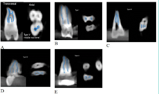
Figure 1: Axial and transverseslices of the scan made on different teeth representing all types of isthmuses: (A) Type V in the coronary part, (B) Type I in the middle part, (C) Type III in the middle part, (D) Type IV in the middle part and (E) Type II in the apical part of the root canal.
For the middle third of the root canals, the data are as follows: Types I (Figure 1B), II, III (Figure 1C), IV (Figure 1D), and V isthmuses in 40.0%, 23.6%, 1.8%, 16.4%, and 18.2% of the samples, respectively. The following data were found for the apical third of the root canals: Types I, II (Figure 1E), III, IV, and V isthmuses in 60.0%, 29.1%, 5.5%, 1.8%, and 3.6% of all samples, respectively. These results are presented in (Table 1). The data from the analyzed extracted teeth show that in the CBCT examination, the highest incidence of an isthmus in all parts of the root canal is in Type I (50.3%) and the lowest in Type III (3.0%). The percentage of both types of such isthmuses is most often found in the apical third of the root canal: 60% for Type I and 5.5% for Type III (Table 1). A statistically significant percentage difference between all types of isthmuses (p = 0.04) was found.
Light Microscopic Examination
The results obtained by the light microscopic examination for the coronary third of the root canals are represented in Table 1: 51.0% Type I isthmus, 16.4% Type II isthmus (Figure 2A), 1.7% Type III isthmus, 7.4% Type IV isthmus and 23.5% Type V isthmus.
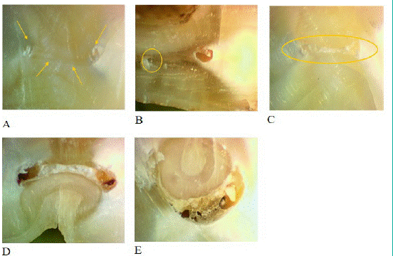
Figure 2: Light microscopic examination (magnification ×40) on transversal slices of teeth representing all types of isthmus: (A) Type II in the coronary part, (B) Type I in the middle part, (C) Type IV in the middle part, (D) Type V in the middle part and (E) Type III in the apical part of the root canal.
For the middle third of the root canals the data are 41.0% Type I isthmus (Figure 2B), 23.7% Type II isthmus, 1.8% Type III isthmus, 16.3% Type IV isthmus (Figure 2C) and 17.2% Type V isthmus (Figure 2D). The following data for the apical third of the root canals are: 59.0% Type I isthmus, 29.2% Type II isthmus, 5.5% Type III isthmus (Figure 2E), 2.8% Type IV isthmus and 3.5% Type V isthmus.
A statistically significant percentage difference between all results obtained by light microscopic examination (p = 0.004) was found.
Figure 2C is of a Type IV isthmus with extensions starting at the main root canals and narrowing to the isthmus area. Figure 2D shows a sample of two root canals with a real communication (a corridor) between them – a Type V isthmus according to Kim et al. [15] Figure 2E shows a Type III isthmus with a C-shaped configuration.
Comparison of the Methods of Imaging Examination
W hen comparing the CBCT and light microscopy results, no statistically significant difference was found between the methods (p = 0.07). The results are presented in detail as percentages for the coronary, middle, and apical parts of the root canals (Table 1). The results show that at all levels of the root canals, the most common isthmus is Type I, and the least common is Type III. A Type III (C-shaped) isthmus is most common in the apical part of the root canal and least common in the coronary one-third. There is a slight percentage difference between the methods of examination.
![]()
%
Type
of isthmusCoronal⅓
Medium ⅓
Apical ⅓
Total
N
CBCT %
LM %
N
CBCT %
LM %
N
CBCT %
LM %
N
CBCT %
LM %
I
28
50.9
51.0
22
40.0
41.0
33
60.0
59.0
83
50.3
50.3
II
9
16.4
16.4
13
23.6
23.7
16
29.1
29.2
38
23.0
23.0
III
1
1.8
1.7
1
1.8
1.8
3
5.5
5.5
5
3.0
3.0
IV
4
7.3
7.4
9
16.4
16.3
1
1.8
2.8
14
8.5
8.5
V
13
23.6
23.5
10
18.2
17.2
2
3.6
3.5
25
15.2
15.2
Total
55
100.0
100.0
55
100.0
100.0
55
100.0
100.0
165
100.0
100.0
N – number of teeth; % – relative share (%); Statistical analysis – Fisher's Exact Test; LM – light microscopic examination
Table 1: Relative part in percent of all types of isthmuses at the coronal, middle, and apical third of the root canal (CBCT and light microscopy observation).
Discussion
CBCT can be a valuable tool to present the morphology of the roots and their canals, especially in cases of doubt [2,16]. The presence of an isthmus in the mesial roots of the lower molars and the mesiobuccal roots of the upper molars was confirmed by CBCT at a resolution of 150 microns. Distinct types of isthmuses have been confirmed in the coronal, middle, and apical parts of the root canal [15]. The analysis of the isthmus in the root canals assists the clinician, allowing more precise endodontic treatment of the complex root canal space. In contemporary endodontic practice, the need to use a dental operating microscope to examine the root canal system is becoming essential [17]. A clinician needs both good equipment and knowledge about the anatomy of the endodontic space to ensure a good prognosis when proceeding with treatment. Digital two-dimensional X-ray diagnostics do not show three-dimensional endodontic anatomy in detail. Furthermore, reading radiographs depends on additional factors, such as the skill of the observer and the quality of the image. Continuous deposition of dentin by odontoblasts throughout the life of the tooth results in small areas that contain pulp tissue. In this way, different types of isthmuses are formed, from Type V to Type II (which are hard-to-reach areas for rotary instrumentation) [15].
This study confirms the high percentage of isthmuses in the mesial roots of the lower molars and the mesiobuccal roots of the upper molars, as the presence of a true type of isthmus (Type V – Figure 2D) is most common in the coronary and middle third of the root canal (23.6% and 18.2% in the CBCT examination and 23.5% and 17.2% in the light microscopic examination, respectively) (Table 1). It has been described in the literature that the incidence of an isthmus (without specifying the type of isthmus) increases on approach to the root apex [15], which differs from the data from our study. Our results indicate that in the apical direction, the root canals are, in most cases, joined into one (59–60%) (Table 1).
The rotary instrumentation is most difficult for a Type III isthmus due to its specific configuration (Figure 2E) [18,19]. Our results confirm that Type III isthmuses increase in frequency from the coronary to the apical direction (Table 1), as the percentages for the coronary, middle, and apical parts are 1.8%, 1.8%, and 5.5%, respectively for CBCT results and 1.7%, 1.8%, and 5.5%, respectively from the microscopic examination. In contrast, Types IV and V isthmuses have the lowest frequency in the apical part of the root canal (8.5% and 15.2%, respectively, in both imaging methods).
There is no statistically significant difference between the examination methods (p = 0.07) to prove the importance of using the CBCT for diagnostic purposes. In the last decade, the use of three-dimensional imaging with the help of CBCT has become increasingly established in contemporary endodontics. A significant disadvantage of conventional radiographs is the representation of three-dimensional data as two-dimensional images. Thus, anatomical structures and tissue changes can remain undiagnosed [7]. In this study, the resolution used in the CBCT study was 150 μm. The size of the CBCT resolution is essential because, at too low a resolution, not all isthmuses and details of the root canal system will be identified, and at too high a resolution, the study will not have sufficient informative value, and the radiation exposure of the patient will be higher. Because of technological developments in recent years, three-dimensional examinations now play a significant role among the methods for diagnosing and detecting isthmuses in complex root canal systems, especially in the apical parts [17,20].
In the limitation of this study, we performed the observation on extracted teeth (n=55) in the laboratory conditions where the influence on the imaging methods of the clinical environment is not taken into account, especially true for CBCT investigations. The quality of the final images is heavily influenced by the specific clinical situation (density of the adjacent tissue, the presence of artifact-generating structures, etc.). Therefore, we believe that subsequent clinical studies in this direction are recommended and necessary to clarify the adequacy of imaging methods in describing the anatomical and morphological features of the endodontic space.
Conclusion
Recent technological developments have led to three-dimensional examinations playing a significant role in diagnosing and detecting isthmuses in complex root canal systems, especially in the apical parts.Isthmuses are parts of the root canal that usually remain untouched by conventional endodontic instruments. These days clinicians rely on contemporary irrigation methods in clinical practice for cleaning and disinfecting these hard-to-reach areas.
Conflicts of Interest
The authors have no conflicts of interest to declare.
References
- Shaikhly B, Stephen K, Mikhail H, Robert AA, Poorya J. Comparison of a Dental Operating Microscope and High-resolution Videoscope for Endodontic Procedures. J Endod. 2020; 46: 688–693.
- Gambarini G, Galli M, Stefanelli LV, Nardo DD, Morese A, et al. Endodontic Microsurgery Using Dynamic Navigation System: A Case Report. J Endod. 2019; 45: 1397–1402.
- Weller RN, Niemczyk SP, Kim S. Incidence and position of the canal isthmus. Part 1. Mesiobuccal root of the maxillary first molar.J Endod. 1995; 21: 380–383.
- Liu Y, Yang W, Wang W, Zhu YN, Lin ZT, et al. Relationship between canal morphology and isthmus in mesiobuccal roots of maxillary first molars in 9- to 12-year-old children: An in-vivo cone-beam computed tomography analysis. Arch Oral Biol. 2020; 112: 104645.
- Nair PN, Henry S, Cano V, Vera J. Microbial status of apical root canal system of human mandibular first molars with primary apical periodontitis after “one-visit” endodontic treatment. Oral Surg Oral Med Oral Pathol Oral Radiol Endod. 2005; 99: 231–252.
- Kang S, Yu H-W, Shin Y, Karabucak B, Kim S, et al. Topographic Analysis of the Isthmus in Mesiobuccal and Mesial Roots of First Molars in a South Korean Population. Sci Rep. 2020; 10: 1247.
- Patel S, Brown J, Pimentel T, Kelly RD, Abella F, et al. Cone beam computed tomography in Endodontics – a review of the literature. Int Endod Jou. 2019; 52: 1138–1152.
- Mishra I, Karjodkar FR, Sansare K, Dora AC, Tambawala SS, et al. Diagnostic Value of Extraoral Periapical Radiograph in Comparison to Intraoral Periapical Radiograph: A Cross-sectional, Institutional Study. Contemp Clin Dent. 2018; 9: 406–409.
- Patel S, Dawood A, Whaites E, Ford TP. The potential applications of cone beam computed tomography in the management of endodontic problems. Int Endod J. 2007; 40: 818–830.
- Cohenca N, Simon JH, Roges R, Morag Y, Malfaz JM. Clinical indications for digital imaging in dento-alveolar trauma. Part 1: Traumatic injuries. Dent Traumatol. 2007; 23: 95–104.
- Yatabe M, Prieto JC, Styner M, Zhu H, Ruellas AC, et al. 3D superimposition of craniofacial imaging – The utility of multicentre collaborations. Orthod Craniofac Res. 2019; 22: 213–220.
- Shah N, Bansal N, Logani A. Recent advances in imaging technologies in dentistry. World J Radiol. 2014; 6: 794–807.
- Zhang R, Wang H, Tian YY, Yu X, Hu T, et al. Use of cone-beam computed tomography to evaluate root and canal morphology of mandibular molars in Chinese individuals. Int Endod J. 2011; 44: 990–999.
- Tu MG, Huang HL, Hsue SS, Hsu JT, Chen SY, et al. Detection of permanent three-rooted mandibular first molars by cone-beam computed tomography imaging in Taiwanese individuals. J Endod. 2009; 35: 503–507.
- Hsu YY, Kim S. The resected root surface. The issue of canal isthmus. Dent Clin North Am. 1997; 41: 529–540.
- Yovchev D, Mihaylova X, Gusiyska A, Traykova N, Yovcheva NNM. An extremely rare case of radiculous premolar coexisting with bilateral four-rooted mandibular second molars. Biomed Res. 2018; 29: 1567–1569.
- Filho C, Cerda R, Filho E, EDG, de Deus AD, Magalhaes KM. The influence of surgical operating microscope in locating the mesiolingual canal orifice: a laboratory analysis. Braz Oral Res. 2006; 20: 59–63.
- Sauáia, TS, Gomes BP, Pinheiro ET, Zaia AA, Ferraz CCR, et al. Thickness of dentine in mesial roots of mandibular molars with different lengths. Int Endod J. 2010; 43: 555–559.
- Yu DC, Tam A, Chen MH. The significance of locating and filling the canal isthmus in multiple root canal systems. A scanning electron microscopy study of the mesiobuccal root of maxillary first permanent molars. Y Can Dent Assoc. 1998; 29: 261–265.
- Borisov R. Radiological templates and CAD/CAM surgical guides. A literature review. J of IMAB. 2016; 22: 1285–1295.