Review Article
J Dent App. 2023; 9(1): 1120.
Recent Development of Laser Assisted Dentistry and Clinical Applications
Anshebo Getachew Alemu*
Department of Physics, College of Natural and Computational Science, Samara Unversity, Ethiopia
*Corresponding author: Anshebo Getachew Alemu Department of Physics, College of Natural and Computational Science, Samara Unversity, Ethiopia. Tel: +251-977-413118 E-mail: agetachew2013alemu@yahoo.com
Received: April 24, 2023 Accepted: May 29, 2023 Published: June 05, 2023
Abstract
Recent advancements in laser applications in clinical and dental medicine have the potential to play a critical role in patient comfort sectors, where laser technology is currently altering human existence. Laser drugs are used in dentistry and clinical therapy to counteract the negative effects of traditional methods, allowing for bloodless surgery with minimal postoperative discomfort and scarring. One of the key goals of laser dentistry is to provide the most comfortable care possible while minimizing risk. The advantages of laser therapy exceed the disadvantages, allowing for straight forward, modern, and safe clinical and dental procedures. This brief study’s main subjects present the current state of the laser in a variety of therapeutic and dental applications
Keywords: Dentistry; Laser; Clinical; Laser safety
Introduction
Due to its many benefits over other conventional current procedures, laser (light amplification by stimulated emission of radiation) applications in dentistry and clinical medicine are now regarded as patient-friendly techniques. Lasers can be employed in a wide range of dental and clinical operations since they are available in a variety of devices and wavelengths [1]. Over the past few years, lasers have increasingly been used in clinical medicine and dentistry [2]. Lasers are recommended for a wide range of treatments due to their various benefits [3-6]. Conventional cavity preparation techniques include low- and high-speed hand pieces, which can be noisy, unpleasant, and stressful for patients [7-10].
Laser Types
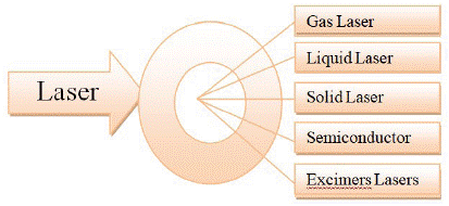
Lasers in dental and clinical applications are classified based on a variety of criteria, including the laser's active medium, which can be gas, liquid, solid, or semi-conductor, and the excimer laser beam that will be emitted (Figure 1).

Figure 1: Types of lasers based source material.
They are invariably classified according to the lasing medium used, as a gas laser or a solid laser. They can also be categorized based on tissue applicability in hard and soft tissue lasers, as well as wavelengths and the danger associated with laser application [11]. In addition, laser classification is based on different criteria such as power, source material (Figure 1), and wave length [12-14]. Another crucial factor in laser classification and treatment is power. Lasers are divided into three classes according to their power, as shown in the Figure 2: Low-power lasers (cold lasers): often less than 250mW cause light stimulation in tissues without having any heat consequences. This process is known as photobiostimulation. High-power lasers (warm or hard lasers) cause heat and improve the flow of energy in tissues to impart their therapeutic effects. The power of high-power lasers, which are usually more than 500mW, has applications in surgery. NdYAG (erbium-doped yttrium aluminum garnet) and Er:YAG (erbium-doped yttrium aluminum garnet) are classified as hard lasers and can be used on both hard and soft tissues, but they have limits due to their high cost and heat harm to the tooth pulp [12-14]. Furthermore, lasers are categorised according to their wavelength range, such as the ultraviolet range (300–400nm) and the visible light range (400–700nm).

Figure 2: Types of lasers based power source.
Additionally, lasers are divided into groups based on their wavelengths, such as the visible light range (400–700nm) and the ultraviolet range (300–400nm). Figure 3 depicts the NIR (near infrared) spectrum from 700 to 1200nm and the infrared (FIR) region of greater than 1200nm [2,12].

Figure 3: Laser classification based on wavelength range.
Gas laser: The first gas laser was the helium neon laser, which had a green wavelength and multiple infrared wavelengths. Common examples of gas lasers are carbon dioxide and argon. A particular sort of gas is produced when noble gases such as argon, krypton, and xenon are mixed with reactive gases [15-17].
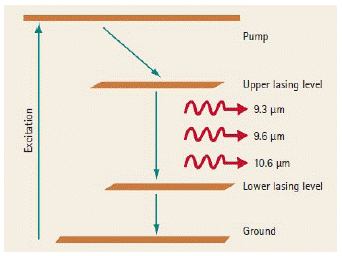
Although the carbon dioxide laser's bulkiness, high price, and destruction of hard tissue are its drawbacks, it is hydrophilic, has quick soft tissue removal, hemostasis with shallow depth penetration, and has maximal absorbency. Applications of the CO2 laser in soft tissue surgery are generally accepted. Despite the use of CO2 lasers for caries prevention, they have not been used in clinical settings as shown in Figure 4. Due to their advantageous optical properties, CO2 wavelengths of 9,300 and 9,600nm can be used to treat dental hard tissue. Technological advances in software and laser settings will aid in the development of new therapeutic applications and techniques.

Figure 4: CO2 laser [18].

Figure 5: Diode Laser [18].
The availability and demand for CO2 lasers as hard tissue lasers will both increase for researchers and medical professionals. Since water efficiently absorbs CO2 lasers, they are absorbed on the surface of soft tissue. The first centimetre of soft tissue is where visible lasers (445–660nm) are most effectively absorbed because pigmented chromophores like melanin and hemoglobin absorb them [15-19].
Liquid laser: A liquid dye is stimulated by liquid lasers to produce light radiation. Most of their "tunable" range is between 550 and 590nm. Their light can be seen. Dye lasers are frequently utilised for vascular reasons, including the treatment of rosacea, spider nevi, and angiomas in children and newborns [20,21]. Other disorders, like psoriasis, where it is beneficial against new lesions, are treated with this type of laser less commonly [22,23].
Solid-state lasers: The medium can be ceramic, glass (neodymium glass), or a crystal (ruby, sapphire, titanium, etc.). There are also laser diodes, such as the ones used in CDs, in a non- exhaustive list.
The erbium laser: The two wavelengths of the erbium laser are the Er:YAG laser and the Er:Cr:YYSGG (yttrium scandium gallium garnet) laser. Because of its high affinity for hydroxyapatite and high water absorption rate, it is the best option for treating both hard and soft tissues that contain a high proportion of water. The erbium laser is a highly flexible laser technology used in dermatology. Because of its ability to be absorbed almost entirely by water, it is an excellent tool for treating a wide range of cutaneous skin problems [24-26]. The Nd:YAG laser is highly absorbed by pigmented tissue and has excellent hemostasis, making it ideal for surgically cutting and coagulating soft tissue.
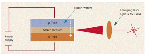
Diode lasers: The size of laser machines has been significantly decreased thanks to the development of micro-structure diode cells that can produce laser light. The range of spectral emissions is now constrained to a very small band (about 400–1,000nm) due to the limitations of the underlying physics. These lasers only use active media made of solid materials. The active medium is placed between silicon wafers in a diode laser (Figure 4). It is possible to selectively polish the ends of the crystal relative to internal refractive indices to form entirely and partially reflecting surfaces, simulating the optical resonators of larger lasers. This is possible because the active media, such as GaAlAs, is crystalline.
Photons are released from the active medium when current is discharged across the active media from one silicon wafer to the next. Current surgically relevant diode lasers use banks of individual diode "chips" connected in parallel to obtain the needed power capability because individual diode "chips" produce relatively low energy output [27-29].
Semiconductor lasers: The semiconductor laser has found widespread use in the medical professions as a result of its benefits, including its compact size, light weight, long lifespan, and great efficiency. The function mechanism of the semiconductor laser have unique applications in the departments of ophthalmology, surgery, cosmetology, and dentistry are examined [30]. In medical applications, semiconductor lasers have two major mechanisms: bio-stimulation and thermal action. The activation of compensation, nutrition, repair, and other immune defense mechanisms in response to the stimulation response is the mobilisation of compensation, nutrition, repair, and other immune defense systems to remove the pathogenic process. The thermal effect is the most important biological effect because it is present in all laser irradiation. The heat consequences of a high-power semiconductor laser are the major reason for its use. The local irradiation from a laser causes a heated, high-temperature increase in the macro performance of the tissue's molecular absorption of photon energy, vibration, and rotation. Because tissue cells include pigments like melanin, hemoglobin, carotenoids, and other substances that can boost light absorption, the heat effect of lasers is more significant [31-33].
Gas lasers
Liquid
Solid
Semiconductor
Excimers
- Argon
- Carbon- dioxide [15-19]
- Dye lasers [20-23]
- Er: YAG laser
- Er:Cr:YYSGG
- Nd:YAG laser [24-26]
- GaAs laser (infrared)
- GaAsP (visible) [34,35]
- Buffer gas (usually neon or helium),
- Halogen gas (fluorine,chlorine, or bromine),
- noble gas (argon,krypton,or xenon [36,37]
Table 1: Types of Laser.
Applications
Reference
Clinical application
Laser Application in Hard Tissues
Caries detection
[22-37]
Cavity Preparation
Carious Lesions Prevention
Etching
Fissure Sealant Therapy
Hard Tissue Cutting
Pulp Therapy
Laser Application in Soft Tissues
Ankyloglossia
Frenectomy
Gingival Remodeling and Gingivectomy
Lesions Removal and Biopsy
Herpetic Lesions
Dentistry
Cellulitis and Spasm treatment
Teeth anesthesia
[38-44]
Anterior dental Teeth Treatment
Temporomandibular Joint (TMJ) Treatment
Surgical Operations and Injuries
Biologic process
Biostimulation
[45-61]
Photodynamic therapy (PDT)
Laser-Assisted Microdissection
The Optical Tweezers
The Laser Ablation
The Utilisation in imagery
- Laser Coherence Tomography
- Photoacoustic imaging (PAI)
- Surface-enhanced Raman scattering (SERS)
Table 2: Applications of laser.
Advantages of laser
Disadvantages of laser
Less time spent the operator’s chair compared to
similar restorative treatmentCosts associated with purchasing and implementing
technology are rather significant.Engage with ailing tissues in a precise and selective manner
Laser operations require specialised knowledge and expertise in a range of clinical applications.
Dry operating room and improved visibility
A change in clinical technique is necessary because dental instruments are typically used for both side and end cutting.
Reduced requirement for antibiotics due to tissue surface sterilisation or decontaminating capabilities of the laser and bactericidal qualities on the tissue
Multiple lasers are required for different procedures since no one wavelength can effectively cure all dental problems.
Excellent homeostasis and a decreased need for sutures
Erbium family lasers are unable to remove faulty restorations made of metal and cast porcelain.
. Reduced oedema, scarring, and heat necrosis of surrounding tissue.
A rotary bur is faster at removing hard tissue than a laser.
Reduced use of local anaesthetics for treating soft tissue
Dangerous to skin as well as to eyes, hence the entire professional team needs to wear particular wavelength eye protection.
Reduced post-operative discomfort and accelerated wound healing with a quicker healing response.
There may be a danger of disease transmission through laser-generated aerosol in immune- compromised patients receiving soft tissue therapy
for viral lesions.Increased acceptance of patients
Little mechanical damage
Compared to electrosurgical tools, lasers induce less thermal necrosis of nearby tissues.
Removal of the high-speed drill and its related
noise, vibrations, smells, and terror
Contouring and removal of ossified tissue
Table 3: Advantages and disadvantages of laser [62-64].
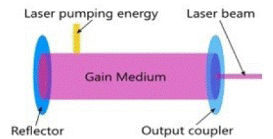
Excimers lasers: A mixture of noble gases and halogens is used as the gain medium in excimer lasers, which are strong ultraviolet lasers as shown in figure6. The power for excimer lasers comes from an electrical current source, like most gas lasers do. A tube that houses the laser medium is filled with three different types of gases, including buffer gas (usually neon or helium), halogen gas (fluorine, chlorine, or bromine), and noble gas (argon, krypton, or xenon). Excimer lasers' main benefit is their ability to generate a very narrow, accurate spot at a very short (UV) wavelength. Due to their ability to accurately obliterate material with minimal to no temperature buildup, excimer lasers are great for removing extra material through laser ablation. Compared to carbon dioxide lasers, which mainly rely on temperature accumulation to "boil off" material during ablation, this is the opposite [34,35].

Figure 6: Diagram of a basic laser.
Laser Applications
Recently Lasers are commonly utilized in clinical medicine and dentistry for cavity and root canal preparations, scaling and root planning, gingival and periodontal procedures, coagulation and haemostasis, biopsies, tongue lesions excision, TMJ issues, implant exposure, and pre-prosthetic surgery. This section of present summarizes the recent advanced application of lasers in the field of clinical, dentistry and biological applications also Low Level Lasers which are used in stimulatory and inhibitory biologic process.
Clinical Application
The laser medicine is considered as a favorable technique in various areas such as clinical applications, dentistry and biological process due to its several advantages for patients compared to other current methods. The major principle in the application of laser is the use of light energy instead of rotation forces and sharp blades [1,22]. Lasers medication can also be classify into hard lasers and soft laser (cold lasers). Hard lasers offer both hard tissue and soft tissue clinical applications, but they are costly and possess a potential for non quantified thermal injury.
Dentistry: There are several different kinds of low level lasers, including the red visible Helium Neon (He-Ne), the invisible infrared Gallium-As, the gallium-aluminum-as, and the indium- gallium-aluminum-phosphode (InGaAlP). Low power lasers affect the target tissues by photochemical and photobiologic processes. Low level lasers have stimulatory and inhibitory effects and have a power output of 50–500mw. Their use in paediatric dentistry includes anaesthesia, the treatment of cellulitis and muscular spasms, the alleviation of temporomandibular joint issues, the attenuation of the gag reflex, and the decrease of postoperative sequelae [38-44].
Biologic Application
The wavelengths of the lasers employed in biological applications are either in the infrared or the ultraviolet range, and they can operate in continuous or pulse mode. The employment of the laser beam for the cutting of biological tissue is suited by its high power density and precise placement. Chemical bridges already present in the tissues will be destroyed by the high photon concentration [45-61].
Advantages and Disadvantages of Laser Applications
As with any application, there are benefits and drawbacks to laser technology. The dangers of laser surgery are similar to those of any form of surgery. Inadequate treatment of the issue, pain, infection, bleeding, scarring, and changes in skin colour are among the risks associated with laser surgery.
Conclusion
One of the greatest inventions of the twenty-first century, laser technology has advanced quickly over the past few decades. Laser is the most prominent innovative technology applicable in various areas, such as clinical, biological, and dental medicine. Lasers can be used to repair and regenerate tissue in addition to being utilised to improve aesthetics, diagnose disease, and remove damaged tissue. The clinician should be familiar with the physical properties, laser wavelengths, and how they interact with biological tissues, in addition to having completed the training program and learning curve at a comfortable speed. Through cutting-edge technologies like laser microdissection and photoablation, which at various levels of expression enable knowledge of the physiological mechanisms in the progression of a disease, the use of lasers in medicine and biology has proven its interest.
Author Statements
Acknowledgement
I would like thank, Journal of Dental Applications for valuable invitation.
References
- Parkins F. Lasers in pediatric and adolescent dentistry. Dent Clin North AM. 2000; 44: 821-30.
- Goldman L, Hornby P, Meyer R, Goldman B. Impact of the Laser on Dental Caries. Nature. 1964; 203: 417.
- Frentzen M, Koort HJ. Lasers in dentistry: new possibilities with advancing laser technology?. Int Dent J. 1990; 40: 323-332.
- Aoki A, Ando Y, Watanabe H, Ishikawa I. In vitro studies on laser scaling of subgingival calculus with an erbium:YAG laser. J Periodontal. 1994 65: 1097-1106.
- Pelagalli J, Gimbel CB, Hansen RT, Swett A, Winn I. Investigational study of the use of Er: YAG laser versus dental drill for caries removal and cavity preparation--phase I. J Clin Laser Med Surg. 1997; 15: 109-115.
- Walsh LJ. The current status of laser applications in dentistry. Aust Dent J. 2003; 48: 146-155.
- Babu B, Uppada UK, Tarakji B, Hussain KA, Azzeghaibi SN, et al. Journal of Orofacial Sciences. 2015; 7: 49-53.
- Lins RDAU, Dantas EM, Lucena KCR, Catão MHCV, Granville-Garcia AF, et al. Efeitos bioestimulantes do laser de baixa potência no processo de reparo. Anais brasileiros de dermatologia. 2010; 85: 849-855.
- Gupta S, Kumar S. Lasers in Dentistry - An Overview. Trends Biomater Artif Organs. 2011; 25: 119-123.
- Dederich DN. Laser/Tissue Interaction: What Happens to Laser Light When it Strikes Tissue?. The Journal of the American Dental Association. 1993; 124: 57-61.
- Verma S, Maheshwari S, Singh R, Chaudhari P. Laser in dentistry: An innovative tool in modern dental practice. Natl J Maxillofac Surg. 2012; 3: 124-132.
- Mokmeli S. Lasers classification, Eslami Faresani R. Rezvan F. Iran, Boshra. 2004.
- Eslami Faresani R, Ashtiani Araghi B, Kamrava K, Rezvan F. Iran, Tehran, Boshra. 2006.
- Mester E, Mester AF, Mester A. The biomedical effects of laser application. Lasers Surg Med. 1985; 5: 31-9.
- Luk K, Zhao IS, Gutknecht N, Chu CH. J Dent Sci. 2019; 3: 1-9.
- Nasim H, Jamil Y. Diode lasers: from laboratory to industry. Opt Laser Technol. 2014; 56: 211-22.
- Fornaini C, Merigo E, Rocca JP, Lagori G, Raybaud H, Selleri S et al. 450 nm Blue Laser and Oral Surgery: preliminary ex vivo Study. J Contemp Dent Pract. 2016; 17: 795-800.
- Parker S. Br Dent J. 2007; 2: 202.
- Sarver DM, Yanosky M. Principles of cosmetic dentistry in orthodontics: Part 2. Soft tissue laser technology and cosmetic gingival contouring. Techno bytes. 2005; 127: 85-90.
- Polla LL, Tan OT, Garden JM, Parrish JA. Tunable pulsed dye laser for the treatment of benign cutaneous vascular ectasia. Dermatologica. 1987; 174: 11-7.
- Scheepers JH, Quaba AA. Treatment of nevi aranei with the pulsed tunable dye laser at 585 nm. J Pediatr Surg. 1995; 30: 101-4.
- Oram Y, Karincaoglu Y, Koyuncu E, Kaharaman F. Pulsed dye laser in the treatment of nail psoriasis. Dermatol Surg. 2010; 36: 377-81.
- Taibjee SM, Cheung ST, Laube S, Lanigan SW. Controlled study of excimer and pulsed dye lasers in the treatment of psoriasis. Br J Dermatol. 2005; 153: 960-6.
- Nisticò SP, Cannarozzo G, Campolmi P, Dragoni F, Moretti S, et al. Erbium Laser for Skin Surgery: A Single-Center Twenty-Five Years’ Experience. Medicines (Basel). 2021; 8: 74.
- Hovenic W, Golda N. Treatment of argyria using the quality-switched 1,064-nm neodymium-doped yttrium aluminum garnet laser: efficacy and persistence of results at 1-year follow-up. Dermatol Surg. 2012; 38: 2031-4.
- Maluki AH, Mohammad FH. Treatment of atrophic facial scars of acne vulgaris by Q-Switched Nd:YAG (Neodymium: Yttrium-Aluminum-Garnet) laser 1064 nm wavelength. J Cosmet Laser Ther. 2012; 14: 224-33.
- Angiero F, Buccianti A, Parma L, Crippa R. Human papilloma virus lesions of the oral cavity: healing and relapse after treatment with 810–980 nm diode laser. Lasers Med Sci. 2015; 30: 747-51.
- Matlach J, Kasper K, Kasper B, Klink T. Successful argon and diode laser photocoagulation treatment of an iris varix with recurrent hemorrhage. Eur J Ophthalmol. 2013; 23: 431-5.
- Golan S, Kurtz S. Diode laser cyclophotocoagulation for nanophthalmic chronic angle closure glaucoma. J Glaucoma. 2015; 24: 127-9.
- Li Y, Kang Z, Hu LJ, Laser J. 2010; 6: 73-5.
- Su H, Li S, Wang L. J Appl Laser. 2006; 2: 125-9.
- Su H, Li S. Information science edition. 2006; 5: 501-6.
- Yang X, Yang J. J Chin Med Equip. 2005; 6: 22-3.
- Hall RN, Fenner GE, Kingsley JD, Soltys TJ, Carlson RO. Coherent Light Emission From GaAs Junctions. Phys Rev Lett. 1962; 9: 366-8.
- N Jr Holonyak, SF Bevacqua. Appl Phys Lett. 1962; 1: 82-83.
- Beggs S, Short J, Rengifo-Pardo M, Ehrlich A. Applications of the Excimer Laser: a Review. Dermatol Surg. 2015; 41: 1201-11.
- Available from: https://www.globalspec.com/learnmore/optical_components_optics/lasers/excimer_lasers.
- Walsh LJ. The current status of low level laser therapy in dentistry. Part 1. Soft tissue applications. Aust Dent J. 1997; 42: 247-54.
- Pinheiro AL, Cavalcanti ET, Pinheiro TI, Alves MJ, Manzi CT. Low-level laser therapy in the management of disorders of the maxillofacial region. J Clin Laser Med Surg. 1997; 15: 181-3.
- Kotlow L. Lasers and soft tissue treatments for the pediatric dental patient. Alpha Omegan. 2008; 101: 140-51.
- Walsh LJ. The current status of low level laser therapy in dentistry. Part 2. Hard tissue applications. Aust Dent J. 1997; 42: 302-6.
- Brullmann D, Schulze RK, d’Hoedt B. The Treatment of Anterior Dental Trauma. Dtsch Ärztebl Int. 2011; 108: 565-70.
- Available from: https://www.hopkinsmedicine.org/health/treatment-tests-and-therapies/laser-surgery-overview.
- Pinheiro AL, Cavalcanti ET, Pinheiro TI, Alves MJ, Manzi CT. Low-level laser therapy in the J. Clin Laser Med Surg. 1997; 15: 181-3.
- Mester E, Mester AF, Mester A. The biomedical effects of laser application. Lasers Surg Med. 1985; 5: 31-9.
- Bihari I, Mester A. The biostimulative effect of low level laser therapy of long standing crural ulcers using HeNe laser, HeNe plus IR laser, and incoherent light: A preliminary report of a randomized double blind comparative study. Laser Ther. 1989; 1: 97-102.
- Colussi VC, Kinsella TJ, Nancy L. Oleinick Journal of Medical and Biological Research. 2000; 33: 869-80.
- Goyal M, Makkar S, Pasricha S. Low level laser therapy in dentistry. International Journal of Laser Dentistry. 2013; 3: 82-8.
- Bertheau P, Meignin V, Janin A. Microdissection on histologic and cytologic preparation: an approach to tissue heterogeneity. Ann Pathol. 1998; 18: 110-9.
- Simone NL, Bonner RF, Gillespie JW, Emmert-Buck MR, Liotta LA. Laser-capture microdissection: opening the microscopic frontier to molecular analysis. Trends Genet. 1998; 14: 272-6.
- Legres LG, Chamot C, Varna M, Janin A. The Laser Technology: New Trends in Biology and Medicine. J Mod Phys. 2014; 05: 267-79.
- Legres LG, Janin A, Masselon C, Bertheau P. Beyond laser microdissection technology: follow the yellow brick road for cancer research. Am J Cancer Res. 2014; 4: 1-28.
- Emmert-Buck MR, Bonner RF, Smith PD, Chuaqui RF, Zhuang Z, et al. Laser capture microdissection. Science. 1996; 274: 998-1001.
- Böhm M, Wieland I, Schütze K, Rübben H. Microbeam MOMeNT: non-contact laser microdissection of membrane-mounted native tissue. Am J Pathol. 1997; 151: 63-7.
- Kölble K. The LEICA microdissection system: design and applications. J Mol Med (Berl). 2000; 78: B24-5.
- Böhm M, Schmidt C, Wieland I, Leclerc N. Onkologie. 1999; 22: 296-30.
- Ashkin A. Optical trapping and manipulation of neutral particles using lasers. Proc Natl Acad Sci USA. 1997; 94: 4853-60.
- Lee MP, Padgett MJ. Optical tweezers: a light touch. J Microsc. 2012; 248: 219-22.
- Hawes C, Osterrieder A, Sparkes IA, Ketelaar T. Optical tweezers for the micromanipulation of plant cytoplasm and organelles. Curr Opin Plant Biol. 2010; 13: 731-5.
- Leitz G, Weber G, Seeger S, Greulich KO. The laser microbeam trap as an optical tool for living cells. Physiol Chem Phys Med NMR. 1994; 26: 69-88.
- Sweeney ST, Hidalgo A, de Belle JS, Keshishian H. Embryonic cell ablation in Drosophila using lasers. Cold Spring Harb Protoc. 2012; 2012: 691-3.
- Rajat B, Kartesh S, Simarpreet VS, Aditya M, Harmandeep K, Arshdeep K et al. Int J Dent Res. 2014; 2: 16-9.
- Donald JC. Fundamentals of dental lasers: science and instruments. Dent Clin N Am. 2004; 48: 751-70.
- Reshma JA, Arathy SL. J Dent Med Sci. 2014; 13: 59-64.