Abstract
While the popularity of implant retained Overdentures has been increased in recent years, root retained Overdentures have been largely disregarded. This clinical report presents the prosthetic treatment of a patient who had abnormally positioned upper canine in the long span anterior edentulous area in the partially edentulous maxillae using an Intraradicular attachment system and a removable partial overdenture and also emphasizes the advantages of root retained Overdentures.
Keywords: Intraradicular attachment; Overdenture; ZAAG; Canine root
Introduction
Although there may be several treatment choices for the management of a prosthetic patient, treatment options are somewhat limited in some unique cases. To overcome the prosthetic challenges, the most logical treatment planning should be selected because the success of prosthesis is primarily depends on a meticulous diagnosis and completion of an accurate treatment plan.
In the case of a long anterior edentulous space, a removable partial denture could lose its direct and indirect retention and support to a great extent. It would be subject to rotational displacement and tipping around the fulcrum. For tooth supported Overdentures preserving the roots in a long edentulous space is crucial for the success of the prosthetic treatment [1,2]. If the remaining roots are canines they become more valuable due to their strategic position [3]. It is widely accepted that the retention of the roots reduces alveolar bone resorption and maintain propriceptive mechanism [4-6]. The roots can be incorporated into the design of removable partial overdenture and further retention and stability can be achieved by the use of various precision attachment systems [7]. Different attachments such as stud attachments, Intraradicular attachments and magnets to anchor the prosthesis in order to increase the retention have been marketed by several manufacturers [4,8]. It is accepted that attachment-retained removable partial Overdentures may provide better aesthetics and improved function. Moreover, compared to the implant treatment, they are relatively more cost-effective treatment option and also a more conservative approach by preserving the patient’s own teeth and surrounding tissues [9]. There are various designs of precision attachment systems for the Overdentures including bars, ball and O-ring, studs and magnets in the market, promoted by several manufacturers. Basically, the selection of the most suitable system depends upon three factors: The number of the remaining natural teeth, the location of the remaining teeth and available space. In some cases, vertical space could be the principal consideration for the selection of an attachment [8,10,11]. In the case of decreased vertical space the use Intraradicular attachments should always be considered [2,9].
Currently, popularity of Overdentures has been increased in the literature with the use of implant-retained attachment systems; however, there are limited numbers of clinical reports about tooth-retained Overdentures. No specific report about the use of Intraradicular attachments in the case of long span anterior edentulous area and limited vertical space has yet been published. The aim of this case report was to describe the prosthetic treatment of a patient with an abnormally positioned maxillary canine tooth in the long span anterior edentulous area in the partially edentulous maxillae using a removable partial overdenture. Furthermore the importance of tooth-retained Intraradicular attachments which were recently ignored due to the increased popularity of implants was emphasized.
Case Presentation
A 57 years old female patient was referred to the Prosthetic Treatment Clinics, Suleyman Demirel University, Faculty of Dentistry, with the complaints of missing anterior teeth and poor aesthetic appearance. Furthermore, the patient stated that she had never been able to achieve an efficient chewing function in her life time. Medical and dental history and clinical examination revealed that the patient had no systemic problems and TMJ complaints.
Intra-oral examination revealed partial edentulism and she had the right canine, right first molar, left canine and left first molar in her maxillae and all teeth from the right first molar to the left first molar in her mandible (second and third molars were previously extracted). Her upper right canine was in markedly palatal position and in reverse articulation (Figure 1). However the tooth was healthy both clinically and radiographically. The upper right first molar had severe coronal damage requiring a post-core restoration and then a full coverage crown. The lower second premolar and first molar teeth had wear on their occlusal surfaces.
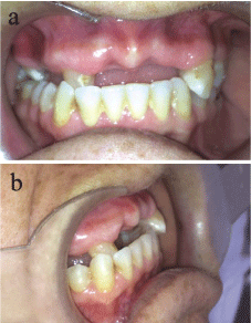
Figure 1: Pre-operative views of the patient. a) Frontal, b) Profile.
Following the clinical examination, preliminary impressions using an irreversible hydrocolloid (CA37; Cavex Holland BV, Haarlem, Netherlands) were made and the diagnostic casts were mounted in a semi-adjustable articulator (Stratos 300, Ivoclar Vivadent AG FL- 9494 Schaan Liechtenstein, Austria) with the aid of maxillary occlusal rim. Having surveyed the casts, diagnostic wax-up was completed on the mounted models and possible prosthetic treatments were evaluated. Examination of the articulated models revealed that the unfavorable position of the upper right canine and the limited vertical space would hinder achievement of proper occlusion and esthetics and therefore incorporation of the canine tooth into the prosthesis design would not be possible. In order to determine the available vertical space in the anterior region, the distance from the gingival margin of the maxillary right canine to the incisal level of the opposite anterior teeth was measured and found to be 2.2 mm. The minimal vertical space restricted the placement of any extra coronal attachment on the upper right canine tooth. The option of extraradicular attachment use was also eliminated due to the palatal cross-bite position of the upper canine; since the thickness of the attachment parts placed in the denture would exceed the thickness of the denture base. Treatment options including “repositioning of the canine with orthodontic treatment” or “extraction of the upper right canine and placement of the implants in the upper edentulous area” were then discussed with the patient. The patient declined those treatments because of the relatively long duration of the treatment phase, the higher costs and the surgical procedures. The restorative treatment plan included crowning the worn teeth, decoronating the maxillary right canine after root-canal treatment for an Intraradicular attachment with the aim of gaining extra retention and making a removable partial overdenture to restore function and esthetics was accepted by the patient. The use of the ZAAG® ST (Zest Anchor Advanced Generation Standard) Intraradicular attachment system (Zest Anchors Inc, CA, USA) which had low emergence profile was thought to be more appropriate for the patient (Figure 2). The attachment selection was made on the basis of available vertical space. Firstly, the maxillary right first molar was restored with post-core and a metal ceramic crown. The lower second premolar and first molar teeth were also restored with metal ceramic crowns.
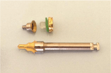
Figure 2: ZAAG ST Cap Male and drill.
Root-canal treatment of the upper right canine was completed using Protaper F2 (Dentsply Maillefer, Ballaigues, Switzerland) and then the canal was filled using gutta-percha and AH plus (Dentsply, Germany). The tooth was then decoronated and the coronal surface of the root was prepared as low as possible at gingival level. For the placement of the ZAAG® ST Female part into the root-canal, the female housing was prepared according to the instructions of the manufacturer. Great care was taken with preparation parallel to the path of insertion of the denture. The metal female was then cemented using resin cement (Multilink Automix; Ivoclar Vivadent, Liechtenstein). When setting of the cement completed, the root surface was contoured and polished. Following the impression procedures, the Co-Cr removable partial overdenture framework was manufactured. The framework included a hole prepared over the canine root surface. After try-in procedures the denture was processed and completed with a hole in the acrylic base. The denture was then adjusted in the mouth (Figure 3). For the placement of the ZAAG ST Denture Cap Male, an auto polymerizing acrylic resin (Meliodent, Heraeus Kulzer, Senden, Germany) was mixed and a small amount was placed in the previously prepared recess in the denture and around the top of the male cap. The denture was then inserted in the mouth in the appropriate position. Patient was guided to close her mouth carefully into occlusion maintaining a proper relationship with the lower arch. The denture was maintained in a passive condition without exerting any excess pressure to the soft tissues during the setting of the acrylic resin. After the polymerization of the acrylic resin, the denture was removed and excess acrylic was relieved over the root surface maintaining no contact between the root and denture base. Thereafter, excess acrylic around the male cap was removed and the denture base was carefully polished (Figure 4). The patient was instructed regarding the inserting insertion and removal of the denture in the path of insertion and also was instructed to achieve the snap into retention of the male part by using only the finger pressure without the aid of the opposing teeth. The denture was then delivered to the patient (Figure 5) and instructions on prosthesis care and oral hygiene maintenance were provided. The patient was given appointments for the routine controls (next day, after one week and then every six months). After one year in the routine control appointment the patient adapted to the prosthesis, reported that she was satisfied with the denture and its retention and had no complaints with regard to the chewing and appearance.
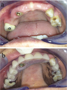
    Figure 3:  Intraoral views of the patient. a) After the placement of the ZAAG
ST female part on the upper right canine root. b) The upper denture before
the placement of the male part into the recess area (hole) prepared in the
denture base.
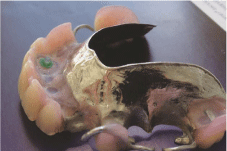
    Figure 4:  The view of the upper denture after the placement of the male part
of the ZAAG ST attachment system into the denture base.
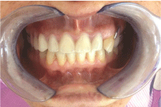
Figure 5: Post-operative intraoral view.
Discussion
In the case of a patient who had limited number of teeth with unfavorable distribution in the jaw to support a removable partial denture, making an attachment-retained overdenture may be an acceptable alternative treatment approach [12]. In some cases vertical space could be one of the principal considerations for the selection of an attachment and is measured from free gingival margin to the marginal ridge of the abutment. Attachment selection should be made after the careful analysis of the occlusal vertical dimension [5,11].Articulated diagnostic casts are important aids for better evaluation of the buccolingual and the vertical space available for the attachment as well as for the remaining teeth and the occlusion. Sufficient space must exist buccolingually and vertically for the selected attachments to be surrounded by a reasonable thickness of acrylic resin without weakening the denture base. In terms of space requirements Intraradicular attachments have a significant advantage that no additional metal casting is required, instead preformed receptacle is placed into the preparation made in the root using the manufacturer’s supplied sizing bur [2]. The other variables to consider when selecting prefabricated post overdenture attachment systems are the length of retained root, the quality and quantity of alveolar bone, the angulations of the root to occlusal plane, the root proximity to other roots, chewing pattern, occlusion and the musculature of the patient [2,5]. Canine teeth are accepted as very crucial abutments for all types of dentures. They are situated in a very strategic position in the dental arch and help keeping the bone level in the anterior region that is more prone to resorption. The high value of the anchorage provided by its bulky root make it an ideal support to sustain partial denture attachments [3]. Maxillary canines are dependable abutments for prosthetic treatments and accepted as the last teeth to be lost for the success of upper partial denture.
The retention of the roots to be used in partial overdenture design and construction is a well-accepted procedure. The retained root may allow placement of the anterior fulcrum line in a position that eliminated the need for tissue support. Removable partial Overdentures tend to rotate tissue-ward during function around the fulcrum line created between the abutment teeth closest to the edentulous area. Repositioning the fulcrum line in more anterior position and reducing of the need for tissue support can prevent these undesirable movements. A retained root in addition to preserving alveolar bone width and height may serve as an important element in the RPD design. Strategically retained roots can help to improve the position of the fulcrum line and to transport tooth-tissue supported denture into tooth supported denture [13]. In the present case when the denture was tried-in before the insertion of the male part, the rotation of the denture around the fulcrum line could be clearly observed (Figure 6). After the insertion of the attachment improved retention in denture and less dislodgement were apparent (Figure 7).
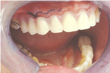
    Figure 6:  Intraoral view before the insertion of the male part of the ZAAG ST
attachment. Note the dislodgement of the denture around the fulcrum line.
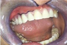
    Figure 7:  Intraoral postoperative view after the insertion of the attachment.
Note the improved retention and decreased dislodgement of the denture.
Zest Anchor Advanced Anchorage Generation (ZAAG) attachment is an Intraradicular attachment system similar to the Zest attachment. Its design has low height for Overdentures and partial dentures on natural teeth. In the present patient attachment selection was made considering the palatal inclination of the canine and the low vertical space. The ZAAG ST attachment offered the following advantages that palatally placed male part did not occupy more than 2 mm thickness in the acrylic, did not block the proper setting of the artificial teeth and did not have a positive intraoral female part which could disturb the tongue. Studies showed that the ZAAG system provided high retentive forces nevertheless the increased retention resulted in higher forces and moments applied on the abutments [14,15]. The most common prosthetic complication with regard to ZAAG system was reported to be the loss of retention over time with the wear of the male part [14,15]. Petropoulos et al. [16] compared various stud attachments and showed that Zest Anchor Advanced Generation Intraradicular attachment system was highly retentive and stable against dislodging forces. Epstein et al. [8] investigated the difference in retention forces among 6 prefabricated attachment systems both at initial placement and over a period of 2000 insertions and removals of the denture and the authors found out that ZAAG was one of the attachment systems showing the least rate of change.
Another alternative to the stud attachments (low profile attachments) are magnetic attachments that they are highly accepted systems as having small dimensions, low profile and favorable load distribution. The degree of stability with magnets can be described as retention without reciprocation and therefore they could be attachment of choice when abutment teeth offer limited support and unpromising prognosis. However, in clinical cases where displacement forces are located far away from the fulcrum, magnetic attachments may not administer satisfactory retention and stability. Stud attachments offer higher retentive and stabilizing forces comparing to magnetic attachments [17].
In the present case the most appropriate attachment for this patient was considered to be the ZAAG Intraradicular attachment system. By using patient’s own biological sources, the root of the upper canine and surrounding tissues, the advantages offered by root-supported Overdentures were gained. Furthermore, the patient received psychological and economical benefits by keeping the root in the oral cavity. The attachment system used in the present case was found to require less complex laboratory procedures and could be easily applied at chair-side. One-year follow up of the patient showed that ZAAG Intraradicular attachment was a reliable system. As a conclusion it can be said that, when indicated, the use of Intraradicular attachments should always be taken into consideration when planning an overdenture.
References
- Brewer AA, Morrow RM. Overdentures. 2nd Edn. St. Louis: CV Mosby Co. 1980.
- Preiskel HW. Overdenture made easy: a guide to implant and root supported prostheses. London: Quintessence Publishing. 1996.
- Auroy P, Lecerf J. Prosthetic restoration of the canine. J Dentofacial Anom Ortho. 2010; 13: 112-132.
- Crum RJ, Rooney GE Jr. Alveolar bone loss in overdentures: a 5-year study. J Prosthet Dent. 1978; 40: 610-623.
- Basker RM, Harrison A, Ralph JP, Watson CJ. Overdentures in general dental practice. 3rd Edn. London: British Dental Association. 1993.
- Epstein DD, Epstein PL. A clinical comparison of three overdenture anchors. Compendium. 1992; 13: 762, 764, 766.
- Langer Y, Langer A. Root-retained overdentures: Part I-Biomechanical and clinical aspects. J Prosthet Dent. 1991; 66: 784-789.
- Epstein DD, Epstein PL, Cohen BI, Pagnillo MK. Comparison of the retentive properties of six prefabricated post overdenture attachment systems. J Prosthet Dent. 1999; 82: 579-584.
- Schuch C, de Moraes AP, Sarkis-Onofre R, Pereira-Cenci T, Boscato N. An alternative method for the fabrication of a root-supported overdenture: a clinical report. J Prosthet Dent. 2013; 109: 1-4.
- Lorencki SF. Planning precision attachment restorations. J Prosthet Dent. 1969; 21: 506-508.
- Zamikoff II. Overdentures-theory and technique. J Am Dent Assoc. 1973; 86: 853-857.
- Jayasree K, Bharathi M, Nag VD, Vinod B. Precision attachment: retained overdenture. J Indian Prosthodont Soc. 2012; 12: 59-62.
- Ben-Ur Z, Gorfil C, Aviv I. Use of roots to establish favorable removable partial denture design: case reports. Quintessence Int. 1994; 25: 173-176.
- Petropoulos VC, Smith W. Maximum dislodging forces of implant overdenture stud attachments. Int J Oral Maxillofac Implants. 2002; 17: 526-535.
- Ibrahim AM. Evaluation of low-profile attachments for implant-retained mandibular overdentures in restoring cases with limited interarch space. Cairo Dental Journal. 2009; 25: 191-203.
- Petropoulos VC, Mante FK. Comparison of retention and strain energies of stud attachments for implant overdentures. J Prosthodont. 2011; 20: 286-293.
- Rutkunas V, Mizutani H. Retentive and stabilizing properties of stud and magnetic attachment retaining mandibular overdenture. Stomatologia. Baltic Dental and Maxillofacial Journal. 2004; 6: 85-90.
