
Case Report
J Dent & Oral Disord. 2024; 10(1): 1184.
Masticatory Muscle Tendon-Aponeurosis Hyperplasia After Orthognathic Surgery: A Case Report and Review of The Literature
Eiji Mitate*; Youta Yamauchi; Taichi Demura; Miho Hasumoto; Satoshi Wada; Hiroyuki Nakano; Noboru Demura
Department of Oral & Maxillofacial Surgery, Faculty of Medicine, Kanazawa Medical University Japan
*Corresponding author: Eiji Mitate, DDS, PhD Department of Oral & Maxillofacial Surgery, Faculty of Medicine, Kanazawa Medical University, 1-1 Daigaku, Uchinada, Kahoku, Ishikawa 920-0293, Japan. Email: mitateeiji@gmail.com
Received: Febryary 01, 2024 Accepted: April 03, 2024 Published: April 05, 2024
Abstract
Mylohyoid Muscular Tendon-Aponeurosis Hyperplasia (MMTAH) is a rare disorder causing progressive mouth opening difficulty due to unexplained overgrowth of tendons and aponeuroses. We report a case of MMTAH following orthognathic surgery for Temporomandibular Joint (TMJ) disorder. Despite initial improvement, mouth opening worsened over time, leading to masseter muscle dissection and myotomy with coronoid process resection.
MMTAH diagnosis is challenging due to similarities with other opening problems. While muscle relaxants during anesthesia may aid diagnosis, surgery remains the primary treatment. This case highlights the potential for MMTAH induction following orthognathic surgery and emphasizes the importance of early intervention and post-surgical mouth opening training. Treatment of masticatory fascia and mandibular angle during surgery might prevent MMTAH in susceptible individuals.
Keywords: Masticatory muscle tendon-aponeurosis hyperplasia (MMTAH); Orthognathic surgery; Trismus; Square Mandible; Muscle tubercle excision
Introduction
Mylohyoid Muscular Tendon-Aponeurosis Hyperplasia (MMTAH) is a rare disorder that causes difficulty in mouth opening due to the unexplained overgrowth of tendons and aponeuroses, such as those in the masseter and temporal muscles [1]. This disorder is typically observed in young individuals and is characterized by a slow, progressive mouth-opening disorder. Cases of MMTAH following orthognathic jaw surgery, although rare, have been reported. In this report, we describe a case of MMTAH that developed after orthognathic surgery. This report presents a case where mylohyoid muscle detachment and muscle tubercle excision procedures were performed in response to a condition affecting the Temporomandibular Joint (TMJ).
Case Presentation
The patient is a woman in her early forties. She first noticed clicking sounds and pain in the right Temporomandibular Joint (TMJ) during her late teens. Despite undergoing splint therapy at a nearby dental clinic, her condition did not improve. As the clicking sounds and pain in the TMJ persisted, and a further leftward deviation of the lower jaw was observed, she sought a referral to our plastic surgery department. Subsequently, she was referred to our department for preoperative orthodontic treatment. She has a history of left-sided degenerative knee joint disease, bilateral hallux valgus, asthma, and bipolar disorder.
At the first visit, facial asymmetry is noted (Figure 2A, 2B). The occlusion on the right side is Angle Class I, and on the left side is Angle Class III (Figure 3). The facial morphology is of the straight type, with overjet and overbite measuring +2mm each (Figure 2C, 2D). The amount of mouth opening was 33 mm, and during mouth opening, pain and clicking sounds were observed in the right TMJ. Orthodontic treatment was administered for the diagnosis of jaw deformity accompanied by facial asymmetry and right-sided temporomandibular disorder.
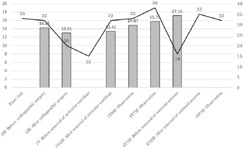
Figure 1: Amount of mouth opening and average transverse width of masseter muscle of both sides.
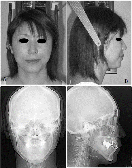
Figure 2: Facial appearance and cephalogram at first visit A. Frontal facial appearance. Facial asymmetry is noted. B. Lateral facial appearance, C. Cephalogram (frontal view) D. Cephalogram (lateral view)
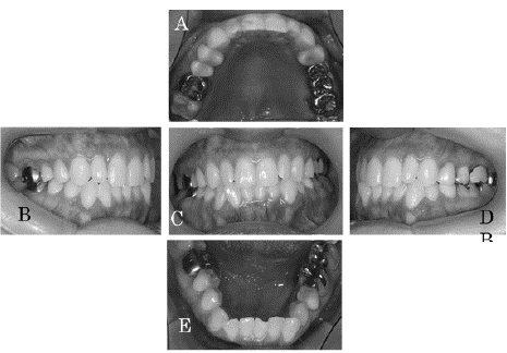
Figure 3: Intraoral photographs at initial examination The occlusion on the right side is Angle Class I, and on the left side is Angle Class III.
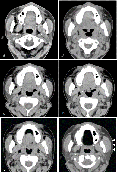
Figure 4: Horizontal sections of computed tomography A. 2 months after first visit (AFV): Before orthognathic surgery B. 2 months AFV: Just after orthognathic surgery. C. 1 year and 4 months AFV: After resection of the articular cartilage. D. 1 year and 6 months AFV: Follow-up observation E. 2 year and 7 months AFV: Follow-up observation F. 4 year and 1 months AFV: Before removal of coronoid process. Hypertrophy of the right masseter muscle is noted (arrowhead).
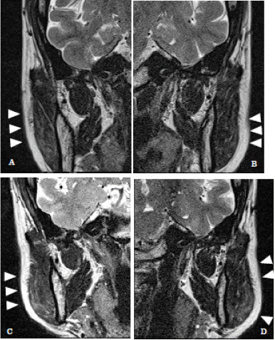
Figure 5: Magnetic resonance imaging (frontal section) Masseter muscles are noted (arrowhead) A. 1 year and 4 months after first visit (AFV): After resection of the right articular cartilage. B. 1 year and 4 months AFV: After resection of the articular cartilage (left side). C. 4 year and 1 months AFV: Before removal of coronoid process (right side). When compared with A, hypertrophy of the masseter muscle is noted. D. 4 year and 1 months AFV: Before removal of coronoid process (left side). When compared with B, hypertrophy of the masseter muscle is noted.
Two months after the initial consultation, Le Fort I osteotomy and sagittal split ramus osteotomy were performed. After the operation, the pain in the right TMJ disappeared, but there was pain in the left TMD. Also at that time, the patient was able to open her mouth by 20 mm.
Subsequently, the amount of mouth opening decreased gradually. Treatment with myomonitoring, pumping manipulation, and other therapies were performed for it, but there was no improvement. Therefore, one year after the first visit, an arthrodiscectomy on the left side and endoscopic dissection passivation on the right side were performed. However, the patient's aperture still did not improve, so a right-sided arthrodesis was performed one year and six months after the first visit. Postoperatively, the patient continued to undergo opening training, and the amount of mouth opening improved to 38 mm. At this point, the patient was given a terminal diagnosis and has not been seen for approximately three years.
The patient was seen again because of a recurrence of opening difficulties. At that time, the amount of mouth opening was 16 mm. As a comparison with the Computed Tomography (CT) images taken over time, the masseter muscle was found to be enlarged after the orthognathic surgery, and a diagnosis of MMTAH was made. Six years and two months after the initial diagnosis, a masseter muscle dissection and myotomy were performed under general anesthesia. Intraoperative findings included an opening of 27 mm after general anesthesia with muscle relaxants. After the masseter muscle was stripped away, the mouth opening was 32 mm, so it was determined that further opening was necessary. Therefore, the coronoid process was resected, and the amount of mouth opening was 43 mm. Early post-operative opening training was given and the amount of mouth opening at discharge was 35 mm.
Discussion
In patients with MMTAH, hard, cord-like fibers are palpable on the anterior margin of the masseter muscle. There is a slowly progressive trismus, but no limitation of mandibular forward or lateral movement. It is often seen in patients with so-called square mandible facial features [1].
The causes of MMTAH are not clear, although congenital factors, functional adaptation (hypertrophy of muscles due to movement), repair of masseter muscle damage, and parafunctional habits (clenching, grinding, mastication on one side) are considered [2].
A wide range of diseases that can cause opening problems include trauma, TMD, tumors, hyperplasia of muscle processes, and rheumatoid arthritis. MMTAH is a rare entity and therefore difficult to diagnose. Katagiri, et al reported a case of diagnosed as polymyositis from trismus and lower limb pain [3]. Elsayed N et al reported a case diagnosed as TMD [4]. In the present case, following orthognathic surgery, the patient was diagnosed with an articular disk disorder of TMD, and the articular disk was treated. However, the patient developed a progressive difficulty in opening her mouth. On the other hand, Morimoto et al reported that if the amount of opening is not increased by the administration of muscle relaxants during the induction of general anesthesia, this may be a useful diagnosis of MMTAH [5]. This finding is helpful, but it can only be checked during the induction of general anesthesia. In the present case, the administration of muscle relaxants did not improve the amount of mouth opening. Treatment involves surgical resection of the tendon and fascia of the muscles involved in closing the mouth, cononoidectomy or coronoidotomy, and resection of the mandibular angle [4,6]. Resection of the mandibular angle reduces the resistance of the masseter and medial pterygoid muscles attached to the inferior border of the mandible, making it easier to open the mouth. Patients with square mandibles have a mandibular angle that compresses the surrounding tissue when opening [7]. Nagao et al. reported that most mouth-opening occurred when the coronoid process was dissected or excised [1]. In our case, the amount of opening increased from 16 mm before resection to 35 mm after resection (Figure 1). Subsequent mouth-opening training is also important. Hayashi et al. reported that patients who underwent mouth opening training for at least 6 months after surgery were able to maintain the amount of mouth opening [8]. The mandibular angle protruded outwards from the initial examination, and we hypothesized that the MMTAH may have been induced by the change in occlusion due to orthognathic surgery and excessive labored hypertrophy caused by open mouth training after articular disk removal. It was thought that treatment of the masticatory fascia and mandibular angle during orthognathic surgery might prevent MMTAH if hyperplasia of the masticatory fascia is observed or if there is a tendency to square-mandibulism.
Author Statements
Acknowledgments
This study was approved by the Research Ethics Review Committee of Kanazawa Medical University Hospital (No. I802).
The findings of this study were presented at the 48th Chubu branch academic meeting of the Japanese Society of Oral and Maxillofacial Surgery (4 June 2023, Gifu).
Conflict of Interest Statement
The authors declare no conflicts of interest.
References
- Nagao F, Habu M, Kiyomiya H, Miyamoto I, Kokuryo S, Uehara M, et al. Effects of surgery and postoperative rehabilitation on masticatory muscle tendon-aponeurosis hyperplasia. J Jpn Soc. TMJ 2014; 26: 108-113.
- Aizawa H, Yamada S, Sakai H, Kondou E, Kurita H. Surgical Treatment for Masticatory Muscle Tendon-Aponeurosis and Coronoid Process Hyperplasia: A Case Report. The Shinshu Medical Journal 2021; 69: 253-259.
- Katagiri W, Saito D, Maruyama S, Ike M, Nisiyama H, Hayashi T, et al. Masticatory muscle tendon-aponeurosis hyperplasia that was initially misdiagnosed for polymyositis: a case report and review of the literature. Maxillofac Plast Reconstr Surg. 2023; 45: 18.
- Elsayed N, Shimo T, Harada F, Takeda S, Hiraki D, Abiko Y, et al. Masticatory muscle tendon-aponeurosis hyperplasia diagnosed as temporomandibular joint disorder: A case report and review of literature. Int J Surg Case Rep. 2021; 78: 120-125.
- Morimoto Y, Sugimura M, Hanamoto H, Niwa H. Limited mouth opening following induction of anesthesia in two patients with masticatory muscle tendon-aponeurosis hyperplasia. J Clin Anesth. 2011; 23: 598-600.
- Yoda T, Sato T, Abe T, Sakamoto I, Tomaru Y, Omura K, et al. Long-term results of surgical therapy for masticatory muscle tendon-aponeurosis hyperplasia accompanied by limited mouth opening. Int J Oral Maxillofac Surg. 2009; 38: 1143-7.
- Inoue N. Treatment for masticatory muscle tendon-aponeurosis hyperplasia. J Jpn Soc TMJ. 2009; 21: 46-50.
- Hayashi N, Sato T, Fukushima Y, Takano A, Sakamoto I, Yoda T. A two-year follow-up of surgical and non-surgical treatments in patients with masticatory muscle tendon-aponeurosis hyperplasia. Int J Oral Maxillofac Surg. 2018; 47: 199-204.