
Case Report
J Dent & Oral Disord. 2024; 10(1): 1185.
Successful Management of Severe Dental Injuries with Pre-existing Chronic Apical Periodontitis: A Case Report
Ieva Vaskelyte*; Greta Lodiene
Department of Dental & Oral Pathology, Faculty of Odontology, Academy of Medicine, Lithuanian University of Health Sciences, Lithuania
*Corresponding author: Ieva Vaskelyte Department of Dental & Oral Pathology, Faculty of Odontology, Academy of Medicine, Lithuanian University of Health Sciences, Eiveniu g.2, 50009 Kaunas, Lithuania. Email: ieva.vask@gmail.com
Received: Febryary 21, 2024 Accepted: March 21, 2024 Published: March 28, 2024
Abstract
Objective: Dental intrusion and lateral luxation are severe dentoalveolar injuries with potential complications, particularly when pre-existing chronic apical periodontitis is involved. Managing such traumatised teeth is challenging, impacting periodontal healing with unpredictable outcomes. Despite the lack of literature on similar case management, this case objective is to report successful treatment outcome of severely traumatised teeth with pre-existing apical periodontitis.
Methods: Following a fall, the patient presented with facial pain and mobility in her front teeth. Clinical and radiographic examinations revealed dental intrusion, lateral luxation with pre-existing chronic apical periodontitis. Initial treatment involved manual repositioning, stabilisation with a wire-composite splint, and subsequent endodontic interventions.
Results: Eighteen months later, clinical and radiographic examination revealed periapical healing in the traumatised teeth, indicating successful management of severe traumatic dental injuries with chronic apical periodontitis.
Conclusion: Despite the initial positive treatment results, future recall visits are necessary to assess the long-term prognosis of the traumatised teeth. Nevertheless, the current case highlights favourable treatment outcomes for severe dental injuries with chronic apical periodontitis. A well-structured treatment plan, close follow-up, and a collaborative approach are very important for achieving positive outcomes in severe dental injuries with pre-existing chronic apical periodontitis.
Keywords: Dental intrusion; Dentoalveolar injuries; Apical periodontitis; Lateral luxation; Traumatic dental injuries
Introduction
Dental intrusion is considered one of the most severe dentoalveolar injuries, causing maximum damage to the pulp and all supporting structures. The management of such traumatised teeth with apical periodontitis can strongly affect periodontal structures healing and could be a challenge with non-predictable outcomes [1]. Meanwhile, lateral luxation of permanent teeth is a common injury, where the degree of displacement of the tooth is a major factor affecting both the pulp [2,3] and periodontal ligament healing [3,4]. However, its prognosis is much better than that of other dental displacement traumas [5]. There is no literature data on the treatment and outcomes of traumatised teeth with pre-existing apical periodontitis. This case report describes the successful management and outcomes of severely traumatised teeth with apical periodontitis.
Case Presentation
A 27-year-old female attended the XXX Hospital, 6 hours after a fall, reporting facial pain and mobility in her front teeth of the upper jaw. The patient's medical history yielded no contributory findings. The clinical and radiographic examination revealed tooth #21 intrusion and chronic apical periodontitis, tooth #11 lateral luxation, and chronic apical periodontitis in tooth #12, no alveolar bone fractures were observed (Figure 1). The initial treatment involved manual repositioning of teeth #11 and #21 under local anaesthesia (Articaine 4% Ubistesin Forte 1.7ml N50 - 3M ESPE Dental AG; Seefeld, Germany), followed by stabilisation using a passive and flexible stainless-steel wire (ø 0,4 mm) and composite splint for four weeks (Figure 2). Postoperative instructions emphasised maintaining good oral hygiene and adhering to a soft diet.
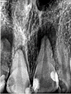
Figure 1: The initial intraoral radiograph.
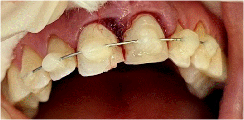
Figure 2: The clinical intraoral image after the splinting.
At the two-weeks follow-up visit, pulp sensitivity test with Endo-Ice (Coltene Group, Altstätten, Switzerland) in tooth #22 responded within normal limits, tooth #11 was slightly positive, while teeth #12 and #21 revealed negative responses. Tooth #21 displayed clinical pocket probing depth until the root apical third, and II° mobility by Miller [6] was recorded. Root canal treatment of teeth #12 and #21 was initiated: the crowns were disinfected with 0.5% Sodium Hypochlorite (NaOCl) before the procedure; the endodontic access cavities were prepared using a round diamond high-speed bur with water cooling under the ZEISS OPMI pico microscope (Carl Zeiss Meditec AG, Oberkochen, Germany); the teeth #12 and #21 canals were chemo-mechanically prepared with rotary Protaper Gold instruments (Dentsply Sirona, Ballaigues, Switzerland), and 0.5% NaOCl was used as an intracanal irrigant. The irrigation was enhanced by ultrasonic activation with size #30 Irrisafe tip (Satelec, Acteon, Merignac, France) for 60s followed by short term intracanal medication with Calcium hydroxide (Ultracal, South Jordan, USA).
Four weeks later, the patient had no complaints, and the wire-composite splint was removed. The pulp sensitivity test yielded consistent results, but II° mobility persisted in tooth #21. Under rubber dam isolation, the canals of teeth #12 and #21 were obturated with Biodentine (Septodont, Saint-Maur-des-Fossés, France).
At the three-month follow-up visit, following a clinical and radiographic examination, pulp necrosis was diagnosed in tooth #11 (Figure 3). Endodontic treatment, following short term calcium hydroxide medication, was initiated. Two weeks later, the canal was obturated with Biodentine, and the patient was referred for crown restorations.
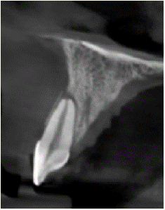
Figure 3: CBCT of tooth #11.
After six months, the patient had no complaints and was satisfied with the aesthetic appearance of her upper front teeth. The clinical examination revealed negative results for percussion and palpation tests, and no mobility was observed. Probing depths measured up to 4 mm on all front teeth, but a periapical radiograph indicated a slight periapical radiolucency around teeth #21 and #11 (Figure 4).
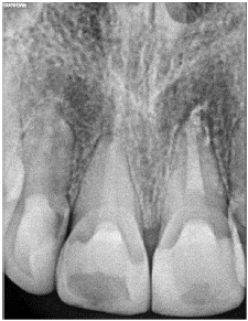
Figure 4: The periapical radiograph after six months.
Eighteen months later, after clinical and radiographic examination, periapical healing was observed in teeth #21 and #11, indicating successful management of the severe traumatic dental injuries with chronic apical periodontitis (Figure 5).
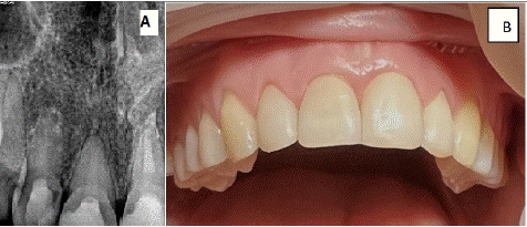
Figure 5: A) The periapical radiograph after eighteen months; B) The clinical image after eighteen months.
Discussion
In this case report, the patient had complex trauma involving dentoalveolar structures caused by a fall accident with leading chronic apical pathology simultaneously.
It has been demonstrated that the stage of root development and the type of luxation are the most critical factors affecting pulp healing in teeth with luxation injury [7]. In this case, the roots were mature, and the initial sensitivity response of tooth #11 was positive. However, three months later, the test became negative and chronic apical periodontitis was diagnosed. The root canal treatment was initiated. This could be explained by the severity of the luxation injury, with lateral displacement of the root apical part within the socket, leading to pulp necrosis and periapical pathology.
In chronic apical periodontitis pathogenesis macrophages are predominant inflammatory cells [8]. Their fusion results in the formation of osteoclasts, specialised for bone resorption [9]. This process can significantly impede the healing of traumatised teeth. Some studies have also revealed that macrophages play a role in promoting tissue repair [10,11].
In dental trauma cases, imaging examinations are essential for diagnosis, planning, executing treatment, and monitoring patients afterward [12]. However, periapical radiography is subject to limitations due to 2-dimensional images, and Cone-Beam Computed Tomographic (CBCT) imaging reduces these problems [13]. The IADT states that CBCT imaging provides better visualisation of dental trauma injuries, particularly in cases of lateral displacements [12]. Also, CBCT imaging has greater sensitivity for diagnosing apical periodontitis [13]. In this regard, three-months after the dental injury, CBCT was performed to refine the diagnosis and precisely plan the treatment of tooth #11.
During root canal obturation with Biodentine, the material extruded beyond the root apexes of teeth #12 and #21. It is known that materials should be packed cautiously to prevent extrusion [14]; however, due to wide root canals, this can occur. According to some studies [15,16], extruded Biodentine should not affect the outcome of endodontic treatment. Similarly, in the present case extruded Biodentine did not impact periapical healing.
Despite the initial positive treatment results, future recall visits are necessary to assess the long-term prognosis of the traumatised teeth. Nevertheless, the current case highlights favourable treatment outcomes for severe dental injuries and chronic apical periodontitis.
Conclusion
The well-structured treatment plan, close follow-up, and a collaborative approach are very important for achieving positive outcomes in severe dental injuries with pre-existing chronic apical periodontitis.
Highlights
• There is no literature data on the successful treatment and outcomes of traumatised teeth with pre-existing apical periodontitis.
• The current case highlights favourable treatment outcomes for severe dental injuries and pre – existing chronic apical periodontitis.
In the present case extruded Biodentine did not impact periapical healing.
References
- Sermet Elbay Ü, Elbay M, Kaya E, Sinanoglu A. Management of an intruded tooth and adjacent tooth showing external resorption as a late complication of dental injury: three-year follow-up. Case Rep Dent. 2015; 2015: 741687.
- Andreasen FM, Zhijie Y, Thomsen BL. Relationship between pulp dimensions and development of pulp necrosis after luxation injuries in the permanent dentition. Endod Dent Traumatol. 1986; 2: 90-8.
- Andreasen JO, Vinding TR, Chtistensen SSA, Predictors for healing complications in the permanent dentition after dental trauma. Endodontic Topics. 2006; 14: 20-27.
- Andreasen FM, Pedersen BV. Prognosis of luxated permanent teeth--the development of pulp necrosis. Endod Dent Traumatol. 1985; 1: 207-20.
- Costa LA, Ribeiro CCC, Cantanhede LM, Santiago Junior JF, de Mendonça MR, Pereira ALP. Treatments for intrusive luxation in permanent teeth: a systematic review and meta-analysis. Int J Oral Maxillofac Surg. 2017; 46: 214–29.
- Miller PD Jr. A classification of marginal tissue recession. Int J Perio Rest Dent. 1985; 5: 9-13.
- Kamali Sabeti A, Golshani S, Vahedi P. Management of a Multiple Dentoalveolar Trauma in Permanent Teeth: A Case Report with an 18-Month Follow-up. J Kermanshah Univ Med Sci. 2022; 26: e129134.
- Azeredo SV, Brasil SC, Antunes H, Marques FV, Pires FR, Armada L. Distribution of macrophages and plasma cells in apical periodontitis and their relationship with clinical and image data. Journal of Clinical and Experimental Dentistry. 2023; 9: e1060–e1065.
- Xu Z, Tong Z, Neelakantan P, Cai Y, Wei X. Enterococcus faecalis immunoregulates osteoclastogenesis of macrophages. Experimental Cell Research. 2018; 362: 152–158.
- Braga TT, Agudelo JS, Camara NO. Macrophages during the fibrotic process: M2 as friend and foe. Frontiers in Immunology. 2015; 6: 602.
- Kurowska-Stolarska M, Stolarski B, Kewin P, Murphy G, Corrigan CJ, Ying S, et al. IL-33 amplifies the polarization of alternatively activated macrophages that contribute to airway inflammation. Journal of Immunology. 2009; 183: 6469–6477.
- Bourguignon C, Cohenca N, Lauridsen E, Flores MT, O’Connell AC, et al. International Association of Dental Traumatology guidelines for the management of traumatic dental injuries: 1. Fractures and luxations. Dent Traumatol. 2020; 36: 314-330.
- Venskutonis T, Plotino G, Juodzbalys G, Mickeviciene L. The importance of cone-beam computed tomography in the management of endodontic problems: a review of the literature. J Endod. 2014; 40: 1895-1901.
- Kanagasingam S, Lim CX, Yong CP, Mannocci F, Patel S. Diagnostic accuracy of periapical radiography and cone beam computed tomography in detecting apical periodontitis using histopathological findings as a reference standard. International Endodontic Journal. 2017; 50: 417–426.
- Sayyad Soufdoost R, Jamali Ghomi A, Labbaf H. Endodontic management of a tooth with apical overfilling and perforating external root resorption: A case report. Clin Case Rep. 2020; 8: 3278-3283.
- Eid AA, Hussein KA, Niu LN, Li GH, Watanabe I, Al-Shabrawey M, et al. Effects of tricalcium silicate cements on osteogenic differentiation of human bone marrow-derived mesenchymal stem cells in vitro. Acta Biomater. 2014; 10: 3327–3334.