
Research Article
J Dent & Oral Disord. 2025; 11(1): 1192.
A Comparative Study of Detection Rates of Different Types of Dental Caries Using CBCT, X-Ray, and Near-Infrared Light Transillumination Imaging: A Cohort Study
Lin M1#, Xie C2#, Lu Z3#, Ren C4#, Jiang JM5#, Tan W6# and Shen R1#*
1Department of Stomatology, Zhongshan Hospital of Xiamen University, School of Stomatology, Xiamen University, Xiamen, China
2Department of Gastroenterology, Zhongshan Hospital of Xiamen University, School of Stomatology, Xiamen University, Xiamen, China
3Fuzhou Hospital of Traditional Chinese Medicine, China
4The 910th Hospital of the Joint Logistics Support Force, China
5Department of Rehabilitation, Zhongshan Hospital of Xiamen University, School of Stomatology, Xiamen University,Xiamen, China
6Department of Spine Surgery, The Third Xiangya Hospital of Central South University, No.138, Tongzipo Road, Changsha 410013, Hunan, P.R. China
#These authors contributed equally to this study and should be considered as co-first authors
*Corresponding author: Renze Shen, Department of Stomatology, Zhongshan Hospital of Xiamen University, Xiamen 361000, China Email: 857811198@qq.com
Received: July 27, 2025 Accepted: August 11, 2025 Published: August 14, 2025
Abstract
Objective: To evaluate and compare detection rates of different types of dental caries using four examination methods.
Methods: Patients who underwent dental caries examination and treatment at the Department of Endodontics and the Department of Prevention, Zhongshan Hospital Affiliated to Xiamen University, were selected. Dental caries was classified according to age, location, and type based on inclusion and exclusion criteria. The examination methods included visual inspection, X-ray, CBCT, and near-infrared light transillumination imaging. Detection rates of various caries types by different examination methods were statistically analyzed.
Results: A total of 266 patients (130 males and 136 females) were included. Among them, 129 patients underwent CBCT scans, and 137 had X-rays taken. Five patients lacked near-infrared light transillumination images. Early caries comprised the majority of included dental caries. In general’ the detection rates by visual inspection and near-infrared transillumination imaging were 93.8% and 87.5%, respectively, which were higher than those obtained by CBCT and X-ray. However, CBCT and X-ray detection rates for primary dental caries approached 100%. Near-infrared transillumination imaging achieved detection rates of 98.5% for occlusal caries, 95.1% for secondary caries, and 100% for cracked tooth, significantly higher than the CBCT and X-ray groups.
Summary: For caries with formed cavities, CBCT showed the highest detection rate. X-ray imaging is suitable for examining non-overlapping adjacent surfaces and root surface caries. Near-infrared transillumination imaging is suitable for detecting early dental caries and cracked teeth on occlusal surfaces.
Keywords: Dental caries; CBCT; X-ray; Near-infrared light transillumination imaging; Caries Prevention.
Introduction
Dental caries is one of the most common diseases in dentistry. If not treated promptly, it can progress to pulpitis, periapical periodontitis, or even tooth extraction. Early detection and treatment of dental caries can effectively reduce societal expenditure on medical resources [1]. Currently, various methods are used for early caries detection, including visual inspection, probing, and auxiliary techniques such as X-ray, CBCT, and near-infrared light transillumination imaging [2- 4]. X-ray and CBCT are commonly used auxiliary methods in clinical practice. Near-infrared light transillumination imaging has recently been proven effective for detecting proximal caries [5,6].
Each detection method has advantages and disadvantages. X-ray imaging is two-dimensional, and factors such as differences in tooth density, tooth structure, and overlapping adjacent teeth may cause inaccuracies in identifying dental caries [7]. CBCT is currently an effective auxiliary method for diagnosing oral diseases involving the jawbone and temporomandibular joint. It provides a three-dimensional assessment of dental caries and has broad application prospects, including artificial intelligence for dental lesion detection [8]. However, CBCT scans have slice thicknesses of 100– 300 micrometers, which can introduce certain errors, particularly in diagnosing cracked teeth. Near-infrared light transillumination imaging is an emerging method for early caries detection. It has advantages of short examination time, real-time imaging, nondestructiveness, and absence of ionizing radiation [9]. However, this method has not been widely adopted in clinical practice, and standardized operational procedures have not yet been established.
Different dental caries types present unique clinical characteristics, resulting in varying detection efficacy among examination methods. Periodontitis and gingival recession often lead to root caries. Visual inspection tends to miss caries in twisted teeth. CBCT imaging may generate high-density shadows around restorations, potentially causing misdiagnosis of secondary caries. X-ray imaging may fail to clearly depict caries adjacent to restorations. Additionally, twisted teeth, shallow caries without cavities, and deep-seated caries often result in unclear visualization or missed diagnosis due to X-ray angulation issues. Near-infrared transillumination imaging provides real-time oral cavity visualization and permits rapid adjustment of imaging angles. However, the penetration of near-infrared light is weaker compared to X-rays, leading to possible missed diagnoses of deep or obstructed caries.
Based on the literature review, different detection methods have specific strengths, limitations, and applicable scopes. We make the following assumption: each caries type has distinctive characteristics, and inappropriate examination methods can increase the risk of missed diagnoses. Currently, no study has systematically classified dental caries types and calculated detection rates for visual inspection, CBCT, X-ray, and near-infrared transillumination imaging accordingly. Thus, evaluating the detection accuracy of these examination methods for different caries types is necessary. This study classified dental caries by age, depth, location, and other relevant features. Detection was performed using X-ray, CBCT, nearinfrared transillumination imaging, and visual inspection. Detection rates for various methods and caries types were compared to provide a theoretical basis for selecting suitable auxiliary examination methods in clinical practice.
Experimental Equipment and Methods
Equipment
Dental X-ray Machine (Sirona Dental systems Germany); CBCT (QRs.r.l, Italy); near-infrared light transillumination images (Guang yu, China).
Inclusion Criteria
(1) Each tooth should ideally have only one cavity. If multiple cavities exist on one tooth, adjacent decayed areas must be at least 2 mm apart to avoid interference with imaging and infrared examinations.
(2) Participants aged between 12 and 71 who are willing to cooperate fully with the examination.
(3) No systemic diseases.
(4) Absence of conditions interfering with imaging and infrared examinations, such as gingival hyperplasia.
(5) Participants voluntarily joined the study, fully understand the research procedure, and signed informed consent forms.
Exclusion Criteria
(1) Caries reaching dental pulp or presenting symptoms of pulpitis.
(2) Limited mouth opening (< 30 mm).
(3) Active periodontitis or poor oral hygiene.
(4) Presence of systemic diseases.
Patients who visited the Endodontics and Preventive Dentistry Departments of Zhongshan Hospital Xiamen University for caries examination or treatment from March 2022 to April 2025 have been selected. The clinical diagnosis was conducted through multimodal approach comprising visual inspection, radiographic examination (employed when visual inspection fails to confirm the presence of caries or requires determination of the caries depth), pulp vitality test (to exclude pulpitis, etc.), probing (for rough occlusal surfaces or when direct observation of the proximal surfaces was not feasible), and dental floss probing (for rough proximal surfaces or when direct observation was not possible). The most comprehensive examination was served as the diagnostic result for caries. In accordance with the inclusion and exclusion criteria, the purpose and procedures of the study were explained to the patients, and informed consent was obtained. The radiographic examination materials of the patients, including X-ray images or cone-beam computed tomography (CBCT) videos, were provided by the attending physician and independently interpreted by two additional physicians. In cases of inconsistent results, an additional physician was involved in the radiographic interpretation. Then, two other physicians performed near-infrared light transillumination (NIR-LT) examination and took photographs of the caries locations. Qualified physicians (holding a practicing physician certificate and possessing independent practice capabilities) were selected through simple random sampling from the department. A blinded method was adopted, where the performing physician was different from the attending physician and remained unaware of the caries status. The information was kept confidential between physicians. This study employed multiple examination approaches including X-ray, CBCT, NIR-LT images, and visual inspection, and pairwise comparisons were conducted for statistical analysis. The diagnostic criteria for caries were defined as follows: (1) shallow caries: white or brown spots on the tooth surface, with rough texture upon probing, limited to the enamel layer, after excluding structural abnormalities caused by congenital or traumatic factors; (2) moderate caries: caries reaching the superficial layer of dentin; (3) deep caries: caries reaching the deep layer of dentin without pulp involvement. Patients were classified and recorded according to the following criteria: cavitation status (presence or absence), crack-related pain (referring to the fine cracks on the crown surface that do not affect the appearance of the tooth), adjacent tooth status (restoration or not), and whether the caries were secondary caries. Teeth with a torsion angle between 15-45° were selected as the affected teeth. Patients were inquired about any discomfort, pain, and willingness to undergo the study, and the information was recorded. SPSS statistical software was used for data analysis. Pairwise comparisons were performed using the least significant difference test (LSD-t), and one-way analysis of variance (ANOVA) was used for comparisons among groups. Chisquare test was used for proportion comparisons, with a significance level of p = 0.05. Its symptoms are similar to those of dental caries, which may include tooth sensitivity or even pain. Sometimes, it tends to occur on carious teeth. Therefore, based on clinical needs, we included cracked teeth in the study.
Results
Case Selection
A total of 266 patients (130 males and 136 females) were included. Among these, 129 underwent CBCT, and 137 underwent X-ray examinations.
(1) Detection of dental caries with cavities (excluding secondary caries, restorations, or fillings in original or adjacent teeth): A total of 216 patients (727 teeth) were included. Among these, 348 teeth from 101 patients were examined by CBCT, and 379 teeth from 115 patients were examined by X-ray. Infrared examinations were performed whenever possible for teeth previously examined by CBCT or X-ray. However, 5 patients did not undergo infrared imaging, and 7 teeth lacked infrared images, we removed this portion of the infrared datary. Salting in 720 teeth included for infrared statistics.
(2) Teeth without cavities: A total of 133 teeth from 80 patients were included, selected from the 216 patients mentioned above. CBCT examination involved 64 teeth from 21 patients, while X-ray examination included 59 teeth from 28 patients. All 133 teeth underwent infrared examination.
(3) Teeth with cracks accompanied by pain or discomfort:50 teeth from 50 patients were included. Among these, CBCT examined 28 teeth, and X-ray examined 22 teeth. All teeth underwent infrared imaging.
(4) Teeth suspected of cracks without discomfort: A total of 149 teeth from 92 patients were selected from the initial 216 patients. 85 cases underwent CBCT, and 64 cases underwent X-ray examinations.
(5) Secondary caries: A total of 83 teeth from 62 patients were selected from the 216 patients mentioned above. CBCT examinations included 43 cases, and X-ray examinations included 40 cases.
(6) Teeth adjacent to restorations or fillings: A total of 74 teeth were selected from the 216 patients. Among these, 38 underwent CBCT examinations, and 36 underwent X-ray examinations.
(7) Twisted teeth: A total of 72 teeth from 39 patients were included, selected from the 216 patients.
(8) CBCT examinations involved 38 teeth, and X-ray examinations involved 34 teeth.
Comparison of the Detection Rates of Dental Caries by Different Examination Methods
Table 1 shows that among the four examination methods, infrared imaging has the highest overall detection rate, followed by X-ray. Visual inspection ranks lower than X-ray, and CBCT has the lowest rate. For caries with cavities formed by primary dental caries, CBCT and X-ray detection rates were highest, while infrared imaging was lowest. Infrared imaging demonstrated the highest detection rates for caries without cavities and secondary caries.
Table 2 shows primary caries with cavity formation classified by age. CBCT effectively detected dental caries in all age groups, achieving a detection rate of 100%. X-ray also had a good detection rate, especially among older age groups. Infrared imaging had a lower detection rate compared to CBCT and X-ray, particularly among older patients, showing an opposite trend compared to X-ray. Visual inspection had a relatively high detection rate across all age groups, with better performance among younger patients.
Table 3 presents primary caries with cavity formation classified by tooth position. CBCT demonstrated effective detection of caries in all tooth positions. X-ray also had high detection rates, particularly for anterior teeth compared to posterior teeth. Infrared imaging showed lower detection rates compared to CBCT and X-ray, with similar rates between anterior and posterior teeth. Visual inspection showed high detection rates overall, particularly better detection for anterior teeth.
Table 4 summarizes primary caries with cavity formation classified by depth. CBCT had consistently high detection rates across different depths of caries. X-ray detection was higher for deep caries compared to shallow caries. Infrared imaging had lower detection rates compared to CBCT and X-ray, particularly for shallow caries. The detection rate of visual inspection was similar to infrared imaging, with higher detection rates observed for deep caries than shallow caries.
Table 5 shows primary dental caries classified by tooth surface location (proximal, root, and occlusal surfaces). CBCT demonstrated very high detection rates for interproximal caries and root surface caries. X-ray had higher detection rates for non-proximal surfaces than proximal surfaces, with particularly high detection rates for root caries. Infrared imaging also showed higher detection rates for nonproximal surfaces compared to proximal surfaces but significantly lower detection rates for root surface caries. Visual inspection similarly showed higher detection rates for non-proximal surfaces, with lower detection rates for root surfaces.
Table 6 shows dental caries without cavity formation, classified according to tooth surface location. CBCT and X-ray exhibited extremely low detection rates. Infrared imaging had higher detection rates than CBCT and X-ray, particularly for non-adjacent surfaces. Visual inspection, exploratory probing, and other comprehensive examinations demonstrated the highest detection rates.
Table 7 indicates that the comfort level of X-ray was lowest, accompanied by the greatest discomfort. CBCT ranked second. Infrared imaging and visual inspection provided the highest comfort levels, with patients expressing greater willingness to undergo these examinations. Infrared imaging had the shortest operation time, followed by visual inspection, while CBCT required the longest.
Table 8 demonstrates that for cracked teeth accompanied by pain or discomfort, the detection rates of CBCT and X-ray were lower than those of infrared imaging and visual inspection. For cracked teeth without discomfort, infrared imaging maintained a high detection rate, followed by visual inspection, whereas CBCT and X-ray detection rates were almost zero. Infrared imaging showed the highest detection rate for secondary caries, followed by X-ray, with CBCT having the lowest detection rate. In detecting dental caries adjacent to restorations, infrared imaging had the highest rate, followed by X-ray, with CBCT again lowest. Conversely, CBCT exhibited the highest detection rate for twisted teeth, and infrared imaging had the lowest.
Figure 1A clearly shows caries on twisted teeth using CBCT. Due to obstruction by adjacent teeth, visual examination and infrared imaging could not clearly reveal the dental caries. Figure 1B illustrates concealed fissures that CBCT did not clearly display. Visual inspection revealed cracks more clearly than CBCT, and infrared imaging provided even clearer detection than visual inspection. Figure 1C shows caries adjacent to teeth with full crowns. Figure 1D Show the cavities that have not developed into cavities. Infrared images can reveal more areas that require dental cavity prevention measures. And Figure 1E depicts caries adjacent to teeth with fillings. The location of caries became unclear due to high-density shadows from crowns or fillings on adjacent teeth. Infrared imaging was unaffected by adjacent teeth and clearly displayed the condition of dental caries.
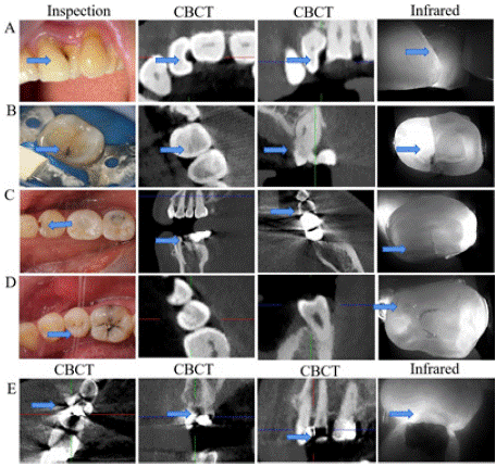
    Figure 1: A. Twisted teeth; B. Cracked teeth; C. Caries adjacent to
prostheses; D. Cavities that have not yet developed into cavities; E. Caries
adjacent to fillings.
Figure 2A shows CBCT images of dental caries at teeth #11 and #21. The patient experienced discomfort from cold and heat stimuli, suggesting secondary caries. Figure 2B, a visual inspection, shows fillings on the palatal side, with margins suspected of secondary caries, and no obvious abnormalities on the labial side. Infrared images of the tooth surfaces in Figures 2D-I and 2D-II appear dark. Figure 2C shows the tooth after filling, and the infrared image in Figure 2D-III shows that the previous dark area has disappeared. Figure 2E compares visual inspection, X-ray, and infrared images of the same anterior tooth position from another case.
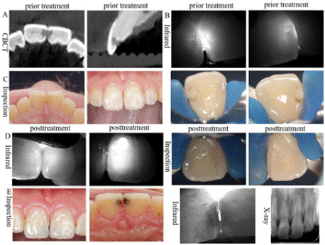
    Figure 2: A. CBCT image; B. Visual infrared before filling; C. before filling;
D. Visual infrared after filling; III.; E. Comparison of visual inspection, X-ray,
and infrared imaging of anterior tooth caries in another case.
Discussion
Dental caries is classified by depth into superficial, moderate, and deep caries. The earlier caries are filled, the lower the probability of filling dropout. Delayed fillings increase the dropout rate. Filling caries early can preserve pulp vitality and prevent root canal treatment, making the detection of early dental caries particularly important. Different auxiliary examination methods vary in effectiveness depending on the type and location of dental caries. In this study, dental caries was investigated according to age and location. Special clinical cases, such as root caries, twisted teeth, secondary caries, and cracked teeth—often challenging to diagnose clinically—were also analyzed. This study aimed to evaluate the detection effectiveness of different examination methods for various types of dental caries.
Table 1 shows that visual inspection and infrared imaging had the highest detection rates, while CBCT had the lowest overall rate. Upon analysis, the higher proportion of secondary caries and caries without cavities in this study population was noted. Researchers involved in early dental caries prevention intentionally selected this population for inclusion. Table 1 further indicates that CBCT achieved a 100% detection rate for primary caries with formed cavities, surpassing infrared and X-ray imaging. Infrared imaging was more effective in detecting secondary caries and caries without cavities.
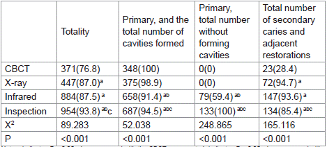
Table 1: Detection rates of dental caries by examination methods.
At age 12, most deciduous teeth have exfoliated, and permanent teeth have erupted, allowing good patient cooperation. Patients were categorized into age groups of ten-year intervals. Table 2 indicates that CBCT demonstrated a consistently high detection rate (100%) for caries across all age groups. X-ray imaging also had high detection rates, especially among older patients. Infrared imaging had lower detection rates compared to CBCT and X-ray, particularly among older patients. Visual inspection showed a relatively high detection rate, especially in younger age groups. Previous studies reported that 85% of elderly subjects had root caries, with a mean root caries index of 36.6±28.5% [10]. It is speculated that the proportion of root caries increases with age, accompanied by interproximal tooth surface wear. This reduces overlap on X-ray images, enhancing detection rates for proximal caries in older patients. Conversely, visual inspection and infrared imaging showed lower detection rates among older patients. It is hypothesized that caries closer to the root surface are less detectable through tooth surfaces, and the penetration ability of infrared imaging through tooth tissues is weaker than that of X-ray and CBCT, reducing its detection rate.

Table 2: Classification by age.
They were classified into incisors, canines, premolars, and molars. Table 3 indicates that CBCT demonstrates a very high detection rate. X-ray also has a high detection rate, with a better detection rate for anterior teeth compared to posterior teeth. Infrared detection rates are lower than those of X-ray and CBCT. Visual examination also has a high detection rate, especially for anterior teeth. Undetected cases were primarily root caries and twisted teeth.

Table 3: Classification by tooth position.
Table 4 categorizes caries according to depth, revealing a 100% detection rate with CBCT. X-ray detection rates are higher for deep caries than for shallow caries. Infrared imaging has lower detection rates than CBCT and X-ray, particularly for shallow caries. Caries was also classified by their location on the tooth surface. Table 5 demonstrates that CBCT detection rates were very high, reaching up to 100%. X-ray showed higher detection rates on non-proximal surfaces compared to proximal surfaces, with particularly high detection for root caries. Previous studies indicate that X-ray, being two-dimensional imaging, is often affected by overlapping adjacent tooth surfaces, leading to decreased clarity or requiring re-imaging [11]. We speculate that overlapping adjacent surfaces accounts for the reduced X-ray detection rates in Table 5. Infrared imaging also shows higher detection rates for non-proximal surfaces than proximal surfaces, with notably reduced detection for root caries. This reduction may explain why infrared detection rates decrease with increasing age, as observed in Table 2. Visual inspection similarly has higher detection rates for non-proximal surfaces, but lower rates for root surface caries compared to the other two methods.

Table 4: Classification by caries depth.

Table 5: Classification by tooth surface location.
CBCT and X-ray utilize radiation with fluorescent, penetrating, and photosensitive effects. Different tissues in the body vary in thickness and density, leading to different degrees of radiation absorption and attenuation [12]. Caries involves loss of organic and inorganic materials, altering radiation absorption at these sites. When caries has not yet formed cavities, probes cannot hold firmly, with only surface color and texture changes occurring. Radiation absorption at these early carious sites differs from sites with cavities. Table 6 indicates that CBCT and X-ray detection rates for caries without cavities are low. This may be because reduced radiation absorption at these locations fails to reach machine resolution thresholds. Nearinfrared technology detects caries based on differing interactions of infrared light with tooth structures. Enamel, dentin, and carious areas differ in their absorption and reflection of infrared rays, producing distinctive reflections from decayed tissues compared to healthy tissues [13-16]. Superficial caries alters infrared reflections. Table 6 demonstrates that infrared imaging is more sensitive than CBCT and X-ray for identifying superficial caries without cavity formation. Figure1D Show the cavities that have not developed into cavities. Infrared images can reveal more areas that require dental cavity prevention measures.
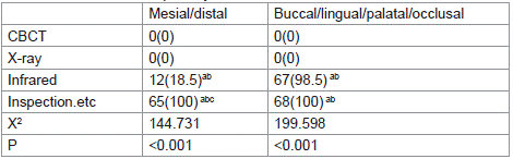
Table 6: Classification of primary unformed caries.
Table 7 indicates that X-ray imaging requires placing film inside the mouth, often causing patient discomfort and pain, leading to patient reluctance. CBCT examinations are costly and involve radiation, further contributing to patient hesitancy. Infrared imaging, conducted chairside, causes minimal discomfort and requires less time, resulting in greater patient preference.
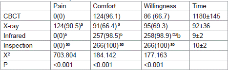
Table 7: Classification by comfort level.
Cracked teeth represent a dental hard-tissue condition that is challenging to diagnose, as cracks often resemble pits and fissures on tooth surfaces. Its symptoms are similar to those of dental caries, which may include tooth sensitivity or even pain. Sometimes, it tends to occur on carious teeth. Therefore, based on clinical needs, we included cracked teeth in the study. The CBCT used in this study scans layers at a thickness of 20 microns, which makes detecting superficial cracks difficult. In clinical practice, visual inspection may thus detect shallow cracks more clearly than CBCT or X-ray. Table 8 indicates that CBCT has a low detection rate for symptomatic cracked teeth and an even lower rate for asymptomatic or minor cracks. X-ray detection of hidden cracks is lower. Infrared imaging demonstrates cracks more clearly than visual inspection and CBCT (Figure 1B).
When adjacent teeth have full crowns or restorations, these materials strongly absorb radiation, resulting in high-density shadows on CBCT images (Figure 1D). Consequently, caries near prostheses often appear unclear on CBCT. Infrared imaging, unaffected by adjacent teeth, clearly displays the condition of dental caries (Figure 1C, D; Table 8). During examination of twisted teeth, visual diagnosis is often impaired by light obstruction, increasing missed diagnoses. CBCT and X-ray maintain high detection rates, whereas infrared imaging detection rates significantly decrease due to tooth torsion (Table 8).

Table 8: Classification of cracked teeth and secondary caries.
Recently, some scholars summarized secondary caries detection methods and found none adequately reliable [17]. CBCT also shows reduced detection rates for secondary caries in isolated teeth compared to primary caries [18]. Table 8 demonstrates that detection rates of secondary caries significantly decrease for both CBCT and X-ray. Visual inspection also has a low detection rate, whereas infrared imaging is the least affected. Figure 2 presents secondary caries filling treatment on proximal surfaces of teeth #11 and #21. The patient experienced cold and heat sensitivity for several weeks, suggesting secondary caries. Figure 2A shows the caries condition on CBCT. In Figure 2C, black lines are visible along the edges of the original filling, with no abnormalities on the labial side. Infrared images in Figures 2B show internal dark areas. Figure 2D shows the tooth after refilling. Figure 2D indicates the dark areas disappeared post-treatment. Figure 2E shows another adjacent caries on teeth #11 and #21, where infrared imaging provided similar results to X-ray, suggesting X-ray as a suitable alternative for detecting caries in this scenario.
Summary
CBCT achieves the highest detection rate for various types of dental caries when not obstructed by high-density shadows from surrounding restorations or fillings. CBCT also effectively detects twisted teeth and root caries. X-ray ranks second but shows reduced effectiveness for caries with overlapping adjacent surfaces. Compared to CBCT and X-ray, infrared imaging and visual inspection show higher detection rates for caries near restorations or fillings obscured by high-density images. Infrared imaging has superior detection rates for cracked teeth and early caries without cavities. Particularly for early-stage caries without cavities, infrared imaging outperforms CBCT, X-ray, and visual inspection.
Authors’ Contribution
All authors read and approved the final manuscript. Availability of data and materials. All data generated or analysed during this study are included in this published article. Declaration of conflicting interests. The author(s) declared no potential conflicts of interest with respect to the research, authorship, and/or publication of this article.
Ethical Approval, Consent to Participate and Publication
The submission reported data collected from animals and human, and all studies were conducted according to the regulations for animal experimentation issued by the State Committee of Science and Technology of the People’s Republic of China. For any patient under the age of 18, a parent or legal guardian provided consent. Ethical approval for this study was obtained from Medical Ethics Committee of Zhangshan Hospital of Xiamen University (No xmzsyyky-2025-579). The submission has reported data collected from animals and humans, and all studies were conducted according to the regulations for animal experimentation issued by the State Committee of Science and Technology of the People’s Republic of China.
Funding
The author(s) disclosed receipt of the following financial support for the research, authorship, and/or publication of this article: This study was supported by Fujian Provincial Natural Science Foundation of China (2022j011347) and Xiamen Science and Technology Program Project(3502Z202374012).
Fujian Key Laboratory of Oral Diseases, School and Hospital of Stomatology, Fujian Medical University (2021kq002).
Data Sharing Statement
All data generated or analyzed during this study are included in this published article.
References
- Moharrami M, Farmer J, Singhal S, Watson E, Glogauer M, Johnson AEW, et al. Detecting dental caries on oral photographs using artificial intelligence: A systematic review. Oral diseases. 2024; 30: 1765-1783.
- Mohammad-Rahimi H, Motamedian SR, Rohban MH, Krois J, Uribe SE, Mahmoudinia E, et al. Deep learning for caries detection: A systematic review. Journal of dentistry. 2022; 122: 104-115.
- Mohamed S, Farha F, Alzubaidi M, Househ M. Teledentistry and Dental Caries Detection - A Scoping Review. Studies in health technology and informatics. 2025; 323: 384-388.
- Ku JC, Mao K, Wang F, Carreiro A, Lam WY, Yu OY. Accuracy of clinical photography for the detection of dental caries: A systematic review and metaanalysis. Journal of dentistry. 2025;157: 105737.
- Cheng JG, Zhang ZL, Wang XY, Zhang ZY, Ma XC, Li G. Detection accuracy of proximal caries by phosphor plate and cone-beam computerized tomography images scanned with different resolutions. Clinical oral investigations. 2012; 16: 1015-1021.
- Zhang ZL, Li JP, Li G, Ma XC. Accuracy of computer aided measurement for detecting dental proximal caries lesions in images of cone-beam computed tomography. Chinese journal of stomatology. 2017; 52: 103-108.
- Zanini LGK, Rubira-Bullen IRF, Nunes F. A Systematic Review on Caries Detection, Classification, and Segmentation from X-Ray Images: Methods, Datasets, Evaluation, and Open Opportunities. Journal of imaging informatics in medicine. 2024; 37: 1824-1845.
- Demir K, Sokmen O, Karabey Aksakalli I, Torenek-Agirman K. Comprehensive Insights into Artificial Intelligence for Dental Lesion Detection: A Systematic Review. Diagnostics (Basel, Switzerland). 2024; 14.
- Lin WS, Alfaraj A, Lippert F, Yang CC. Performance of the caries diagnosis feature of intraoral scanners and near-infrared imaging technology-A narrative review. Journal of prosthodontics: official journal of the American College of Prosthodontists. 2023; 32: 114-124.
- Lundgren M, Emilson CG, Osterberg T. Caries prevalence and salivary and microbial conditions in 88-year-old Swedish dentate people. Acta odontologica Scandinavica. 1996; 54: 193-209.
- Mauriello SM, Broome AM, Platin E, Mol A, Inscoe C, Lu J, et al. The role of stationary intraoral tomosynthesis in reducing proximal overlap in bitewing radiography. Dento maxillo facial radiology. 2020; 49: 20190504.
- Chan EK, Wah YY, Lam WY, Chu CH, Yu OY. Use of Digital Diagnostic Aids for Initial Caries Detection: A Review. Dentistry journal. 2023; 11.
- Fried D, Glena RE, Featherstone JD, Seka W. Nature of light scattering in dental enamel and dentin at visible and near-infrared wavelengths. Applied optics. 1995; 34: 1278-1285.
- Jones R, Huynh G, Jones G, Fried D. Near-infrared transillumination at 1310- nm for the imaging of early dental decay. Optics express. 2003; 11: 2259- 2265.
- Marinova-Takorova M, Panov V, Anastasova R. Effectiveness of near-infrared transillumination in early caries diagnosis. Biotechnology & Biotechnological Equipment. 2016; 30:1207-1211.
- Baltacioglu IH, Orhan K. Comparison of diagnostic methods for early interproximal caries detection with near-infrared light transillumination: an in vivo study. BMC oral health. 2017; 17: 130.
- Ku JC, Lam WY, Li KY, Hsung RT, Chu CH, Yu OY. Accuracy of detection methods for secondary caries around direct restorations: A systematic review and meta-analysis. Journal of dentistry. 2025; 153: 105541.
- Shahidi S, Zadeh NK, Sharafeddin F, Shahab S, Bahrampour E, Hamedani S. An in vitro comparison of diagnostic accuracy of cone beam computed tomography and phosphor storage plate to detect simulated occlusal secondary caries under amalgam restoration. Dental research journal. 2015; 12: 161-166