
Case Report
J Dent & Oral Disord. 2016;2(3): 1014.
Surgical Management of Endo-Perio Lesion with Demineralised Bone Matrix in Combination of Platelet Rich Fibrin: A Case Report
Kale P¹, Talesara K², Patil KP² and Nayyar AS³*
¹Department of Conservative Dentistry & Endodontics, Dr. D.Y. Patil Dental College & Hospital, India
²Department of Periodontics and Oral Implantology, Dr. D.Y. Patil Dental School, India
³Department of Oral Medicine and Radiology, Saraswati- Dhanwantari Dental College and Hospital and Post- Graduate Research Institute, India
*Corresponding author: Nayyar AS, Department of Oral Medicine and Radiology, Saraswati-Dhanwantari Dental College and Hospital and Post-Graduate Research Institute, India
Received: February 17, 2016; Accepted: May 02, 2016; Published: May 03, 2016
Abstract
Periodontal disease is an inflammatory disease leading to periodontal attachment loss and bone destruction. The objective of periodontal therapy is to regenerate the lost periodontal supporting tissues. Platelets are rich in growth factors that may contribute to an accelerated process of tissue regeneration. Demineralised Bone Matrix (DMBM) Xenograft is a bone inductive sterile bioresorbable Xenograft composed of Type I collagen. Given the unique properties of autologous Platelet Rich Fibrin (PRF) and already demonstrated regenerative capacity of commercially available bone grafts, application of a combination approach was attempted. We present here a case report with an eight month follow-up report of a combined endo-perio lesion, which was treated by means of combination of autologous PRF with bovine derived xenograft, and assessed clinically and radiographically.
Keywords: Periodontal disease; Periodontal therapy; Demineralised bone matrix (DMBM); Xenograft; Platelet rich fibrin (PRF); Bone grafts
Introduction
Periodontal disease is an inflammatory disease leading to periodontal attachment loss and bone destruction. The objective of periodontal therapy is to regenerate the lost periodontal supporting tissues. However, periodontal regeneration requires a sequence of biological events including cell adhesion, migration, proliferation and differentiation [1]. A combination of growth factors may more effectively stimulate formation of mineralized as well as non mineralized tissues [2]. Platelets are rich in growth factors that may contribute to an accelerated process of tissue regeneration. During the early stages of wound healing, platelets released growth factors, including platelet derived growth factor, insulin like growth factor-1 and transforming growth factor-β, initiate a cascade of cellular and molecular events that result in wound healing in a highly regulated and coordinated manner [2]. The application of these growth factors to bone and periodontal regeneration has been investigated using numerous in-vitro and in-vivo models with promising results [3- 6]. Xenografts used in the treatment of infrabony defects can be both bovine bone and natural coral, these are also referred to as anorganic bone, since they are suggested to remove all cells and proteinaceous material leaving behind an inert absorbable bone scaffolding upon which revascularization, osteoblasts migration, and woven bone formation supposedly occurs [7]. Demineralised Bone Matrix (DMBM) Xenograft is a bone inductive sterile bioresorbable Xenograft composed of Type I collagen. It is prepared from bovine cortical samples, resulting in non-immunogenic flowable particles of approximately 250 μm dimensions that are completely replaced by host bone in 4-24 weeks [8]. Platelet-Rich fibrin (PRF) described by Choukroun et al. is a second-generation platelet concentrate which allows one to obtain fibrin membranes enriched with platelets and growth factors, after starting from an anticoagulant-free blood harvest without any artificial biochemical modification [9]. The PRF clot forms a strong natural fibrin matrix, which concentrates almost all the platelets and growth factors of the blood harvest and shows a complex architecture as a healing matrix, including mechanical properties no other platelet concentrate offers [10-11]. It is an autologous biomaterial which has found numerous clinical applications which have been described in the specializations of oral maxillofacial surgery [12-14] and in dental implantology [15- 16]. Given the unique properties of autologous PRF and already demonstrated regenerative capacity of commercially available bone grafts, application of a combination approach was attempted. We present here a case report with a 12 month follow-up report of a combined endo-perio lesion, which was treated by means of combination of autologous PRF with bovine derived xenograft, and assessed clinically and radiographically.
Case Presentation
A 35 year old patient reported to the department with a chief complaint of swelling in the upper front tooth region since 2 months. The patient had reported trauma in the same region 7 years back. The nature of the pain was intermittent in nature. The patient used to take analgesics to relieve the pain. The past medical history of the patient did not reveal any significant finding and the patient was systemically healthy and was not on any medication. Patient revealed no history of smoking and alcohol and other deleterious habits. On examination, there was a swelling of about 3cm x 3cm with Ellis class IV fracture in relation to 11. The tooth appeared to be discolored (Figure 1). The pulp vitality tests revealed that the tooth was nonvital while the radiographic examination of the concerned area in the form of an Intra-Oral Peri-Apical Radiograph (IOPAR) revealed that it had a large peri-apical radiolucency (Figure 2). Periodontal probing revealed a deep periodontal pocket measuring about 15mm in depth. (Figure 3). The provisional diagnosis arrived-at was a combined endo-perio lesion in relation to 11 region.
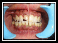
Figure 1: Showing discolored, non-vital 11 with Ellis class IV fracture.
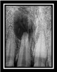
Figure 2: Showing IOPAR revealing a large peri-apical radiolucency.
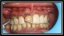
    Figure 3: Showing probing depth of around 15mm as measured with the help
of a periodontal probe.
Endodontic therapy
At the initial visit, immediate emergency access opening was done and the pus was drained-out. The access was kept open and only a thick cotton pledget was used to close the access for a day. Next day, the patient was recalled and the cotton pledget was removed and the canal was re-irrigated with saline. The swelling had reduced. Patient was recalled for irrigation and dressing after 4 days till which time, the swelling had completely resolved. Working length and biomechanical preparation were completed with the tooth (Figure 4). Calcium hydroxide dressing was given for 7 days and the access was closed with a temporary restoration (Figure 5). A dressing was given again and the patient was recalled after 7 days for the follow-up and then, the patient was transferred to Department of Periodontics for opinion and needful. A thorough oral prophylaxis was done in the concerned region (Figure 6) and the patient was recalled for completion of endodontic treatment (Figure 7).
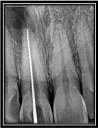
Figure 4: Showing working length determination as seen on IOPAR.
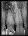
    Figure 5: Showing calcium hydroxide dressing with access opening closed
with a temporary restoration as seen on IOPAR.
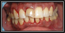
Figure 6: Showing oral prophylaxis done in relation to maxillary arch.
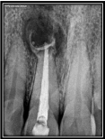
Figure 7: Showing completed endodontic treatment in relation to 11.
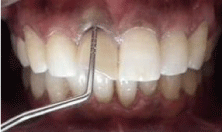
    Figure 8: Showing pre-surgical photographs with periodontal pocket depth
reduced to 7mm.
Periodontal therapy
A periodontal surgery was planned after 1 month of the completion of the endodontic treatment. Routine blood investigations were advised for which the reports were found to be normal. Presurgical periodontal pocket depth was reduced to 7mm (Figure 8). After proper isolation of the surgical field, the operative site was anaesthetized using 2% xylocaine hydrochloride with adrenaline (1:200000). A crevicular incision was given covering 12, 11, 21 region and a full thickness mucoperiosteal flap was raised (Figure 9). The infected, necrosed bone was removed and a continuous apicomarginal defect was observed along with buccal wall with dehiscence. 3mm of root was resected for apicoectomy and 3mm gutta percha was removed from the apex with the help of a heated probe (Figure 10).
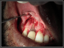
    Figure 9: Showing crevicular incision given in relation to 12, 11, 21 region
with raised full thickness mucoperiosteal flap.
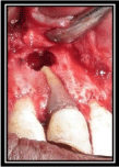
    Figure 10: Showing removed infected, necrosed bone with a continuous
apico-marginal defect being seen along with buccal wall with dehiscence and
3mm of root resected for apicoectomy with 3mm gutta percha removed from
the apex with the help of a heated probe.
PRF preparation
Blood sample of the patient was drawn in 10mL test tubes without an anti-coagulant and centrifuged immediately. Blood was centrifuged using a tabletop centrifuge (REMY Laboratories, Chennai, Tamilnadu, India) for 12 min at 3000 rpm. The resultant product consisted of the following three layers (Figure 11).
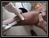
    Figure 11: Showing blood sample of the patient being drawn in 10mL test
tubes without an anti-coagulant for centrifugation.
• The upper layer of acellular PPP (Platelet-Poor Plasma);
• PRF clot in the middle; and
• Red blood cells at the bottom.
Platelet rich fibrin was then separated by cutting with a sterile surgical scissor and collected using a dappen dish. (Figure 12). PRF clot was placed inside the periapical defect (Figure 13). A Demineralised Bone Matrix (DBM) xenograft was then placed over the peri-apical defect and also, the entire root length devoid of bone (Figure 14). A Collagen membrane was finally placed over the defect. The Collagen membrane was cut according to requirement and then placed over the defect using 4-0 vicryl resorbable suture (Figure 15). The flap was sutured with 3-0 black braided silk (Figure 16). A perio pack was then mixed and placed over the sutured area so that the repositioned flap be immobile till healing ensued (Figure 17). Immediate Post-Operative Radiograph (IOPAR) was advised (Figure 18). Antibiotics and analgesics were prescribed for one week. After 15 days, the pack was removed. The sutures were removed and the healing was found to be uneventful. The crown preparation with 11 was done (Figure 19) and elastomeric impression was recorded (Figure 20). All Ceramic Emax crown was fabricated and cemented (Figure 21). The patient was asked for regular follow-up visits and the 12 months post-operative radiograph was taken which revealed apparent bone fill with resolution of the osseous defect (Figure 22). Clinical evaluation of the periodontal pocket depth revealed a successful outcome of the procedure with a considerable reduction in pocket depth that got reduced from 15mm to 3mm depth (Figure 23).
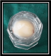
Figure 12: Showing PRF clot in the dappen dish.
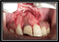
Figure 13: Showing PRF clot placed inside the peri-apical defect.
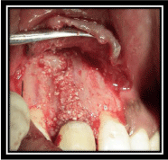
    Figure 14: Showing Demineralised Bone Matrix (DBM) xenograft placed over
the peri-apical defect and the entire root length devoid of bone.
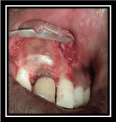
Figure 15: Showing collagen membrane placed over the defect.
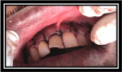
Figure 16: Showing sutured flap with 3-0 black braided silk.
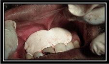
    Figure 17: Showing perio pack over the sutured area to make the repositioned
flap immobile till it heals.
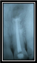
Figure 18: Showing immediate post-operative radiograph.
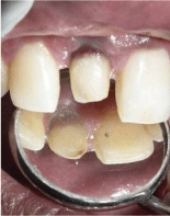
Figure 19: Showing crown preparation in relation to 11.
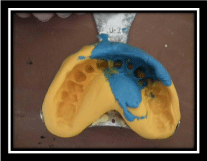
Figure 20: Showing elastomeric impression registered in relation to 11.
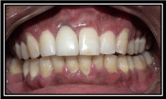
Figure 21: Showing all Ceramic Emax crown cemented in relation to 11.
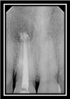
    Figure 22: Showing 12 months’ post-operative radiograph showing apparent
bone fill with resolution of the osseous defect.
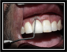
Figure 23: Showing pocket depth reduced from 15mm to 3mm.
Discussion
The treatment of combined endo-perio lesions requires both endodontic therapy and periodontal regenerative procedures, as discussed in the above case report. The goal of peri-radicular surgery is to remove all necrotic tissues from the surgical site to completely seal the entire root canal system, and to facilitate the regeneration of hard and soft tissues including the formation of a new attachment apparatus [17]. It is interesting to note that there was no radiographic or clinical evidence of a pre-existing deep decay in the tooth, and no cracks were evident. The most common clinical/ radiographic features of these endo-perio lesions reported were the periapical radiolucency and deep pocket depths with a nonvital pulp status. Traditional approaches to treat endo-perio defects include non-surgical debridement of root surfaces or root canals, as well as surgical approaches that provide better access to clean the root surfaces and apical lesions and to reshape the surrounding bone/root apex. Bone loss caused by pulpal disease is reversible, whereas advanced bone loss caused by periodontal disease is usually irreversible [18]. The necessity of periodontal surgical therapy most likely was because the periodontal bone loss was more advanced and was less likely to resolve after non surgical root canal therapy alone [19]. Generally, partial apical root resection has been suggested for all endodontic surgeries advised for extensive involvements [20]. In this case report, we had planned a regeneration therapy for bone as well as the periodontal ligament as it would lead to a better prognosis. Hence, here we performed root canal debridement with subsequent retrograde filling and removal of granulation tissue around the root and apex. Apicoectomy was done to prevent recurrence of infection as the maximum number of canal variations and complexities are seen in the apical third [21]. PRF i.e. Platelet Rich Fibrin, is in the form of a platelet gel and can be used in conjunction with bone grafts, which offers several advantages including promoting wound healing, bone growth and maturation, graft stabilization, wound sealing and hemostasis, and improving the handling properties of graft materials. It is saturated with growth factors which expediates the regenerative process during healing [12]. Also here, using the Guided Tissue Regeneration (GTR) membrane technique, combined with bone graft, the result was clinically successful after a 1-year follow-up period. The role of bone graft in the above case was for space-making and also for inducing bone formation and attachment gain. The rationale for using GTR barrier membranes in above case with bone grafting materials is to encourage the growth of key surrounding tissues, while excluding unwanted cell types such as epithelial cells [22]. GTR therapy has been implemented in the endodontic surgeries as a concomitant treatment during the management of the combined endo-perio lesions. However, from clinical and radiographic findings, the result of this combined technique was quite impressive, resulting in a significant reduction of probing depth and bone fill. Although traditional non-surgical periodontal therapy and regular endodontic therapy can be predictably used to arrest mild to moderate periodontal bone defects in combined endo-perio lesions, it might be inadequate for the treatment of disease characterized by deep pockets or apicomarginal defects. Currently, regenerative techniques are widely available in terms of their predictability to regenerate the lost tissue/ bone in all types of defects or for all situations.
Conclusion
A careful pre-operative diagnosis, appropriate case selection and knowledge of the factors that can negatively affect regeneration outcomes can help to optimize successful regenerative attempts. Treatment strategies used in this case report suggests that combined endo-perio lesions can be successfully managed with multiple regenerative procedures.
References
- [No authors listed]. The potential role of growth and differentiation factors in periodontal regeneration. J Periodontol. 1996; 67: 545-553.
- Lynch SE. The role of growth factors in periodontal repair and regeneration. Periodontal Regeneration: Current status and directions. 1994: 179-198.
- Pierce GF, Mustoe TA, Altrock BW, Deuel TF, Thomason A. Role of platelet-derived growth factor in wound healing. J Cell Biochem. 1991; 45: 319-326.
- Kaigler D, Avila G, Wisner-Lynch L, Nevins ML, Nevins M, Rasperini G, et al. Platelet-derived growth factor applications in periodontal and peri-implant bone regeneration. Expert Opin Biol Ther. 2011; 11: 375-385.
- Hollinger JO, Hart CE, Hirsch SN, Lynch S, Friedlaender GE. Recombinant human platelet-derived growth factor: biology and clinical applications. J Bone Joint Surg Am. 2008; 90: 48-54.
- Kao DW, Fiorellini JP. Regenerative periodontal therapy. Front Oral Biol. 2012; 15: 149-159.
- Sowmya NK, Kumar ABT, Mehta DS. Clinical evaluation of regenerative potential of type I collagen membrane along with Xenographic bone graft in the treatment of periodontal intra-bony defects assessed with surgical re-entry and radiographic linear and densitometric analysis. J Indian Soc Periodontal. 2009; 14: 23-29.
- Blumenthal N, Sabe T, Barrington E. Healing responses to grafting of combined collagen-decalcified bone in periodontal defects in dogs. J Periodontol. 1986; 57: 84-90.
- Choukroun J, Adda F, Schoeffer C, Vervelle A. PRF: an opportunity in perio implantology. Implantodontie 2000; 42: 55-62.
- Dohan DM, Choukroun J, Diss A, Dohan SL, Dohan AJ, Mouhyi J, et al. Platelet-rich fibrin (PRF): A second-generation platelet concentrate. Part II: Platelet-related biologic features. Oral Surg Oral Med Oral Pathol Oral Radiol Endod. 2006; 101: 45-50.
- Dohan DM, Choukroun J, Diss A, Dohan SL, Dohan AJ, Mouhyi J, et al. Platelet-rich fibrin (PRF): A second-generation platelet concentrate. Part III: Leucocyte activation: A new feature for platelet concentrates?. Oral Surg Oral Med Oral Pathol Oral Radiol Endod. 2006; 101: 51-55.
- Choukroun J, Diss A, Simonpieri A, Girard MO, Schoeffler C, Dohan SL, et al. Plateletrich fibrin (PRF): A second-generation platelet concentrate. Part IV: Clinical effects on tissue healing. Oral Surg Oral Med Oral Pathol Oral Radiol Endod. 2006; 101: 56-60.
- Choukroun J, Diss A, Simonpieri A, Girard MO, Schoeffler C, Dohan SL, et al. Plateletrich fibrin (PRF): A second-generation platelet concentrate. Part V: Histologic evaluations of PRF effects on bone allograft maturation in sinus lift. Oral Surg Oral Med Oral Pathol Oral Radiol Endod. 2006; 101: 299-303.
- Zhao JH, Tsai CH, Chang YC. Clinical and histologic evaluations of healing in an extraction socket filled with platelet-rich fibrin: A case report. J Dent Sci. 2011; 6: 116-122.
- Diss A, Dohan DM, Mouhyi J, Mahler P. Osteotome sinus floor elevation using Choukrouna platelet-rich fibrin as grafting material: A 1-year prospective pilot study with micro-threaded implants. Oral Surg Oral Med Oral Pathol Oral Radiol Endod. 2008; 105: 572-579.
- Mazor Z, Horowitz RA, Del Corso M, Prasad HS, Rohrer MD, Dohan Ehrenfest DM. Sinus floor augmentation with simultaneous implant placement using Choukrouna PRF (Platelet-Rich Fibrin) as sole grafting material: A radiological and histological study at 6 months. J Periodontol. 2009; 80: 2056-2064.
- Karabucak B, Setzer FC. Conventional and surgical retreatment of complex periradicular lesions with periodontal involvement. J Endod. 2009; 35: 1310-1315.
- Law AS, Beaumont RH. Resolution of furcation bone loss associated with vital pulp tissue after non-surgical root canal treatment of three rooted mandibular molars: A case report of identical twins. J Endod. 2004; 30: 444-447.
- Meng HX. Periodontic-endodontic lesions. Ann Periodontol. 1999; 4: 84-90.
- Bashutski JD, Wang HL. Periodontal and endodontic regeneration. J Endod. 2009; 35: 321-328.
- Siqueira JF Jr, Araújo MC, Garcia PF, Fraga RC, Dantas CJ. Histological evaluation of the effectiveness of five instrumentation techniques for cleaning the apical third of root canals. J Endod. 1997; 23: 499-502.
- Oh SL, Fouad AF, Park SH. Treatment strategy for guided tissue regeneration in combined endodontic-periodontal lesions: case report and review. J Endod. 2009; 35: 1331-1336.